. 2015 Aug;58(8):1877-86.
doi: 10.1007/s00125-015-3627-3. Epub 2015 May 30.

Insulin regulates Rab3-Noc2 complex dissociation to promote GLUT4 translocation in rat adipocytes

Affiliations

Insulin regulates Rab3-Noc2 complex dissociation to promote GLUT4 translocation in rat adipocytes

Francoise Koumanov et al. Diabetologia. 2015 Aug.

Abstract

Aims/hypothesis: The glucose transporter GLUT4 is present mainly in insulin-responsive tissues of fat, heart and skeletal muscle and is translocated from intracellular membrane compartments to the plasma membrane (PM) upon insulin stimulation. The transit of GLUT4 to the PM is known to be dependent on a series of Rab proteins. However, the extent to which the activity of these Rabs is regulated by the action of insulin action is still unknown. We sought to identify insulin-activated Rab proteins and Rab effectors that facilitate GLUT4 translocation.

Methods: We developed a new photoaffinity reagent (Bio-ATB-GTP) that allows GTP-binding proteomes to be explored. Using this approach we screened for insulin-responsive GTP loading of Rabs in primary rat adipocytes.

Results: We identified Rab3B as a new candidate insulin-stimulated G-protein in adipocytes. Using constitutively active and dominant negative mutants and Rab3 knockdown we provide evidence that Rab3 isoforms are key regulators of GLUT4 translocation in adipocytes. Insulin-stimulated Rab3 GTP binding is associated with disruption of the interaction between Rab3 and its negative effector Noc2. Disruption of the Rab3-Noc2 complex leads to displacement of Noc2 from the PM. This relieves the inhibitory effect of Noc2, facilitating GLUT4 translocation.

Conclusions/interpretation: The discovery of the involvement of Rab3 and Noc2 in an insulin-regulated step in GLUT4 translocation suggests that the control of this translocation process is unexpectedly similar to regulated secretion and particularly pancreatic insulin-vesicle release.


Figures
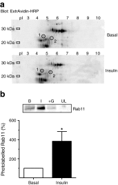
Fig. 1
Fig. 1
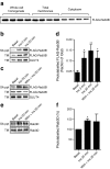
Identification of Rab3B as an insulin-sensitive GTPase in rat adipocytes. (a) Two-dimensional gel analysis of Bio-ATB-GTP-labelled total membranes. Biotinylated proteins were blotted with ExtrAvidin–HRP (Sigma, St. Louis, MO, USA). Circled spots 1 (Rab3B) and 2 (Rab11) indicate proteins for which the biotin signal was identified as increasing upon insulin stimulation. (b) Bio-ATB-GTP photolabelling of Rab11 in total-membrane preparations from rat adipocytes. Biotinylated proteins were precipitated with streptavidin and immunoblotted with antibodies against Rab11. Data are means ± SEM from at least three independent experiments. *p < 0.05 vs basal. B, basal; I, insulin; +G, excess GTP; UL, unlabelled (without Bio-ATB-GTP)
Fig. 2
Fig. 2
FLAG-Rab3B and endogenous Rab3D are activated upon insulin stimulation. (a) Distribution of expressed FLAG-Rab3B between the total-membrane and the cytoplasmic fractions of rat adipocytes. (b) Insulin stimulation (Ins) of the Bio-ATB-GTP loading state of FLAG-Rab3B. Streptavidin-precipitated proteins were detected with anti-FLAG antibody. (c) Effect of wortmannin (200 nmol/l for 10 min) (Wtm) on insulin-stimulated FLAG-Rab3B GTP loading. (d) Quantification of the data presented in (b) and (c). Data are means ± SEM from three independent experiments. *p < 0.05 vs basal. (e) Effect of insulin stimulation on endogenous Rab3D GTP loading. Total-membrane preparations (300 μg/condition) were labelled with Bio-ATB-GTP and streptavidin-precipitated proteins immunoblotted with anti-Rab3D antibody. (f) Quantification of the data shown in (e). Data are means ± SEM from three independent experiments. *p < 0.05 vs basal. SA ppt, streptavidin precipitation; TM, total-membrane loading control
Fig. 3
Fig. 3
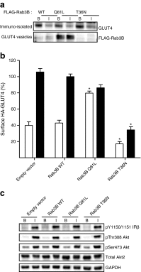
Rab3B modulates GLUT4 vesicle translocation. (a) Isolated GLUT4 vesicles from rat adipocytes expressing FLAG-Rab3B-WT, FLAG-Rab3B-Q81L or FLAG-Rab3B-T36N cDNA were separated by SDS-PAGE, and immunoblotted with anti-GLUT4 or with anti-FLAG antibodies. A representative immunoblot from three independent experiments is shown. B, basal; I, insulin. (b) Effect of constitutively active Rab3B-Q81L on HA-GLUT4 present at the cell surface. Rat adipocytes were co-transfected with HA-Glut4 cDNA in combination with pcDNA3.1 empty vector, Rab3B-WT, Rab3B-Q81L or Rab3B-T36N cDNA. Data are means ± SEM from four independent experiments. *p < 0.05 vs empty vector control. White bars, basal; black bars, insulin. (c) Insulin signalling in rat adipocytes electroporated with Rab3B constructs. Representative immunoblots from two experiments are shown of phosphorylated-state specific antibodies against insulin receptor β subunit (pY1150/1151 IRβ) and Akt (pThr308 Akt and pSer473 Akt) in basal (B) and insulin-stimulated (I) cells
Fig. 4
Fig. 4
Rab3 silencing in 3T3-L1 adipocytes decreases glucose uptake and GLUT4 translocation. (a) Immunoblot of Rab3A and Rab3D siRNA silencing and insulin-signalling components (as described, Fig. 3c) in 3T3-L1 adipocytes. 3T3-L1 adipocytes (day 3 of differentiation) were transfected with siRNA against Rab3A, Rab3B and Rab3D (separate or combined). Representative immunoblots from three experiments are shown. Control cells were transfected with equal amounts of non-targeting siRNA. (b) Effect of siRNA silencing of Rab3 isoforms on insulin-stimulated 2-deoxy-d-glucose (2-DG) uptake. Data are means ± SEM from three independent experiments. *p < 0.05 vs non-targeting siRNA. White bars, basal; black bars, insulin. (c) Effect of siRNA silencing of Rab3 isoforms on GLUT4 translocation to the cell surface. 3T3-L1 adipocytes (basal or 100 nmol/l insulin for 30 min) were labelled with Bio-ATB-BGPA. Solubilised and streptavidin-precipitated proteins were immunoblotted for GLUT4. SA ppt, streptavidin precipitation, B, basal; I, insulin, UL, no photolabel. (d) Quantification of the data shown in (c). Data are means ± SEM from three independent experiments. *p < 0.05 vs control. White bars, basal; black bars, insulin
Fig. 5
Fig. 5
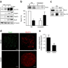
Noc2 is an insulin-responsive Rab3 effector that localises to PMs in rat adipocytes. (a) 3T3-L1 fibroblasts and differentiating adipocytes were lysed and immunoblotted for Rab3A, Rab3D, Noc2, GLUT4 and β-tubulin. Representative immunoblots from three independent experiments. (b) Subcellular distribution of Noc2 in rat adipocytes. Data are means ± SEM from five independent experiments. *p < 0.05 vs basal. B, basal; I, insulin. White bars, basal; black bars, insulin. (c) Effect of wortmannin (W) on insulin-stimulated Noc2 dissociation from PMs. Representative immunoblots from two experiments. (d) Noc2 and GLUT4 distribution at the cell surface of rat adipocytes. Confocal images are from single adipose cells representative of the cell populations from at least three separate experiments. Scale bar, 10 μm. (e) Comparison of Noc2 immunofluorescent intensity at the cell surface of rat adipocytes. Data are mean intensity/area ± SEM from three independent experiments. *p < 0.05 vs basal. B, basal; I, insulin
Fig. 6
Fig. 6
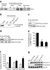
Noc2 is a negative regulator of Rab3B and GLUT4 translocation. (a) FLAG-Rab3BT36N and FLAG-Rab3BQ81L recombinant proteins were immobilised on anti-FLAG-antibody–agarose beads and incubated with cell lysate from basal (B) or insulin-stimulated (I) rat adipocytes. Interacting proteins were eluted and immunoblotted for Noc2. A representative immunoblot from three independent experiments is shown. (b) FLAG-Rab3B was expressed in rat adipocytes and loaded with GTPγS and then binding to WT Noc2 (GST-Noc2WT) and the Rab-domain-deficient mutant (GST-Noc2AAA) was examined. A representative immunoblot from three independent experiments is shown. (c) Pull-down of FLAG-Rab3B from lysates of basal and insulin-stimulated cells with GST-Noc2WT bound to glutathione beads. Bound FLAG-Rab3B was detected by immunoblotting with anti-FLAG antibody (IB). Representative immunoblots from two independent experiments are shown. (d) Identical experiment as in (c) performed with MBP-Noc2. W + I, 200 nmol/l wortmannin before insulin stimulation. (e) Quantification of the data in (d). Data are means ± SEM from three independent experiments. *p < 0.05 vs basal. (f) Effect of overexpression of WT Noc2 on GLUT4 translocation. WT Noc2 (FLAG-Noc2 WT) and the Rab3-binding-deficient mutant (FLAG-Noc2 AAA) were co-transfected with HA-Glut4 cDNA in rat adipocytes and the surface HA signal was monitored. Data are means ± SEM from four independent experiments. *p < 0.05 vs HA-Glut4-only control. White bars, basal; black bars, insulin. (g) Immunoblots of expression of HA-GLUT4, FLAG-Noc2WT and FLAG-Noc2AAA used in the experiments in (f). NT, non-transfected cells; B, basal; I, insulin
Fig. 7
Fig. 7
Rab3 modulation of the final stages of insulin-regulated GLUT4 vesicle exocytosis. In the basal state AS160 is active and maintains Rab10 in an inactive state. Noc2 is bound to PMs and maintains Rab3B in its GDP/inactive state (1). Upon insulin stimulation AS160 is phosphorylated, Rab10 is activated and interacts with MyoVA to facilitate the movement of GLUT4 vesicles to the PM. Insulin stimulation leads to Rab3B GTP loading and an associated Noc2 release from the PM (2). This allows Rab3B to interact with GLUT4 vesicles, which then dissociate from Rab10–MyoVA-associated actin and engage with SNAREs (3)

Similar articles

Cited by

References

    1. Stenmark H. Rab GTPases as coordinators of vesicle traffic. Nat Rev Mol Cell Biol. 2009;10:513–525. doi: 10.1038/nrm2728. - DOI - PubMed
    1. Zerial M, McBride H. Rab proteins as membrane organizers. Nat Rev Mol Cell Biol. 2001;2:107–117. doi: 10.1038/35052055. - DOI - PubMed
    1. Pfeffer SR. Rab GTPase regulation of membrane identity. Curr Opin Cell Biol. 2013;25:414–419. doi: 10.1016/j.ceb.2013.04.002. - DOI - PMC - PubMed
    1. Chen Y, Wang Y, Zhang J, et al. Rab10 and myosin-Va mediate insulin-stimulated GLUT4 storage vesicle translocation in adipocytes. J Cell Biol. 2012;198:545–560. doi: 10.1083/jcb.201111091. - DOI - PMC - PubMed
    1. Chadt A, Immisch A, de Wendt C, et al. Deletion of both Rab-GTPase-activating proteins TBC1D1 and TBC1D4 in mice eliminates insulin- and AICAR-stimulated glucose transport. Diabetes. 2015;64:746–759. doi: 10.2337/db14-0368. - DOI - PubMed

Publication types

LinkOut - more resources