. 2016 Jan;23(1):76-88.
doi: 10.1038/cdd.2015.70. Epub 2015 May 29.

Characterization of RIPK3-mediated phosphorylation of the activation loop of MLKL during necroptosis

Affiliations

Characterization of RIPK3-mediated phosphorylation of the activation loop of MLKL during necroptosis

D A Rodriguez et al. Cell Death Differ. 2016 Jan.

Abstract

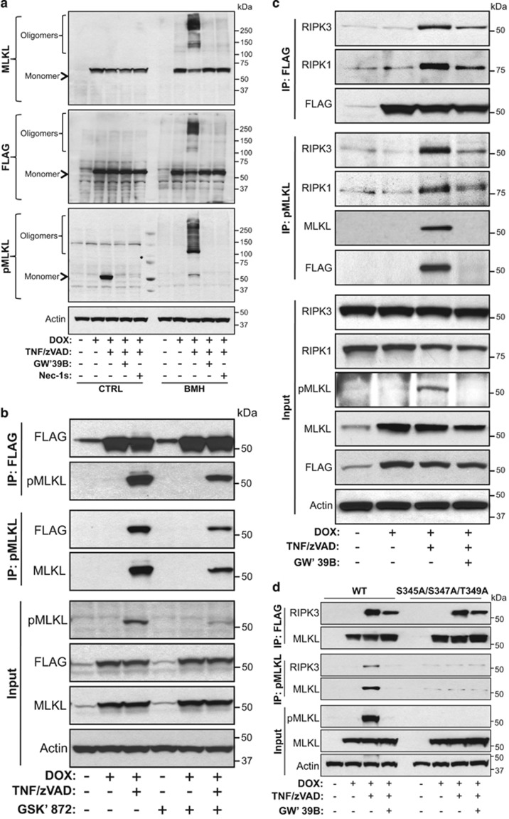
Mixed lineage kinase domain-like pseudokinase (MLKL) mediates necroptosis by translocating to the plasma membrane and inducing its rupture. The activation of MLKL occurs in a multimolecular complex (the 'necrosome'), which is comprised of MLKL, receptor-interacting serine/threonine kinase (RIPK)-3 (RIPK3) and, in some cases, RIPK1. Within this complex, RIPK3 phosphorylates the activation loop of MLKL, promoting conformational changes and allowing the formation of MLKL oligomers, which migrate to the plasma membrane. Previous studies suggested that RIPK3 could phosphorylate the murine MLKL activation loop at Ser345, Ser347 and Thr349. Moreover, substitution of the Ser345 for an aspartic acid creates a constitutively active MLKL, independent of RIPK3 function. Here we examine the role of each of these residues and found that the phosphorylation of Ser345 is critical for RIPK3-mediated necroptosis, Ser347 has a minor accessory role and Thr349 seems to be irrelevant. We generated a specific monoclonal antibody to detect phospho-Ser345 in murine cells. Using this antibody, a series of MLKL mutants and a novel RIPK3 inhibitor, we demonstrate that the phosphorylation of Ser345 is not required for the interaction between RIPK3 and MLKL in the necrosome, but is essential for MLKL translocation, accumulation in the plasma membrane, and consequent necroptosis.


Figures

Figure 1
Figure 1
Combined mutations in the activation loop of MLKL abrogate RIPK3-mediated activation upon TNF plus zVAD stimulation. (a and b) Mlkl−/− MEF expressing DOX-inducible WT, single (S345A, S347A and T349A), double (S345A/S347A, S345A/T349A and S347A/T349A) or triple (S345A/S347A/T349A) MLKL-FLAG (C-Term) were stimulated with 1 μg/ml DOX for 8 h. (c and d) Mlkl−/− MEF expressing DOX-inducible WT, single (S360A and T361A) or double (S345A/S347A and S360A/T361A) MLKL-FLAG (C-Term) were stimulated with 1 μg/ml DOX for 8 h. (a and c) Western blots for MLKL expression. Actin was used as loading control. (b and d) After DOX treatment, cells were stimulated or not with 10 ng/ml TNF and 25 μM zVAD. Cell death was monitored by Sytox Green uptake using an Incucyte Kinetic Live Cell Imager. Data are representative of three independent experiments. Error bars, s.d. (e) Mlkl−/− or ripk3−/−mlkl−/− MEF expressing WT, S345A/S347A or S345D/S347D MLKL-FLAG (C-Term) were stimulated with 1 μg/ml DOX and cell death was assessed by propidium iodide uptake via flow cytometry (PI+ cells (%)) after 16 h. Data are representative of two independent experiments. Error bars, s.d.
Figure 2
Figure 2
Western blot detection of RIPK3-mediated phosphorylation of MLKL at Ser345. (a) The reactivity of the purified antibody from the hybridoma clone pS345-7C6.1 against specific phosphorylated residues was assessed by ELISA, using different peptides (nonphosphorylated peptide (CTRL), pS345, pS347 or pT349 – see Materials and Methods for details). Data are expressed as absorbance at 640 nm (A640). (bh) Total cell lysates were tested for total MLKL and pSer345-MLKL (pMLKL) by western blot using anti-MLKL (AP14272b, Abgent) and anti-pMLKL (clone pS345-7C6.1), respectively. Actin was used as a loading control. (b) Mlkl−/− MEF expressing WT MLKL-FLAG (C-Term) were incubated 16 h with 1 μg/ml DOX followed by a time course (0, 60, 120 and 240 min) of 10 ng/ml TNF plus 25 μM zVAD stimulation. (c) Mlkl−/− MEF expressing WT, S345A, S347A, T349A or S345A/S347A/T349A (Triple) FLAG-MLKL (N-Term) were incubated 16 h in presence or absence of 1 μg/ml DOX and treated or not with 10 ng/ml TNF plus 25 μM zVAD. (d) Immortalized WT MEF were stimulated for the specified periods with 10 ng/ml TNF plus 25 μM zVAD. (e and f) NIH-3T3+RIPK3-2xFV cells were stimulated for the specified periods with (e) 10 ng/ml TNF plus 25 μM zVAD or (f) 10 nM Dimerizer (AP 20187). (g) Primary WT MEF were preincubated for 16 h in the presence or absence of 25 U/ml IFNβ and then stimulated with 10 ng/ml TNF plus 25 μM zVAD for the specified time points. (h) Comparison of pMLKL staining kinetics among three independently generated lines of immortalized WT MEF (A, B and C) as well as NIH-3T3+RIPK3-2xFV stimulated with 10 ng/ml TNF plus 25 μM zVAD. The asterisk (*) indicates nonspecific bands, whereas the arrowhead indicates the specific band for pMLKL
Figure 3
Figure 3
The novel RIPK3 inhibitor GW440139B (GW'39B) blocks the phosphorylation of MLKL at Ser345. (a–l) Total cell lysates were tested for total MLKL and pSer345-MLKL (pMLKL) by western blot using anti-murine MLKL (AP14272b, Abgent) or anti-human MLKL (M6697, Sigma) and anti-pS345-MLKL (clone pS345-7C6.1) or anti-human pS358-MLKL (Ab187091, Abcam), respectively. Actin was used as a loading control. (a) Mlkl−/− MEF expressing WT FLAG-MLKL (N-Term) was stimulated for 16 h with different combinations of 1 μg/ml DOX, 10 ng/ml TNF, 25 μM zVAD, 30 μM Nec-1s, 0.5 μM GSK'872 or increasing concentrations of GW'39B, as indicated. (b) NIH-3T3+RIPK3-2xFV or (c) immortalized WT MEF were incubated for 2 h with different combinations of 10 ng/ml TNF, 25 μM zVAD, 30 μM Nec-1s, 0.5 μM GW'39B or 0.5 μM GSK'872, as indicated. (d) Mouse L929 cells were stimulated for 4 h with 50 μM zVAD in the absence or presence of 0.5 μM GW'39B or 30 μM Nec-1s. (e) Primary WT MEF were primed 16 h with 25 U/ml IFNβ and incubated 2 h with or without 10 ng/ml TNF and 25 μM zVAD in the presence or absence of 0.5 μM GW'39B. (f) HT-29 or (g) RIPK3-expressing HeLa cells were incubated for 2 h with different combinations of 5 ng/ml hTNF-α, 25 μM zVAD, 5 μM LCL-161, 30 μM Nec-1s, 1.0 μM GW'39B or 1.0 μM GSK'840, as indicated. (h) Mlkl−/− MEF expressing FLAG-MLKL (N-Term) pretreated for 16 h with 1 μg/ml DOX or (i) immortalized WT MEF were incubated for 2 h in the presence of 100 μg/ml poly I:C and/or 25 μM zVAD and/or 0.5 μM GW'39B or 30 μM Nec-1s. (j) Immortalized macrophages (iMACs) were cultured for 2 h with 50 ng/ml LPS in the absence or presence 25 μM zVAD and/or 0.5 μM GW'44B. (k) WT or ripk1−/− MEF or (l) NIH-3T3s (RIPK3-2xFV) were incubated for 2 h in the presence of 10 ng/ml TNF plus 25 μM zVAD or alternatively, 10 ng/ml TNF plus 0.5 μg/ml CHX. 0.5 μM GW'39B was added to some samples. After the treatments, cell lysates were analyzed by western blot. Cleaved Caspase 3 (cCasp 3) was assessed as an indicator of apoptosis. Where indicated, the asterisk (*) indicates nonspecific bands, whereas the arrowhead indicates the specific band for pMLKL
Figure 4
Figure 4
RIPK1/RIPK3/MLKL complex formation does not depend on the phosphorylation of MLKL at Ser345. (a) Mlkl−/− MEF expressing FLAG-MLKL (N-Term) were cultured 16 h with 1 μg/ml DOX followed by 4 h treatment with 10 ng/ml TNF plus 25 μM zVAD in the absence or presence of 0.5 μM GW'39B or 30 μM Nec-1s. Cell lysates were incubated in the absence or presence of 125 μM BMH cross-linker and the formation of higher molecular complexes was analyzed by western blot. (b and c) Mlkl−/− MEF expressing FLAG-MLKL (N-Term) were cultured 16 h with 1 μg/ml DOX followed by 2 h treatment with 10 ng/ml TNF plus 25 μM zVAD in the absence or presence of (b) 0.5 μM GSK'872 or (c) 0.5 μM GW'39B. Total MLKL or pMLKL was immunoprecipitated by beads coupled with anti-FLAG or anti-pS345-MLKL, respectively (see Materials and Methods). Independent blots were probed with antibodies to RIPK1, RIPK3, FLAG, MLKL, pMLKL and actin as loading control. (d) Mlkl−/− MEF expressing WT or the triple mutant (S345A/S347A/T349A) FLAG-MLKL (N-Term) were cultured 16 h with 1 μg/ml DOX followed by 2 h treatment with 10 ng/ml TNF plus 25 μM zVAD in the absence or presence of 0.5 μM GW'39B. After treatment, the cell lysates were analyzed by western blot
Figure 5
Figure 5
Phosphorylated MLKL at Ser345 is localized at the plasma membrane. (a) Mlkl−/− MEF expressing WT FLAG-MLKL (N-Term) were cultured 16 h with 1 μg/ml DOX followed by 4 h treatment with 10 ng/ml TNF plus 25 μM zVAD in the absence or presence of 0.5 μM GW'39B. Cells were washed, fixed, permeabilized and stained using anti-FLAG (green) or anti-pS345-MLKL (red). F-actin (white) and nuclei (DAPI) were stained with 0.1 μM phalloidin and 1 μg/ml Hoechst, respectively. Images were acquired by confocal microscopy (see Material and Methods). Merged images illustrate DAPI/pMLKL/FLAG fluorescence. (b) Representative histogram for the pMLKL (red line) and FLAG (green line) intensities along a line drawn across an area encompassing cytoplasm bounded by plasma membrane. (c) Pearson's correlation for pMLKL and FLAG between cytosol and membrane. (d) Western blot analysis of mlkl−/− MEF expressing WT, double (S345A/S347A) or triple mutant (S345A/S347A/T349A) FLAG-MLKL (N-Term). Cells were treated 16 h with 1 μg/ml DOX followed treatment of 4 h with 10 ng/ml TNF and 25 μM zVAD in the absence or presence of 0.5 μM GW'39B. Western blots were probed with antibodies to pMLKL, total MLKL and actin as loading control. (e and f) MEF expressing different MLKL mutants were treated as in a and the treatment condition with DOX plus TNF and zVAD is shown (refer to Supplementary Figure 5 for additional treatment conditions). The distribution of pMLKL, MLKL and actin were quantified for individual pixels along a line drawn across an area encompassing cytoplasm bounded by plasma membrane and shown as histograms (right panel). Scale bars in a and e represent 20 μm and in f 10 μm

Similar articles

Cited by

References

    1. Zhao J, Jitkaew S, Cai Z, Choksi S, Li Q, Luo J et al. Mixed lineage kinase domain-like is a key receptor interacting protein 3 downstream component of TNF-induced necrosis. Proc Natl Acad Sci USA 2012; 109: 5322–5327. - PMC - PubMed
    1. Sun L, Wang H, Wang Z, He S, Chen S, Liao D et al. Mixed lineage kinase domain-like protein mediates necrosis signaling downstream of RIP3 kinase. Cell 2012; 148: 213–227. - PubMed
    1. Murphy JM, Czabotar PE, Hildebrand JM, Lucet IS, Zhang JG, Alvarez-Diaz S et al. The pseudokinase MLKL mediates necroptosis via a molecular switch mechanism. Immunity 2013; 39: 443–453. - PubMed
    1. Li J, McQuade T, Siemer AB, Napetschnig J, Moriwaki K, Hsiao YS et al. The RIP1/RIP3 necrosome forms a functional amyloid signaling complex required for programmed necrosis. Cell 2012; 150: 339–350. - PMC - PubMed
    1. Green DR, Oberst A, Dillon CP, Weinlich R, Salvesen GS. RIPK-dependent necrosis and its regulation by caspases: a mystery in five acts. Mol Cell 2011; 44: 9–16. - PMC - PubMed

Publication types

MeSH terms