Identification of a distinct population of CD133(+)CXCR4(+) cancer stem cells in ovarian cancer
- PMID: 26020117
- PMCID: PMC4650662
- DOI: 10.1038/srep10357
Identification of a distinct population of CD133(+)CXCR4(+) cancer stem cells in ovarian cancer
Abstract
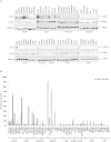
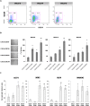
CD133 and CXCR4 were evaluated in the NCI-60 cell lines to identify cancer stem cell rich populations. Screening revealed that, ovarian OVCAR-3, -4 and -5 and colon cancer HT-29, HCT-116 and SW620 over expressed both proteins. We aimed to isolate cells with stem cell features sorting the cells expressing CXCR4(+)CD133(+) within ovarian cancer cell lines. The sorted population CD133(+)CXCR4(+) demonstrated the highest efficiency in sphere formation in OVCAR-3, OVCAR-4 and OVCAR-5 cells. Moreover OCT4, SOX2, KLF4 and NANOG were highly expressed in CD133(+)CXCR4(+) sorted OVCAR-5 cells. Most strikingly CXCR4(+)CD133(+) sorted OVCAR-5 and -4 cells formed the highest number of tumors when inoculated in nude mice compared to CD133(-)CXCR4(-), CD133(+)CXCR4(-), CD133(-)CXCR4(+) cells. CXCR4(+)CD133(+) OVCAR-5 cells were resistant to cisplatin, overexpressed the ABCG2 surface drug transporter and migrated toward the CXCR4 ligand, CXCL12. Moreover, when human ovarian cancer cells were isolated from 37 primary ovarian cancer, an extremely variable level of CXCR4 and CD133 expression was detected. Thus, in human ovarian cancer cells CXCR4 and CD133 expression identified a discrete population with stem cell properties that regulated tumor development and chemo resistance. This cell population represents a potential therapeutic target.
Figures


Similar articles
-
Microenvironment-Modulated Metastatic CD133+/CXCR4+/EpCAM- Lung Cancer-Initiating Cells Sustain Tumor Dissemination and Correlate with Poor Prognosis.Cancer Res. 2015 Sep 1;75(17):3636-49. doi: 10.1158/0008-5472.CAN-14-3781. Epub 2015 Jul 3. Cancer Res. 2015. PMID: 26141860
-
Epigenetic regulation of CD133 and tumorigenicity of CD133+ ovarian cancer cells.Oncogene. 2009 Jan 15;28(2):209-18. doi: 10.1038/onc.2008.374. Epub 2008 Oct 6. Oncogene. 2009. PMID: 18836486
-
CD133+ subpopulation of the HT1080 human fibrosarcoma cell line exhibits cancer stem-like characteristics.Oncol Rep. 2013 Aug;30(2):815-23. doi: 10.3892/or.2013.2486. Epub 2013 May 23. Oncol Rep. 2013. PMID: 23708735
-
The role of cancer stem cells and the side population in epithelial ovarian cancer.Histol Histopathol. 2010 Jan;25(1):113-20. doi: 10.14670/HH-25.113. Histol Histopathol. 2010. PMID: 19924647 Review.
-
Evidence for cancer stem cells contributing to the pathogenesis of ovarian cancer.Front Biosci (Landmark Ed). 2011 Jan 1;16(1):368-92. doi: 10.2741/3693. Front Biosci (Landmark Ed). 2011. PMID: 21196176 Review.
Cited by
-
Prognostic and clinicopathological significance of Cacna2d1 expression in epithelial ovarian cancers: a retrospective study.Am J Cancer Res. 2016 Sep 1;6(9):2088-2097. eCollection 2016. Am J Cancer Res. 2016. PMID: 27725913 Free PMC article.
-
Mechanisms of tumor cell resistance to the current targeted-therapy agents.Tumour Biol. 2016 Aug;37(8):10021-39. doi: 10.1007/s13277-016-5059-1. Epub 2016 May 7. Tumour Biol. 2016. PMID: 27155851 Review.
-
Cancer Stem Cells in Carcinogenesis and Potential Role in Pancreatic Cancer.Curr Stem Cell Res Ther. 2024;19(9):1185-1194. doi: 10.2174/1574888X19666230914103420. Curr Stem Cell Res Ther. 2024. PMID: 37711007 Review.
-
First Experimental Evidence for Reversibility of Ammonia Loss from Asparagine.Int J Mol Sci. 2022 Jul 28;23(15):8371. doi: 10.3390/ijms23158371. Int J Mol Sci. 2022. PMID: 35955504 Free PMC article.
-
Chemoresistance in ovarian cancer: exploiting cancer stem cell metabolism.J Gynecol Oncol. 2018 Mar;29(2):e32. doi: 10.3802/jgo.2018.29.e32. J Gynecol Oncol. 2018. PMID: 29468856 Free PMC article. Review.
References
-
- Visvader J.E. & Lindeman G.J. Cancer stem cells in solid tumors: accumulating evidence and unresolved questions. Nat. Rev. Cancer 8, 755–768 (2008). - PubMed
-
- Bhatia M., Bonnet D., Murdoch B., Gan O.I. & Dick J.E. A newly discovered class of human hematopoietic cells with SCID-repopulating activity. Nat. Med. 4, 1038–1045 (1998). - PubMed
-
- Scopelliti A. et al. Therapeutic implications of Cancer Initiating Cells. Expert Opin. Biol. Ther. 9, 1005–1016 (2009). - PubMed
-
- Hermann P.C., Bhaskar S., Cioffi M. & Heeschen C. Cancer stem cells in solid tumors. Semin. Cancer Biol. 20, 77–84 (2010). - PubMed
Publication types
MeSH terms
Substances
LinkOut - more resources
Full Text Sources
Other Literature Sources
Medical
Research Materials

