. 2015;7(6):572-83.
doi: 10.1159/000381915. Epub 2015 May 19.

Processing Body Formation Limits Proinflammatory Cytokine Synthesis in Endotoxin-Tolerant Monocytes and Murine Septic Macrophages

Affiliations

Processing Body Formation Limits Proinflammatory Cytokine Synthesis in Endotoxin-Tolerant Monocytes and Murine Septic Macrophages

Clara McClure et al. J Innate Immun. 2015.

Abstract

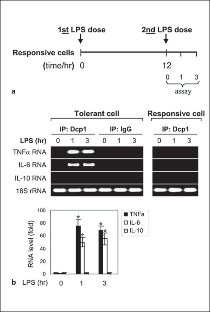
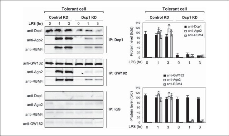
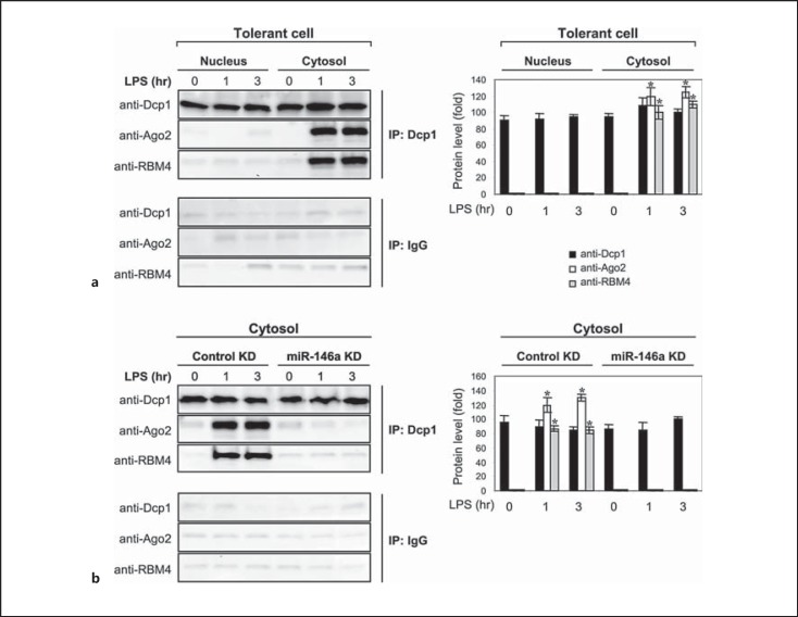
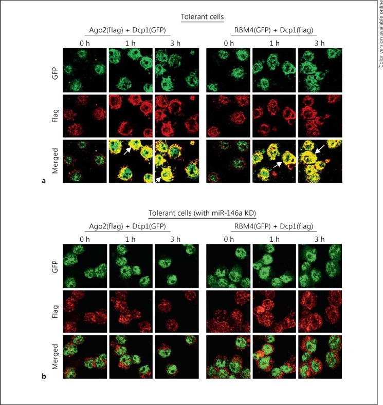
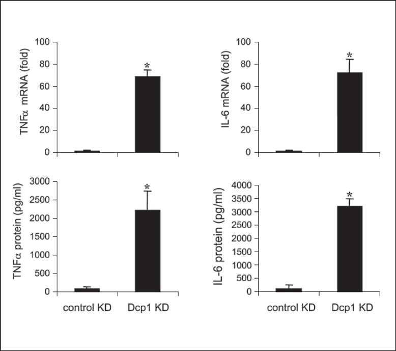
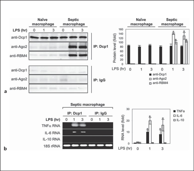
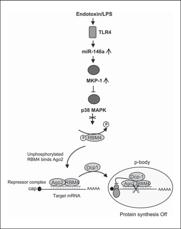
An anti-inflammatory phenotype with pronounced immunosuppression develops during sepsis, during which time neutrophils and monocytes/macrophages limit their Toll-like receptor 4 responses to bacterial lipopolysaccharide (LPS/endotoxin). We previously reported that during this endotoxin-tolerant state, distinct signaling pathways differentially repress transcription and translation of proinflammatory cytokines such as TNFα and IL-6. Sustained endotoxin tolerance contributes to sepsis mortality. While transcription repression requires chromatin modifications, a translational repressor complex of Argonaute 2 (Ago2) and RNA-binding motif protein 4 (RBM4), which bind the 3'-UTR of TNFα and IL-6 mRNA, limits protein synthesis. Here, we show that Dcp1 supports the assembly of the Ago2 and RBM4 repressor complex into cytoplasmic processing bodies (p-bodies) in endotoxin-tolerant THP-1 human monocytes following stimulation with LPS, resulting in translational repression and limiting protein synthesis. Importantly, this translocation process is reversed by Dcp1 knockdown, which restores TNFα and IL-6 protein levels. We also find this translational repression mechanism in primary macrophages of septic mice. Because p-body formation is a critical step in mRNA translation repression, we conclude that Dcp1 is a major component of the translational repression machinery of endotoxin tolerance and may contribute to sepsis outcome.


Figures

Fig. 1
Fig. 1
The mRNAs of proinflammatory cytokines TNFα and IL-6 but not the anti-inflammatory IL-10 associates with Dcp1 protein in endotoxin-tolerant but not endotoxin-responsive THP-1 cells after LPS stimulation. a THP-1 cells were made tolerant through the pretreatment of responsive (normal) cells overnight with 1 μg/ml of LPS, followed by RelB knockdown for 24 h, which restores mRNAs of TNFα, IL-6 and IL-β, but protein synthesis remains repressed (see first paragraph of Results). b The tolerant THP-1 cells with RelB knockdown, along with responsive (normal) THP-1 cells (as a control), were stimulated (2nd LPS dose) with 1 μg/ml of LPS for the indicated times. Whole cell lysates were prepared and immunoprecipitated with an anti-Dcp1 or IgG isotype (control) antibody. RNA was then extracted from the immunoprecipitated protein complexes and analyzed by RT-PCR to detect the presence of TNFα, IL-6 or IL-10 RNA in the protein complexes using primers that amplify the 3′-UTR sequences. RNA was also extracted from a portion of the cell lysate before the immunoprecipitation and was used to measure the level of the 18S ribosomal RNA as an internal (input) control. The quantitation of RNA levels is shown below. Values were normalized to the 18S rRNA and are presented as the fold change relative to the IgG-immunoprecipitated samples (set at 1-fold). Data are expressed as the mean ± SD from three experiments. * p < 0.05, compared with 0 h.
Fig. 2
Fig. 2
Dcp1 forms a protein complex with Ago2 and RBM4 after LPS stimulation in endotoxin-tolerant cells. Endotoxin-tolerant THP-1 cells were prepared by pretreatment of responsive (normal) cells overnight with 1 μg/ml of LPS. Tolerant cells were transfected with a pool of Dcp1-specific or scrambled siRNA (control KD). After 24 h, cells were washed and restimulated with 1 μg/ml of LPS for the indicated times. Whole cell lysates were prepared and immunoprecipitated with an anti-Dcp1, GW182 or IgG isotype antibody. The immunoprecipitated protein complexes were resolved by SDS-10% PAGE and then immunoblotted with a Dcp1, Ago2 or RBM4 antibody. The quantitation of protein levels is shown on the right. Values are presented as the fold change relative to the IgG-immunoprecipitated samples (set at 1-fold). Data are expressed as the mean ± SD from three experiments. * p < 0.05, compared with 0 h. KD = Knockdown.
Fig. 3
Fig. 3
The Dcp1 protein binding to Ago2 and RBM4 is detected in the cytoplasm in tolerant cells after LPS stimulation and is disrupted by miR-146a knockdown (KD). Endotoxin-tolerant THP-1 cells were prepared by pretreatment of responsive (normal) cells overnight with 1 μg/ml of LPS. a Tolerant cells were washed and restimulated with 1 μg/ml of LPS for the indicated times. Cytoplasmic and nuclear proteins were isolated and immunoprecipitated with an anti-Dcp1 or IgG isotype antibody. The immunoprecipitated protein complexes were resolved by SDS-10% PAGE and then immunoblotted with a Dcp1, Ago2 or RBM4 antibody. b Tolerant cells were transfected with anti-miR-146a oligonucleotides (antagomiRs) or scrambled anti-miRNA oligonucleotides (control KD). After 24 h, cells were washed and stimulated with 1 μg/ml of LPS for the indicated times. Cytoplasmic proteins were isolated, immunoprecipitated and immunoblotted as in a. The quantitation of protein levels is shown on the right. Values are presented as the fold change relative to the IgG-immunoprecipitated samples (set at 1-fold). Data are expressed as the mean ± SD from three experiments. * p < 0.05, compared with 0 h.
Fig. 4
Fig. 4
Dcp1 colocalizes with Ago2 and RBM4 in tolerant but not responsive cells after LPS stimulation and miR-146a knockdown (KD) disrupts this colocalization. a Endotoxin-tolerant cells were cotransfected with Flag-tagged Ago2 plus GFP-tagged Dcp1 expression constructs or GFP-tagged RBM4 plus Flag-tagged Dcp1 expression constructs for 36 h and then stimulated with 1 μg/ml of LPS for the indicated times. Arrows indicate colocalized protein complexes (yellow; colors refer to the online version only). b miR-146a was knocked down in endotoxin-tolerant cells for 24 h. Cells were then transfected and stimulated as in a. Cytospin preparations were readied and cells were fixed, permeabilized and costained with anti-Flag (red) and anti-GFP (green) antibodies. Images were captured at 40× magnification. The results are representative of two experiments.
Fig. 5
Fig. 5
Knockdown (KD) of Dcp1 in endotoxin-tolerant THP-1 cells restores the mRNA and protein levels of TNFα and IL-6. Endotoxin-tolerant THP-1 cells were prepared by pretreatment of responsive (normal) cells overnight with 1 μg/ml of LPS. Tolerant cells were transfected with pools of RelB- plus Dcp1-specific or scrambled siRNAs (control KD). After 24 h, cells were washed and restimulated with 1 μg/ml of LPS. After 1 h in LPS, a portion of the cells was harvested and RNA was isolated and used to measure TNFα and IL-6 mRNA levels by real-time RT-PCR. After an additional 5 h, the cells and supernatants were harvested and used to measure TNFα and IL-6 protein levels by ELISA. Data are the mean ± SD from three experiments. * p < 0.05.
Fig. 6
Fig. 6
The Dcp1 protein forms a complex with Ago2 and RBM4 in primary macrophages from septic mice and is associated with TNFα and IL-6 but not IL-10 mRNA. Sepsis was induced by CLP. Peritoneal macrophages were harvested from sham and septic mice that were moribund and sacrificed at days 10–28 (i.e. during late sepsis response). Cells were stimulated with 100 ng/ml of LPS for the indicated times. a Whole cell lysates were prepared and immunoprecipitated with anti-Dcp1 or IgG isotype antibody, resolved by SDS-10% PAGE, and then immunoblotted with a Dcp1, Ago2 or RBM4 antibody. The quantitation of protein levels is shown on the right. Values are presented as the fold change relative to the IgG-immunoprecipitated samples (set at 1-fold). Data are expressed as the mean ± SD from three experiments. * p < 0.05, compared with 0 h. b RNA was extracted from the immunoprecipitated protein complexes and then analyzed by RT-PCR for the presence of TNFα, IL-6 or IL-10 RNA. RNA was also extracted from a portion of the cell lysate before the immunoprecipitation and was used to measure the level of the 18S rRNA as an internal (input) control. Cells were pooled from 8 sham mice and 6 late septic mice. The quantitation of RNA levels is shown on the right. Values were normalized to the 18S rRNA and are presented as the fold change relative to the IgG-immunoprecipitated samples (set at 1-fold). Data are expressed as the mean ± SD from three experiments. * p < 0.05.
Fig. 7
Fig. 7
A model depicting Dcp1-mediated translational repression in endotoxin-tolerant cells. Based on our present and previous findings, LPS stimulation in endotoxin-tolerant cells elevates miR-146a, which inactivates p38 MAPK. In the absence of activated p38, RBM4 accumulates in the cytosol in an unphosphorylated form, where it binds Ago2 and Dcp1 and forms a translational repressor complex at the target mRNA, which will then be deposited into p-bodies where translation repression occurs. Note RBM4 phosphorylation is disrupted in tolerant cells due to induction of the MAPK phosphatase MKP-1, which is controlled by miR-146a [17, 19]. Not shown is the RelB-induced cytokine gene transcription repression pathway, which is also induced in parallel as described in the first paragraph of Results.

Similar articles

Cited by

References

    1. Bianchi ME. Editorial: A recipe for inflammation. J Leukoc Biol. 2009;86:471–472. - PubMed
    1. Biswas SK, Lopez-Collazo E. Endotoxin tolerance: new mechanisms, molecules and clinical significance. Trends Immunol. 2009;30:475–487. - PubMed
    1. Hotchkiss RS, Karl IE. The pathophysiology and treatment of sepsis. N Engl J Med. 2003;348:138–150. - PubMed
    1. McCall CE, Yoza B, Liu T, El Gazzar M. Gene-specific epigenetic regulation in serious infections with systemic inflammation. J Innate Immun. 2010;2:395–405. - PMC - PubMed
    1. Remick DG. Pathophysiology of sepsis. Am J Pathol. 2007;170:1435–1444. - PMC - PubMed

Publication types

MeSH terms