. 2015 May 21;6(5):e1768.
doi: 10.1038/cddis.2015.131.

The nucleoprotein of influenza A virus induces p53 signaling and apoptosis via attenuation of host ubiquitin ligase RNF43

Affiliations

The nucleoprotein of influenza A virus induces p53 signaling and apoptosis via attenuation of host ubiquitin ligase RNF43

H Nailwal et al. Cell Death Dis. .

Abstract

The interplay between influenza virus and host factors to support the viral life cycle is well documented. Influenza A virus (IAV) proteins interact with an array of cellular proteins and hijack host pathways which are at the helm of cellular responses to facilitate virus invasion. The multifaceted nature of the ubiquitination pathway for protein regulation makes it a vulnerable target of many viruses including IAV. To this end we conducted a yeast two-hybrid screen to search for cellular ubiquitin ligases important for influenza virus replication. We identified host protein, RING finger protein 43 (RNF43), a RING-type E3 ubiquitin ligase, as a novel interactor of nucleoprotein (NP) of IAV and an essential partner to induce NP-driven p53-mediated apoptosis in IAV-infected cells. In this study, we demonstrate that IAV leads to attenuation of RNF43 transcripts and hence its respective protein levels in the cellular milieu whereas in RNF43 depleted cells, viral replication was escalated several folds. Moreover, RNF43 polyubiquitinates p53 which further leads to its destabilization resulting in a decrease in induction of the p53 apoptotic pathway, a hitherto unknown process targeted by NP for p53 stabilization and accumulation. Collectively, these results conclude that NP targets RNF43 to modulate p53 ubiquitination levels and hence causes p53 stabilization which is conducive to an enhanced apoptosis level in the host cells. In conclusion, our study unravels a novel strategy adopted by IAV for utilizing the much conserved ubiquitin proteasomal pathway.


Figures

Figure 1
Figure 1
IAV NP interacts with RNF43. (a) Tabular representation of the yeast two-hybrid screening of lung cDNA library using NP as the bait protein. The filter β-gal assay for the clone 13-4 is shown as the last column of the table. (b) pHybLexA/Zeo-NP and pYESTrp2-RNF43 were cotransformed in L-40 yeast strain and their interaction was verified by β-galactosidase assay. (c) Lung epithelial A549 cells were transfected with either empty vector pCDNA3.1 (Mock) or pCDNA3.1-Myc-NP (NP-Myc). Forty-eight hours post transfection, cells were lysed and the lysates were subjected to co-immunoprecipitation with anti-RNF43 antibody followed by Western blotting with anti-Myc antibody. (d) A549 cells were infected with PR8 virus and X-31 virus at 1 MOI for 24 h and the whole-cell lysates were used for co-immunoprecipitation assays. Lysates (5%) from the same experiment were subjected to Western blotting with anti-NP (panel 6), anti-M2 (panel 7), anti-RNF43 (panel 8) and anti-βactin (panel 9) antibodies to show the cellular levels of these proteins
Figure 2
Figure 2
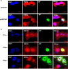
IAV NP and RNF43 co-localize in the nucleus of mammalian cells. (a) A549 cells were transfected with pEGFPN1 control plasmid or pEGFP-NP. Cells were fixed after 48 h and stained with DAPI for nucleus and anti-goat secondary antibody conjugated to Alexa-594 for RNF43 (red) and observed under confocal microscope. GFP-tagged NP is shown in green. (b) A549 cells were infected with PR8 IAV with 5 MOI and were fixed at the indicated time points. NP was stained using anti-NP monoclonal primary antibody and anti-mouse Alexa488 conjugated secondary antibody (green). RNF43 was stained using specific primary antibody and anti-rabbit Alexa-594 conjugated secondary antibody (red). Panels are labeled for their respective staining, RNF43 (red), NP (green) and nucleus (blue)
Figure 3
Figure 3
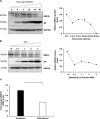
IAV infection decreases abundance of RNF43 at both mRNA and protein levels. (a) Lung epithelial A549 cells were infected with PR8 virus at an MOI of 1 and cells were harvested at indicated time intervals post infection and the whole-cell lysate was resolved on SDS-PAGE for Western blot analysis of RNF43, NP and GAPDH. (b) A549 cells were infected with PR8 virus at indicated MOIs and harvested at 24-h post infection for Western blot analysis of RNF43, NP and GAPDH. Quantitative representation of the immunoblots of both the experiments is shown as the line diagram after normalization with GAPDH (extreme right). (c) A549 cells were mock infected or infected with PR8 virus for 24 h and total RNA was extracted followed by rnf43 mRNA estimation with qRT-PCR. Results are shown as mean of ±S.D. of three independent experiments. * indicates statistically significant difference at P<0.05
Figure 4
Figure 4
Host factor RNF43 decreases IAV replication. (a–c) HEK293T cells were transiently transfected with plasmid pCDNA3.1 (Mock) or pCDNA3.1-RNF43-Flag-HA (RNF43-HA) (1 and 2 μg) followed by PR8 infection and incubated for 24 h. Total RNA was isolated and NP mRNA (a), NP vRNA (b) and RNF43 mRNA(c) levels were estimated through qRT PCR. (d) HEK293T cells were treated with NT siRNA or RNF43 siRNA for 24 h followed by PR8 IAV infection for next 24 h. Cells were processed for qRT-PCR analysis of mRNA levels of NP and RNF43. (e) A549 cells, pretreated with either NT or RNF43 siRNA were transfected with plasmids-encoding polymerase complex components (PA, PB1, PB2 and NP) derived from PR8 (H1N1 virus) were co-transfected alongside a reporter plasmid containing noncoding sequence from the NS1 segment of influenza A virus and luciferase gene driven by the Pol 1 promoter. The relative luciferase units were calculated after normalization with plasmid pRL-TK (Promega, Madison, WI, USA), which expresses Renilla luciferase that was transfected along. Results are shown as mean±S.D. of three independent experiments. * and # indicate statistically significant difference at P<0.05 and P<0.01, respectively
Figure 5
Figure 5
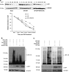
NP interacts with RNF43 to stabilize p53 through compromised ubiquitination of p53 by RNF43. (a) A549 cells were transiently transfected with plasmids pEGFPN1 (Mock), pEGFP-NP (NP-GFP) or pCDNA3.1-RNF43-Flag-HA (RNF43-HA) for 48 h. The transfectants were treated with cycloheximide at 50 μg/ml for the indicated times (minutes) post treatment (mpt) and subjected to Western blot analysis using the indicated antibodies. The quantitative data of immunoblots is shown as line diagram after normalization with GAPDH expression levels. (b) HEK293T cells were transiently transfected with a combination of indicated plasmids and incubated for 48 h. The transfectants were treated with 20 μg/ml MG132, 5 h before harvest. The cell lysates were subjected to Western blot analysis using the indicated antibodies. (c) HEK293T cells were transiently transfected with a combination of indicated plasmids and incubated for 48 h. The transfectants were treated with 20 μg/ml MG132, 5 h before harvest. Ubiquitinated p53 (Ub-p53) was pulled down using Ni2+–NTA–agarose beads and analyzed by Western blotting with indicated antibodies
Figure 6
Figure 6
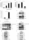
RNF43 decreases NP driven increased p53 transcriptional activity and signaling in the cells. (a) A549 cells were transfected with p21-Luc reporter plasmid, with or without growing amounts of pEGFP-NP (500 and 750 ng, and 1 μg) in conjunction with pcDNA-p53 and a control plasmid, Renilla luciferase pRL-TK. Luciferase activity of cell lysates of transfectants was analyzed. (b) A549 cells were transfected with p21-Luc reporter plasmid, pcDNA-p53 and a control plasmid, Renilla luciferase pRL-TK along with plasmids pEGFPN1 (mock), pEGFP-NP (NP-GFP) and pCDNA3.1-RNF43-Flag-HA (RNF43-HA) in the indicated combinations. Luciferase activity was measured. (c) HEK293T cells were treated with NT siRNA or RNF43 siRNA for 24 h followed by PR8 IAV infection at 1 MOI. Cells were harvested at 24-h post infection and total RNA was extracted followed by p21 mRNA estimation with qRT-PCR. (d) A549 cells were transiently transfected with pEGFP-NP with or without pCDNA3.1-RNF43-Flag-HA and were harvested at 48 h followed by SDS-PAGE. Western blotting was done using indicated antibodies. (e) A549 cells were seeded in a 6-well plate and were infected with PR8 virus at 1 MOI. Cells were harvested at 0, 4, 8, 16 and 24-h post infection, and subjected to Western blotting with indicated antibodies. (f) A549 cells were transfected with pCDNA3.1 (mock) and pCDNA3.1-RNF43-Flag-HA (RNF43-HA) plasmids followed by PR8 infection, 24-h post transfection. The cells were harvested after 24 h followed by Western blot analysis with anti-acetyl p53, HA, NP and β-actin antibodies. (g) A549 cells were transiently transfected with pCDNA3.1 (mock) or pCDNA3.1-RNF43-Flag-HA (RNF43-HA) plasmid constructs and after 24 h of incubation, cells were infected with PR8 virus at an MOI of 1 for 24 h. (h) Similarly, A549 cells were transiently transfected with pEGFP-NP with or without pCDNA3.1-RNF43-Flag-HA and were incubated for 48 h. (g and h) Cells were harvested and processed for Western blot analysis with indicated antibodies. Results in ac are shown as mean±S.D. three independent experiments. * and # indicate statistically significant difference at P<0.05 and P<0.01, respectively
Figure 7
Figure 7
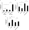
RNF43 attenuates IAV NP-induced cell death. (a) A549 cells were treated with NT siRNA or RNF43 siRNA for 24 h followed by X-31 IAV infection at 1 MOI. Cells were harvested at 24 h.p.i, stained with Annexin V FITC and subjected to flow cytometry. The percentage of Annexin V positive population is plotted on the graph. (b) A549 cells were transiently transfected with the indicated combinations of plasmids and 48 h post-transfection cells were harvested and processed for flow cytometric analysis of Annexin V FITC stained population which is plotted on the graph. (c) Same experiment was conducted in p53−/− HCT116 cells. All graphs represent mean±S.D. of three independent experiments. * and # indicate statistically significant difference at P<0.05 and P<0.01, respectively, whereas NS refers to non-significant difference
Figure 8
Figure 8
The proposed model for the role of NP/RNF43 interaction in regulation of p53-mediated cell death by IAV NP. The proposed model for the regulation of p53-mediated cell death by IAV NP. p53 is proposed to be regulated by RNF43 through ubiquitylation resulting in its destabilization. NP interacts with RNF43 thereby preventing ubiquitination of p53 and causing its stabilization and accumulation inside the infected cell resulting into activation of p53 signaling cascade including p21, Bax, Puma and eventually results in cell death in IAV-infected cell

Similar articles

Cited by

References

    1. Nailwal H, Kamra K, Lal SK. H7N9: a killer in the making or a false alarm? Can J Microbiol 2014; 60: 425–429. - PubMed
    1. Kawaoka Y, Cox NJ, Haller O, Hongo S, Kaverin N, Klenk HD et al. Orthomyxoviridae. In: Fauquet CM, Mayo MA, Maniloff J, Desselberger U, Ball LA (eds), Virus taxonomy: 8th report of the international committee on taxonomy of viruses. Elsevier Academia Press: London, UK, 2005; pp 681–693.
    1. Portela A, Digard P. The influenza virus nucleoprotein: a multifunctional RNA-binding protein pivotal to virus replication. J Gen Virol 2002; 83: 723–734. - PubMed
    1. Sharma S, Mayank AK, Nailwal H, Tripathi S, Patel JR, Bowzard JB et al. Influenza A viral nucleoprotein interacts with cytoskeleton scaffolding protein α-actinin-4 for viral replication. FEBS J 2014; 281: 2899–2914. - PMC - PubMed
    1. Gabriel G, Herwig A, Klenk HD. Interaction of polymerase subunit PB2 and NP with importin α1 is a determinant of host range of influenza A virus. PLoS Pathogen 2008; 4: e11. - PMC - PubMed

MeSH terms