. 2015 May 25;209(4):539-48.
doi: 10.1083/jcb.201502033. Epub 2015 May 18.

Ltc1 is an ER-localized sterol transporter and a component of ER-mitochondria and ER-vacuole contacts

Affiliations

Ltc1 is an ER-localized sterol transporter and a component of ER-mitochondria and ER-vacuole contacts

Andrew Murley et al. J Cell Biol. .

Abstract

Organelle contact sites perform fundamental functions in cells, including lipid and ion homeostasis, membrane dynamics, and signaling. Using a forward proteomics approach in yeast, we identified new ER-mitochondria and ER-vacuole contacts specified by an uncharacterized protein, Ylr072w. Ylr072w is a conserved protein with GRAM and VASt domains that selectively transports sterols and is thus termed Ltc1, for Lipid transfer at contact site 1. Ltc1 localized to ER-mitochondria and ER-vacuole contacts via the mitochondrial import receptors Tom70/71 and the vacuolar protein Vac8, respectively. At mitochondria, Ltc1 was required for cell viability in the absence of Mdm34, a subunit of the ER-mitochondria encounter structure. At vacuoles, Ltc1 was required for sterol-enriched membrane domain formation in response to stress. Increasing the proportion of Ltc1 at vacuoles was sufficient to induce sterol-enriched vacuolar domains without stress. Thus, our data support a model in which Ltc1 is a sterol-dependent regulator of organelle and cellular homeostasis via its dual localization to ER-mitochondria and ER-vacuole contact sites.


Figures

Figure 1.
Figure 1.
Ylr072w/Ltc1 is localized to ER–mitochondria and ER–vacuole contact sites. (A) Deletion of YLR072W causes no significant growth defect on fermentable or nonfermentable carbon sources. Cells were grown to mid-log phase and plated on synthetic complete media containing 2% dextrose (SD) or 3% glycerol + 2% ethanol (SEG). (B) Deletion of YLR072W causes no significant change in mitochondrial morphology. Cells expressing mitochondria-targeted DsRed were grown to mid-log phase and imaged as described in “Fluorescence microscopy.” (C) Deletion of YLR072W causes a synthetic sick/lethal phenotype in Δmdm34 cells. Expression of Ylr072w-yEGFP restores Δmdm34 growth phenotype. Yeast diploids with the indicated genotypes were sporulated, subjected to tetrad dissection, and the resulting spore colonies were genotyped based on segregation pattern of markers. Red circles indicate inviable Δmdm34 Δylr072w cells. Green circles indicate viable Δmdm34 YLR072W-yEGFP cells. (D–F) Ylr072w localization was assessed in WT cells using Ylr072w-yEGFP integrated at its endogenous locus relative to ERMES marked by Mdm34-mCherry (D), ER and mitochondria marked by DsRed-HDEL and mitochondrial-targeted mtBFP, respectively (E), and ER marked by DsRed-HDEL and vacuoles marked by Pho8-3XBFP (F). Cells were grown to mid-log phase and imaged as described in “Fluorescence microscopy.” Yellow arrowheads in E mark ER–mitochondria contact sites, open white arrowheads in E and F mark NVJs, and closed white arrowheads in F mark ER–vacuole contacts. (G) Quantification of Ltc1 foci localization from D–F. Dashed lines in B and D demarcate cell boundaries. Dashed lines in E and F indicate enlarged regions shown as separate grayscale images to the right. Bars, 2 µm.
Figure 2.
Figure 2.
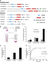
Ylr072w/Ltc1 is a member of a conserved protein family and catalyzes sterol transport between membranes. (A) Ltc1 family members in budding yeast and humans, also identified by Khafif et al. (2014). Members were identified by BLAST searches with the amino acid sequence of Ylr072w/Ltc1. Proteins with significant sequence similarity were queried for shared predicted secondary structure and domains with Phyre2 and i-TASSER (Kelley and Sternberg, 2009; Yang et al., 2015). In yeast, the paralogues Ltc1/Ltc2(Yfl042c) and Ltc3(Yhr080c)/Ltc4(Ysp2) arose from a genome duplication event. (B–D) Ltc1 selectively transports sterol between membranes (B; n = 4 for cholesterol and n = 2 for triolein and phospholipids) in a concentration (C; n = 4)- and time (D; with protein n = 3; control without protein n = 1)-dependent manner and were performed as depicted in the schematic in B and as described in Materials and methods. Errors bars represent standard deviation.
Figure 3.
Figure 3.
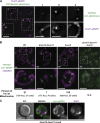
Localization of Ltc1 to ER–mitochondria and ER–vacuole contact sites requires Tom70/71 and Vac8, respectively. (A) Ltc1, marked by Ltc1-yEmCherry, colocalizes with Tom71, marked by Tom71-yEGFP, in foci on the mitochondrial outer membrane. Cells expressing Ltc1-yEmCherry and Tom71-yEGFP were grown to mid-log phase and imaged as described in “Fluorescence microscopy.” Dashed lines and numbers denote enlarged regions shown as separate grayscale images of each channel to the right of the merged image. (B) Vac8 and Tom70/71 are required for Ltc1 foci localization to vacuoles and mitochondria, respectively. Cells of the indicated genotypes expressing Ltc1-yEGFP and mtDsRed were grown to mid-log phase and imaged as described in “Fluorescence microscopy.” Images represent a maximum intensity z-projection. Dashed lines denote enlarged regions shown below. (C) Ltc1 is an ER membrane–associated protein. Δtom70 Δtom71 Δvac8 cells expressing Ltc1-yEGFP were grown to mid-log phase and treated with 1 µg/ml DAPI for 20 min to label the nucleus, washed twice in SD media, and imaged as described in “Fluorescence microscopy.” Extranuclear DAPI-stained structures are likely mtDNA. Bars, 2 µm.
Figure 4.
Figure 4.
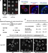
The distribution of Ltc1 to ER–mitochondria and ER–vacuolar contacts regulates Ltc1-dependent separable mitochondrial and vacuolar functions. (A) The localization of Ltc1 to ER–mitochondria, but not to ER–vacuole, contacts is essential in Δmdm34 cells. Diploid yeast heterozygous for the indicated deletions were sporulated and analyzed by tetrad dissection as described in “Fluorescence microscopy.” The green circle indicates viable Δvac8 Δmdm34 cells (the colonies immediately above and below are Δvac8 and WT, respectively). Red circles indicate nonviable Δtom70 Δtom71 Δmdm34 cells. (B) The Ltc1 GRAM domain is required for the mitochondrial localization of Ltc1. Cells expressing Ltc1(ΔGRAM)-yEGFP, Vph1-yEmCherry (vacuoles), and mtBFP (mitochondria) were grown to mid-log phase and imaged as described in “Fluorescence microscopy.” Open arrowheads indicate the localization of Ltc1 to Vph1-depleted regions on vacuoles. Dashed lines denote the enlarged region shown as two-color images to the right of the merged, three-color image. (C) The mitochondrial localization of Ltc1 is essential in Δmdm34 mutants. Diploid yeast heterozygous for Δmdm34 and Δltc1 and harboring a yeast centromeric plasmid pRS313 containing Ltc1-yEGFP or Ltc1(ΔGRAM) were sporulated and analyzed by tetrad dissection as described in “Fluorescence microscopy.” The expected frequency of viable Δmdm34 Δltc1 mutants for pRS313 plasmids containing a WT Ltc1 is 1/8 spore colonies. The observed frequency for WT Ltc1 was 12/104 (∼1/8), whereas the observed frequency for Ltc1(ΔGRAM) and an empty vector control were 1/108 and 2/92, respectively. (D) Ltc1 is required for vacuole membrane domain formation in response to cycloheximide and glucose starvation. WT and Δltc1 cells expressing Vph1-yEGFP were grown exponentially in synthetic complete media for at least 12 generations and then subjected to the indicated treatments and imaged as described in “Fluorescence microscopy.” Quantification represents triplicate biologically independent experiments. (E) Shifting the proportional localization of Ltc1 to ER–vacuole contacts is sufficient to induce vacuole membrane domain formation under normal growing conditions. WT, Δtom70 Δtom71, and Δltc1 Δtom70 Δtom71 cells were grown for at least 12 generations in nutrient-replete media, and domain formation was assessed using Vph1-yEGFP as described in “Fluorescence microscopy.” Quantification represents triplicate biologically independent experiments. The arrowhead indicates a vacuole with Vph1-GFP–labeled domains. Bars, 2 µm.

Similar articles

Cited by

References

    1. Altmann K., and Westermann B.. 2005. Role of essential genes in mitochondrial morphogenesis in Saccharomyces cerevisiae. Mol. Biol. Cell. 16:5410–5417. 10.1091/mbc.E05-07-0678 - DOI - PMC - PubMed
    1. Bartlett G.R. 1959. Colorimetric assay methods for free and phosphorylated glyceric acids. J. Biol. Chem. 234:469–471. - PubMed
    1. Begley M.J., Taylor G.S., Kim S.A., Veine D.M., Dixon J.E., and Stuckey J.A.. 2003. Crystal structure of a phosphoinositide phosphatase, MTMR2: insights into myotubular myopathy and Charcot-Marie-Tooth syndrome. Mol. Cell. 12:1391–1402. 10.1016/S1097-2765(03)00486-6 - DOI - PubMed
    1. Berchtold D., Piccolis M., Chiaruttini N., Riezman I., Riezman H., Roux A., Walther T.C., and Loewith R.. 2012. Plasma membrane stress induces relocalization of Slm proteins and activation of TORC2 to promote sphingolipid synthesis. Nat. Cell Biol. 14:542–547. 10.1038/ncb2480 - DOI - PubMed
    1. Böckler S., and Westermann B.. 2014. Mitochondrial ER contacts are crucial for mitophagy in yeast. Dev. Cell. 28:450–458. 10.1016/j.devcel.2014.01.012 - DOI - PubMed

Publication types

MeSH terms

Substances