

Process of the Functional Reorganization of the Cortical Centers for Movement in GBM Patients: fMRI Study
- PMID: 25986127
- PMCID: PMC8076113
- DOI: 10.1007/s00062-015-0398-7
Process of the Functional Reorganization of the Cortical Centers for Movement in GBM Patients: fMRI Study
Abstract
Purpose: The aim of this study was to verify whether the functional reorganization of motor cortex is associated with the increase in the size of WHO type IV glioma lesion, that is, disease duration and development, and whether surgical treatment has an impact on cerebral plasticity.
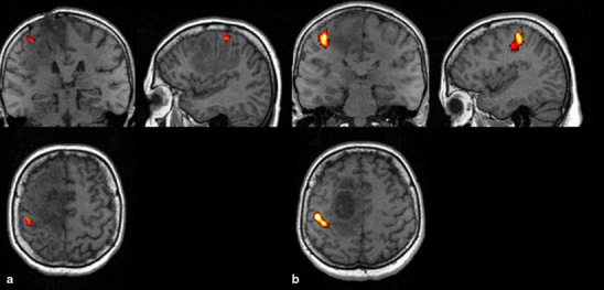
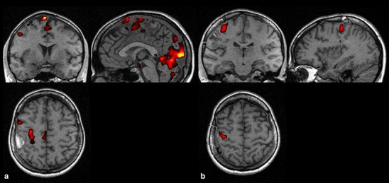
Methods: The study included 16 patients with primary tumors of the brain located at the region of central sulcus. The clinical status of patients and tumor volume was determined. Functional magnetic resonance imaging examinations were performed before and 3 months after operation.
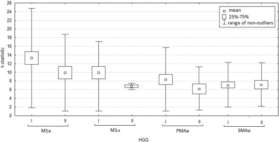
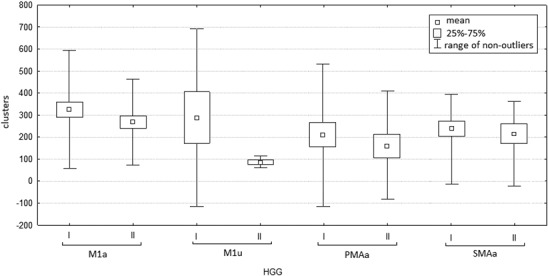
Results: The activity of all cortical centers, both contralateral and ipsilateral, was observed in a group of small as well as large tumors. The intensity of activation and the number of activated clusters of small tumors were almost always higher as compared with the large tumors. The frequency of the activity of contralateral areas was similar during the first and the second examination. In the case of ipsilateral centers, the frequency of activation during the second examination was lower. Mean values of t-statistics during the first examination were higher than during the second examination. Supplementary motor area (SMAa) was the only center for which the mean values of activation intensity remained similar.
Conclusions: SMAa seems to play the most important role in the processes of motor cortex plasticity in high-grade glioma patients. Surgery seems not having a significant influence on the pattern of functional reorganization of the cortical centers for movement. Identification of the individual patterns of the reorganization of motor centers plays an important role in clinical practice.
Keywords: Brain plasticity; Brain tumor; Movement centers; fMRI.
Conflict of interest statement
On behalf of all authors, the corresponding author states that there is no conflict of interest.
Figures
Similar articles
-
Intraoperative mapping during repeat awake craniotomy reveals the functional plasticity of adult cortex.J Neurosurg. 2016 May;124(5):1460-9. doi: 10.3171/2015.5.JNS142833. Epub 2015 Nov 6. J Neurosurg. 2016. PMID: 26544767
-
Functional rearrangement of the primary and secondary motor cortex in patients with primary tumors of the central nervous system located in the region of the central sulcus depending on the histopathological type and the size of tumor: Examination by means of functional magnetic resonance imaging.Pol J Radiol. 2012 Jan;77(1):12-20. doi: 10.12659/pjr.882576. Pol J Radiol. 2012. PMID: 22802861 Free PMC article.
-
Cortical plasticity catalyzed by prehabilitation enables extensive resection of brain tumors in eloquent areas.J Neurosurg. 2017 Apr;126(4):1323-1333. doi: 10.3171/2016.2.JNS152485. Epub 2016 May 20. J Neurosurg. 2017. PMID: 27203145
-
Integrated technology for evaluation of brain function and neural plasticity.Phys Med Rehabil Clin N Am. 2004 Feb;15(1):263-306. doi: 10.1016/s1047-9651(03)00124-4. Phys Med Rehabil Clin N Am. 2004. PMID: 15029909 Review.
-
Cortical Plasticity in the Setting of Brain Tumors.Top Magn Reson Imaging. 2016 Feb;25(1):25-30. doi: 10.1097/RMR.0000000000000077. Top Magn Reson Imaging. 2016. PMID: 26848558 Free PMC article. Review.
Cited by
-
Direct Evidence of Plasticity within Human Primary Motor and Somatosensory Cortices of Patients with Glioblastoma.Neural Plast. 2020 Sep 22;2020:8893708. doi: 10.1155/2020/8893708. eCollection 2020. Neural Plast. 2020. PMID: 33029127 Free PMC article.
-
Motor Functional Reorganization Is Triggered by Tumor Infiltration Into the Primary Motor Area and Repeated Surgery.Front Hum Neurosci. 2020 Aug 14;14:327. doi: 10.3389/fnhum.2020.00327. eCollection 2020. Front Hum Neurosci. 2020. PMID: 32922279 Free PMC article.
-
Brain imaging and morphological plasticity in glioblastoma: a literature review.J Med Life. 2023 Mar;16(3):344-347. doi: 10.25122/jml-2022-0201. J Med Life. 2023. PMID: 37168303 Free PMC article. Review.
-
Diffusion Tensor Imaging Shows Corpus Callosum Differences between High-Grade Gliomas and Metastases.J Neuroimaging. 2018 Mar;28(2):199-205. doi: 10.1111/jon.12478. Epub 2017 Oct 24. J Neuroimaging. 2018. PMID: 29064137 Free PMC article.
-
What do we know about pre- and postoperative plasticity in patients with glioma? A review of neuroimaging and intraoperative mapping studies.Neuroimage Clin. 2020;28:102435. doi: 10.1016/j.nicl.2020.102435. Epub 2020 Sep 14. Neuroimage Clin. 2020. PMID: 32980599 Free PMC article. Review.
References
-
- Peck KK, Bradbury MS, Hou BL, Brennan NP, Holodny AI. The role of the supplementary motor area (SMA) in the execution of primary motor activities in brain tumor patients: functional MRI detection of time-resolved differences in the hemodynamic response. Med Sci Monit. 2009;15:55–62. - PubMed
Publication types
MeSH terms
LinkOut - more resources
Full Text Sources
Medical
