. 2015 Aug;89(15):7506-20.
doi: 10.1128/JVI.00013-15. Epub 2015 May 13.

The Nuclear DNA Sensor IFI16 Acts as a Restriction Factor for Human Papillomavirus Replication through Epigenetic Modifications of the Viral Promoters

Affiliations

The Nuclear DNA Sensor IFI16 Acts as a Restriction Factor for Human Papillomavirus Replication through Epigenetic Modifications of the Viral Promoters

Irene Lo Cigno et al. J Virol. 2015 Aug.

Abstract

The human interferon-inducible IFI16 protein, an innate immune sensor of intracellular DNA, was recently demonstrated to act as a restriction factor for human cytomegalovirus (HCMV) and herpes simplex virus 1 (HSV-1) infection by inhibiting both viral-DNA replication and transcription. Through the use of two distinct cellular models, this study provides strong evidence in support of the notion that IFI16 can also restrict human papillomavirus 18 (HPV18) replication. In the first model, an immortalized keratinocyte cell line (NIKS) was used, in which the IFI16 protein was knocked down through the use of small interfering RNA (siRNA) technology and overexpressed following transduction with the adenovirus IFI16 (AdVIFI16) vector. The second model consisted of U2OS cells transfected by electroporation with HPV18 minicircles. In differentiated IFI16-silenced NIKS-HPV18 cells, viral-load values were significantly increased compared with differentiated control cells. Consistent with this, IFI16 overexpression severely impaired HPV18 replication in both NIKS and U2OS cells, thus confirming its antiviral restriction activity. In addition to the inhibition of viral replication, IFI16 was also able to reduce viral transcription, as demonstrated by viral-gene expression analysis in U2OS cells carrying episomal HPV18 minicircles and HeLa cells. We also provide evidence that IFI16 promotes the addition of heterochromatin marks and the reduction of euchromatin marks on viral chromatin at both early and late promoters, thus reducing both viral replication and transcription. Altogether, these results argue that IFI16 restricts chromatinized HPV DNA through epigenetic modifications and plays a broad surveillance role against viral DNA in the nucleus that is not restricted to herpesviruses.

Importance: Intrinsic immunity is mediated by cellular restriction factors that are constitutively expressed and active even before a pathogen enters the cell. The host nuclear factor IFI16 acts as a sensor of foreign DNA and an antiviral restriction factor, as recently demonstrated by our group for human cytomegalovirus (HCMV) and herpes simplex virus 1 (HSV-1). Here, we provide the first evidence that IFI16 inhibits HPV18 replication by repressing viral-gene expression and replication. This antiviral restriction activity was observed in immortalized keratinocytes transfected with the religated genomes and in U2OS cells transfected with HPV18 minicircles, suggesting that it is not cell type specific. We also show that IFI16 promotes the assembly of heterochromatin on HPV DNA. These changes in viral chromatin structure lead to the generation of a repressive state at both early and late HPV18 promoters, thus implicating the protein in the epigenetic regulation of HPV gene expression and replication.


Figures

FIG 1
FIG 1
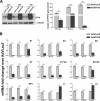
The IFI16 protein is a negative regulator of HPV replication. (A) Genomic DNA was extracted from pooled NIKS-HPV18 cells and examined by Southern blotting. All samples contained 5 μg of genomic DNA and were digested with DpnI to remove any residual input DNA and with HindIII (D/H) (with no restriction site in HPV18) or DpnI and EcoRI (D/E) (with a single restriction site in HPV18) to linearize HPV DNA. The positions of open-circle (oc), linear (lin), and supercoiled (sc) forms of HPV DNA are indicated on the right of the blots. A standard (St) (pBluescript-HPV18 plasmid digested with EcoRI) corresponding to 50 copies of the HPV18 genome per cell was included in the left lane. (B) NIKS-HPV18 cells were electroporated with a mixture of four different small interfering RNAs (siIFI16) or scrambled control siRNA (siCtrl). After 24 h, the cells were transferred into methylcellulose medium for 72 h to allow differentiation. (Top left) The efficiency of siRNA depletion was analyzed by Western blotting with anti-IFI16 polyclonal antibody, and α-tubulin was included as a loading control. One representative blot of 3 independent experiments is shown. (Top right) Densitometric analysis predicting the fold change in expression (means ± SD) from three independent experiments is represented by histograms. (Bottom) To measure viral replication, total cellular DNA was analyzed by qPCR after DpnI digestion. The levels of glyceraldehyde 3-phosphate dehydrogenase were used to normalize the HPV18 levels. The data shown are the means of the results of three experiments ± SD (*, P < 0.05; **, P < 0.01; unpaired t test). (C) NIKS-HPV18 cells were infected for 24 h with AdVIFI16 or AdVLacZ and then transferred into methylcellulose medium for 72 h to allow differentiation. The efficiency of IFI16 overexpression was analyzed by both qRT-PCR and Western blotting using anti-IFI16 polyclonal antibody and α-tubulin (included as a loading control). (Top left) Total RNA was extracted and analyzed by qRT-PCR to measure the efficiency of IFI16 overexpression. The values were normalized to glyceraldehyde 3-phosphate dehydrogenase and are shown as fold changes relative to AdVLacZ-infected cells. The data shown are the averages of the results of three experiments ± SD (**, P < 0.01; unpaired t test). (Top middle) One representative Western blot is shown. (Top right) Densitometric analysis predicting the fold change in expression (means ± SD) from three independent experiments is represented by histograms (*, P < 0.05; unpaired t test). (Bottom) To measure viral replication, total cellular DNA was analyzed by qPCR after DpnI digestion. The levels of glyceraldehyde 3-phosphate dehydrogenase were used to normalize the HPV18 levels. The data shown are the means of the results of three experiments ± SD (**, P < 0.01; unpaired t test).
FIG 2
FIG 2
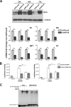
IFI16 overexpression impairs HPV18 replication by inhibiting both early and late viral-gene expression. (A) U2OS cells were electroporated with HPV18 minicircle DNA and, at passage 1, infected with AdVIFI16 or AdVLacZ. (Left) Total protein extracts were harvested at 24, 48, and 72 h after AdV infection (p.i.) and analyzed for IFI16 expression by Western blotting. α-Tubulin was included as a loading control. Experiments were repeated at least three times, and one representative result is shown. (Right) Total DNA was extracted at 24, 48, and 72 h after AdV infection to measure replicated (DpnI-resistant) viral DNA by qPCR. Levels of glyceraldehyde 3-phosphate dehydrogenase were used to normalize the HPV18 levels. The data shown are the averages of the results of three experiments ± SD (*, P < 0.05; **, P < 0.01; unpaired t test). (B) Total RNA was extracted at 24, 48, and 72 h after AdV infection and analyzed by real-time RT-PCR to measure the viral mRNAs. The values were normalized to glyceraldehyde 3-phosphate dehydrogenase and are shown as fold changes relative to AdVLacZ-infected cells for each time point. The data shown are the averages of the results of three experiments ± SD (*, P < 0.05; **, P < 0.01; ***, P < 0.001; unpaired t test).
FIG 3
FIG 3
Effects of IFI16 overexpression on HPV18 promoter activity. (A) HeLa cells were infected with AdVIFI16 or AdVLacZ. (Top) Total protein extracts were harvested at 24, 48, and 72 h after AdV infection to demonstrate IFI16 overexpression by Western blotting with anti-IFI16 polyclonal antibody. α-Tubulin was included as a loading control. Experiments were repeated at least three times, and one representative result is shown. (Bottom) Total RNA was extracted at 24, 48, and 72 h after AdV infection and analyzed by real-time RT-PCR to measure the viral mRNAs. The values were normalized to glyceraldehyde 3-phosphate dehydrogenase and are shown as fold changes relative to AdVLacZ-infected cells for each time point. The data shown are the averages of the results of three experiments ± SD (*, P < 0.05; **, P < 0.01; unpaired t test). (B) C33A and U2OS cells were transfected with luciferase reporter plasmids containing either the wild-type HPV18 LCR promoter (pGL4-LCR18wt) or the promoter with a specific mutation in the Sp1 binding site (pGL4-LCR18Sp1mut). Twenty-four hours later, the cells were infected with AdVIFI16 or AdVLacZ at an MOI of 30 PFU/cell, and luciferase activity was assayed after 48 h. Luciferase activities are depicted relative to pGL4.20. The data represent the means of the relative activities from three independent experiments, each performed in duplicate, ± SD (**, P < 0.01; ***, P < 0.001; unpaired t test). (C) Nuclear protein extracts from HeLa and NIKS-HPV18 cells infected with AdVIFI16 or AdVLacZ at an MOI of 30 PFU/cell for 24 h or left uninfected were incubated with a radiolabeled oligonucleotide containing the consensus Sp1 binding site. Competition was done with 100-fold excess of cold specific oligonucleotide. Each experiment was repeated three times, and one representative result is shown.
FIG 4
FIG 4
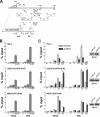
IFI16 promotes heterochromatin association with viral DNA. (A) Linear depiction of the HPV18 genome with the early (p55 and p102) and late (p811) regulatory regions expanded. (B) Extracts were prepared from HeLa, U2OS-mcHPV18, and NIKS-HPV18 cells, and ChIP was carried out using antibodies specific to unmodified histone H3 (H3), AcH3, dimethylated lysine 4 of H3 (H3K4me2), dimethylated lysine 9 of H3 (H3K9me2), or IgG as a control. Immunoprecipitated early and late promoter sequences were measured by qPCR, and CT values for the samples were equated to input CT values to give the percentages of input values for comparison. The data shown are the averages of the results of three experiments ± SD. (C) HeLa, U2OS-mcHPV18 at 2nd passage (#2), and NIKS-HPV18 cells at 6th passage (#6) were infected with AdVIFI16 or AdVLacZ. Cell extracts were prepared at 24 h after AdV infection, and ChIP was carried out as described for panel B. The data shown are the averages of the results of three experiments ± SD (*, P < 0.05; **, P < 0.01; unpaired t test). (Right) Lysate samples taken prior to immunoprecipitation (10 μl) were used for Western blot analysis (IB) with the antibodies for H3 to monitor equal loading.
FIG 5
FIG 5
Stable knockout of the IFI16 gene in U20S cells enhances HPV18 replication and inhibits viral-gene expression through histone modifications. (A) U2OS IFI16-knockout cells and the parental cell line were electroporated with HPV18 minicircle DNA and, at the indicated passage (#0, #2, #3, #5, or #6), total cellular DNA was extracted. Viral replication was measured by qPCR of the DpnI-digested DNA. The levels of glyceraldehyde 3-phosphate dehydrogenase were used to normalize the HPV18 levels. The data shown are the averages of the results of three experiments ± SD (*, P < 0.05; **, P < 0.01; unpaired t test). (B) U2OS IFI16-knockout cells at the 2nd passage after HPV18 minicircle DNA electroporation and the parental cell line were infected with AdVIFI16 or AdVLacZ, and at 24 and 48 h after AdV infection, total protein extracts were prepared for Western blot analysis with anti-IFI16 polyclonal antibody or α-tubulin as a loading control. Experiments were repeated at least three times, and one representative result is shown. (C) U2OS IFI16-knockout cells at the 2nd passage after HPV18 minicircle DNA electroporation were infected with AdVIFI16 or AdVLacZ, and at 24 and 48 h after AdV infection, total DNA was extracted and used to measure replicated (DpnI-resistant) viral DNA by qPCR. Levels of glyceraldehyde 3-phosphate dehydrogenase were used to normalize the HPV18 levels. The data shown are the averages of the results of three experiments ± SD (**, P < 0.01; unpaired t test). (D) U2OS IFI16-knockout cells at the 2nd passage after HPV18 minicircle DNA electroporation were infected with AdVLacZ (lanes 1 and 3) or AdVIFI16 (lanes 2 and 4), and 48 h later, total DNA extracts were prepared for Southern blot analysis. All samples contained 5 μg of genomic DNA and were digested with DpnI to remove any residual input DNA and with HindIII (with no restriction site in HPV18) (lanes 1 and 2) or DpnI and EcoRI to linearize HPV DNA (with one site in the HPV genome and one in the linker, which gave two bands of 5,113 bp and 2,835 bp) (lanes 3 and 4). The positions of HPV open-circle (oc) and oligomer forms are indicated on the left of the blots. Standards (HPV18 minicircles digested with EcoRI) corresponding to 10, 100, and 1,000 copies of the HPV18 genome per cell are included on the right (lanes 5, 6, and 7, respectively). The ∼8,000-bp band corresponds to partially digested minicircle DNA. The Southern blot was hybridized using the complete HPV18 genome as a probe. (E) Total RNA was extracted at 24 and 48 h after AdV infection in U2OS IFI16-knockout cells at the 2nd passage after HPV18 minicircle DNA electroporation and analyzed by real-time RT-PCR to measure the viral mRNAs. The values were normalized to glyceraldehyde 3-phosphate dehydrogenase and are shown as fold changes relative to AdVLacZ-infected cells for each time point. The data shown are the averages of the results of three experiments ± SD (*, P < 0.05; **, P < 0.01; unpaired t test). (F) ChIP assay performed with formalin-fixed extracts obtained from U2OS IFI16-knockout cells, at the 2nd passage after HPV18 minicircle electroporation, infected with AdVIFI16 or AdVLacZ for 24 h. ChIP was carried out using antibodies specific to unmodified histone H3 (H3), AcH3, H3K4me2, H3K9me2, or IgG as a control. Immunoprecipitated early and late promoter sequences were measured by qPCR; CT values for the samples were equated to input CT values to give the percentages of input values for comparison. The data shown are the averages of the results of three experiments ± SD (*, P < 0.05; **, P < 0.01; unpaired t test). (Right) Lysate samples taken prior to immunoprecipitation (10 μl) were used for Western blot analysis with the antibodies for H3 to monitor equal loading.
FIG 6
FIG 6
IFI16 does not colocalize with the HPV18 genome, and its viral restriction activity is independent of IFN response. (A) U2OS IFI16-knockout cells at the 2nd passage after HPV18 minicircle DNA electroporation or nonelectroporated cells were grown at high density, infected with AdVIFI16 for 8 h, and then fixed in 4% paraformaldehyde and subjected to combined FISH with HPV18 probe (green dots) and immunofluorescence analysis with anti-IFI16 antibodies (red). The images were taken by confocal microscopy; the far-left image shows IFI16 expression in nonelectroporated cells (scale bar = 40 μm), while all the other images were taken in U2OSIFI16null-mcHPV18 cells; the far-right image shows cross-sectional analysis of a z-stack image series. Ten fields were digitally analyzed, and one representative image is shown. Scale bars = 10 μm. (B) U2OS IFI16-knockout cells at the 2nd passage after HPV18 minicircle DNA electroporation were infected with AdVLacZ or AdVIFI16 in the presence of anti-IFNAR antibodies or an isotype control, as detailed in Materials and Methods. At 48 h after AdV infection, total DNA was extracted and used to measure replicated (DpnI-resistant) viral DNA by qPCR. The levels of glyceraldehyde 3-phosphate dehydrogenase were used to normalize the HPV18 levels. The data shown are the averages of the results of three experiments ± SD (*, P < 0.05; unpaired t test). (C) Total RNA was also extracted from U2OS IFI16-knockout cells at the 2nd passage after HPV18 minicircle DNA electroporation infected with AdVLacZ or AdVIFI16 (24 h) or treated with poly(I·C) (8 h) in the presence of anti-IFNAR antibodies or isotype control, as detailed in Materials and Methods. Representative expression of a set of ISGs is shown. The values were normalized to glyceraldehyde 3-phosphate dehydrogenase and are shown as fold changes relative to AdVLacZ-infected cells for each time point. The data shown are the averages of the results of three experiments ± SD (*, P < 0.05; **, P < 0.01; unpaired t test).

Similar articles

Cited by

References

    1. Stremlau M, Owens CM, Perron MJ, Kiessling M, Autissier P, Sodroski J. 2004. The cytoplasmic body component TRIM5alpha restricts HIV-1 infection in Old World monkeys. Nature 427:848–853. doi:10.1038/nature02343. - DOI - PubMed
    1. Bieniasz PD. 2004. Intrinsic immunity: a front-line defense against viral attack. Nat Immunol 5:1109–1115. doi:10.1038/ni1125. - DOI - PubMed
    1. Duggal NK, Emerman M. 2012. Evolutionary conflicts between viruses and restriction factors shape immunity. Nat Rev Immunol 12:687–695. doi:10.1038/nri3295. - DOI - PMC - PubMed
    1. Yan N, Chen ZJ. 2012. Intrinsic antiviral immunity. Nat Immunol 13:214–222. doi:10.1038/ni.2229. - DOI - PMC - PubMed
    1. Takeuchi H, Matano T. 2008. Host factors involved in resistance to retroviral infection. Microbiol Immunol 52:318–325. doi:10.1111/j.1348-0421.2008.00040.x. - DOI - PubMed

Publication types

MeSH terms

LinkOut - more resources