. 2015 May 12;6(3):e00553-15.
doi: 10.1128/mBio.00553-15.

Dengue Virus NS Proteins Inhibit RIG-I/MAVS Signaling by Blocking TBK1/IRF3 Phosphorylation: Dengue Virus Serotype 1 NS4A Is a Unique Interferon-Regulating Virulence Determinant

Affiliations

Dengue Virus NS Proteins Inhibit RIG-I/MAVS Signaling by Blocking TBK1/IRF3 Phosphorylation: Dengue Virus Serotype 1 NS4A Is a Unique Interferon-Regulating Virulence Determinant

Nadine A Dalrymple et al. mBio. .

Abstract

Dengue virus (DENV) replication is inhibited by the prior addition of type I interferon or by RIG-I agonists that elicit RIG-I/MAVS/TBK1/IRF3-dependent protective responses. DENV infection of primary human endothelial cells (ECs) results in a rapid increase in viral titer, which suggests that DENV inhibits replication-restrictive RIG-I/interferon beta (IFN-β) induction pathways within ECs. Our findings demonstrate that DENV serotype 4 (DENV4) nonstructural (NS) proteins NS2A and NS4B inhibited RIG-I-, MDA5-, MAVS-, and TBK1/IKKε-directed IFN-β transcription (>80%) but failed to inhibit IFN-β induction directed by STING or constitutively active IRF3-5D. Expression of NS2A and NS4B dose dependently inhibited the phosphorylation of TBK1 and IRF3, which suggests that they function at the level of TBK1 complex activation. NS2A and NS4B from DENV1/2/4, as well as the West Nile virus NS4B protein, commonly inhibited TBK1 phosphorylation and IFN-β induction. A comparative analysis of NS4A proteins across DENVs demonstrated that DENV1, but not DENV2 or DENV4, NS4A proteins uniquely inhibited TBK1. These findings indicate that DENVs contain conserved (NS2A/NS4B) and DENV1-specific (NS4A) mechanisms for inhibiting RIG-I/TBK1-directed IFN responses. Collectively, our results define DENV NS proteins that restrict IRF3 and IFN responses and thereby facilitate DENV replication and virulence. Unique DENV1-specific NS4A regulation of IFN induction has the potential to be a virulence determinant that contributes to the increased severity of DENV1 infections and the immunodominance of DENV1 responses during tetravalent DENV1-4 vaccination.

Importance: Our findings demonstrate that NS2A and NS4B proteins from dengue virus serotypes 1, 2, and 4 are inhibitors of RIG-I/MDA5-directed interferon beta (IFN-β) induction and that they accomplish this by blocking TBK1 activation. We determined that IFN inhibition is functionally conserved across NS4B proteins from West Nile virus and DENV1, -2, and -4 viruses. In contrast, DENV1 uniquely encodes an extra IFN regulating protein, NS4A, that inhibits TBK1-directed IFN induction. DENV1 is associated with an increase in severe patient disease, and added IFN regulation by the DENV1 NS4A protein may contribute to increased DENV1 replication, immunodominance, and virulence. The regulation of IFN induction by nonstructural (NS) proteins suggests their potential roles in enhancing viral replication and spread and as potential protein targets for viral attenuation. DENV1-specific IFN regulation needs to be considered in vaccine strategies where enhanced DENV1 replication may interfere with DENV2-4 seroconversion within coadministered tetravalent DENV1-4 vaccines.


Figures

FIG 1
FIG 1
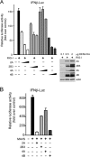
NS2A and NS4B antagonize RIG-I/MDA5-directed type I IFN induction. (A) Schematic of DENV polyprotein, indicating structural and nonstructural (NS) proteins produced after cleavage by host and viral proteases. Full-length (uncleaved) and pro (active) forms of the viral 2B3 protease are depicted. UTR, untranslated region. (B and C) HEK293T cells were cotransfected with identical amounts of total DNA, including IFN-β promoter or ISRE-driven firefly luciferase (Luc) reporters, Renilla luciferase plasmid, and the indicated RIG-I-CARD or MDA5 expression vectors in the presence (+) or absence (−) of plasmids expressing Flag-tagged DENV4 NS2A, NS2B3, NS4A, NS4B, or empty control plasmid (11). Luciferase activity was measured 24 h posttransfection, normalized to Renilla luciferase activity, and reported as fold increase compared to controls lacking RIG-I-CARD or MDA5. Assays were performed in duplicate with similar results from at least three separate experiments. Expression of dengue virus nonstructural proteins and inducers was assessed by Western blot analysis using anti-Flag (α-Flag) and anti-RIG-I or anti-MDA5. β-Actin serves as a loading control. Asterisks indicate statistical significance (P < 0.05) as determined by Student’s t test.
FIG 2
FIG 2
NS2A and NS4B dose dependently inhibit RIG-I- and MAVS-directed IFN induction. HEK293T cells were cotransfected as described in the legend to Fig. 1 with IFN-β promoter firefly luciferase reporter, Renilla luciferase plasmid, and RIG-I-CARD (A) or MAVS (B) expression vectors in the presence or absence of the indicated DENV NS expression plasmids (0.1, 0.5, or 2 µg [indicated by the black triangle] [A]) or 2 µg [B]) or control empty plasmid (2 µg). Luciferase activity was assessed and evaluated as described in the legend to Fig. 1, and expression of DENV4 NS proteins was determined by Western blot analysis using anti-HA with β-actin as a loading control (11).
FIG 3
FIG 3
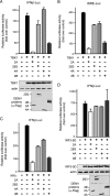
NS2A and NS4B inhibit TBK1- and IKKε-directed IFN-β transcription. HEK293T cells were cotransfected as described in the legend to Fig. 1 in the presence or absence of TBK1 (A and B), IKKε (C), or IRF3-5D (D) and the indicated DENV4 NS expression plasmids. Luciferase activity was measured 24 h posttransfection and reported as described in the legend to Fig. 1. Assays were performed in duplicate with similar results from at least three separate experiments. Expression of DENV NS proteins and inducers was assessed by Western blot analysis using anti-Flag and anti-TBK1, anti-IKKε, or anti-IRF3 antibodies, respectively, with β-actin as a loading control.
FIG 4
FIG 4
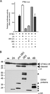
NS2A and NS4B fail to inhibit the induction of IFN-β by STING. (A) HEK293T cells were cotransfected as described in the legend to Fig. 1 with luciferase reporters and the STING-V5 expression plasmid as activator in the presence or absence of indicated plasmids expressing DENV4 NS2A, NS2B3pro (active, processed form), NS2B3 (full length), NS4A, or NS4B. Luciferase activity was measured 24 h posttransfection and reported as fold increase over the value for the empty plasmid control. Assays were performed in duplicate with similar results from at least three separate experiments. (B) HEK293T cells were transfected with plasmid expressing STING-V5 and either empty plasmid (−) or plasmid expressing the indicated DENV4 NS proteins. Cell lysates were analyzed 24 h posttransfection by Western blot analysis of STING-V5 expression using anti-V5 and DENV proteins using anti-HA. The positions of the full-length (36 kDa) and cleaved forms (32 kDa) of STING-V5 are indicated by the arrows to the right of the blots. Asterisks to the right of the Western blot denote expression of each DENV NS protein.
FIG 5
FIG 5
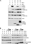
NS2A and NS4B block RIG-I-induced IRF3 phosphorylation. (A) HEK293T cells were cotransfected with IRF3-T7 expression plasmid, and as indicated, RIG-I-CARD expression plasmid, plasmids expressing DENV4 NS2A, NS4A, or NS4B, or empty vector. Cells were harvested 24 h posttransfection and analyzed by Western blotting for pIRF3 (anti-pIRF3 Ser396), total IRF3 (anti-IRF3), β-actin (anti-β-actin), and DENV4 NS protein expression (anti-Flag). IRF3 levels were similarly analyzed in identical samples that were treated with MG132 (20 µM) 5 h prior to harvest. β-Actin serves as a loading control. (B) Cells were cotransfected and analyzed as described in the legend to Fig. 6A with increasing amounts of indicated NS protein expression plasmid (0.5, 1, or 2 µg).
FIG 6
FIG 6
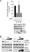
NS2A and NS4B dose dependently inhibit TBK1 phosphorylation. (A) Plasmids expressing TBK1 and DENV4 NS2A, NS4A, and NS4B, as indicated, were cotransfected into HEK293T cells. Cells were harvested 24 h posttransfection and analyzed for pTBK1 (anti-pTBK1 Ser172), total TBK1 (anti-TBK1), β-actin (anti-β-actin), and DENV4 NS protein (anti-Flag) expression by Western blot analysis. Phosphorylated TBK1 expression levels were measured from three replicate Western blot analyses and graphed as a percentage of control levels in the absence of DENV protein expression. (B) Increasing amounts of NS2A or NS4B plasmid (0.5, 1, or 2 µg) were cotransfected with TBK1 expression plasmid and analyzed as described above for panel A.
FIG 7
FIG 7
The N-terminal domain of DENV NS4B inhibits RIG-I and TBK1 induction of IFN-β. HEK293T cells were cotransfected as described in the legend to Fig. 1 with RIG-I-CARD or TBK1, IFN-β promoter luciferase reporter, Renilla luciferase plasmid, and as indicated, plasmids expressing HA-tagged DENV4 wild-type NS4B, NS4BΔ118-260, or NS4BΔ1-117. Changes in IFN-β transcriptional responses are reported as fold increase compared to the values for controls lacking RIG-I or TBK1. Asterisks indicate statistical significance (P < 0.05) as determined by Student’s t test.
FIG 8
FIG 8
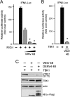
West Nile virus NS4B inhibits RIG-I- and TBK1-directed IFN-β induction. (A and B) HEK293T cells were cotransfected with IFN-β reporters as described in the legend to Fig. 7A and RIG-I-CARD (A) or TBKI (B) expression plasmids in the presence of constant or increasing amounts of plasmid expressing WNV (NY99 strain) NS4B (0.5, 1, or 2 µg) or empty vector. Luciferase activity was measured and analyzed as described in the legend to Fig. 1 and reported as fold induction over the value for the control lacking RIG-I or TBK1. Asterisks indicate statistical significance (P < 0.05) as determined by Student’s t test. (C) HEK293 cells were cotransfected with TBK1 plasmid in the presence or absence of plasmid expressing the NS4B protein from WNV or DENV4 as indicated. Cells were harvested 24 h posttransfection. Phosphorylation of TBK1 was analyzed by Western blot analysis using anti-pTBK1 (Ser172). WNV NS4B, DENV4 NS4B, and total TBK1 levels were detected using anti-FLAG and anti-TBK1 antibodies, respectively, with β-actin expression as a loading control.
FIG 9
FIG 9
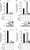
Comparison of DENV1 and DENV2 NS protein inhibition of RIG-I/TBK1 signaling. (A to D) HEK293T cells were cotransfected as described in the legend to Fig. 1 with RIG-I-CARD (A and B) or TBK1 (C and D) plasmids, IFN-β luciferase reporter, and Renilla luciferase plasmids, and as indicated, plasmids expressing NS2A, NS4A and NS4B from DENV1 (DV1) (A and C) or DENV2 (DV2) (B and D). Luciferase activity was measured 24 h posttransfection, normalized to Renilla luciferase activity, and reported as fold increase compared to the control values. Results are reported from duplicate samples and were similar in at least three independent experiments.
FIG 10
FIG 10
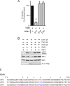
DENV1 NS4A uniquely inhibits TBK1 signaling and IRF3 phosphorylation. (A) HEK293T cells were cotransfected as described in the legend to Fig. 1 with TBK1 plasmid, IFN-β luciferase reporter, and Renilla luciferase plasmids, and plasmids expressing NS4A from DENV1 (DV1), DENV2 (DV2), or DENV4 (DV4). Luciferase activity was measured as described in the legend to Fig. 9. Results are reported from duplicate samples and were similar in at least three independent experiments. Asterisks indicate statistical significance (P < 0.05) as determined by Student’s t test. (B) Cells were cotransfected as described in the legend to Fig. 5 with IRF3-T7 expression plasmids in the presence or absence of RIG-I-CARD and plasmids expressing Flag-tagged NS4A proteins from DENV1, DENV2, or DENV4 as indicated. Phosphorylation of IRF3 was measured 24 h posttransfection by Western blot analysis as described in the legend to Fig. 5. (C) Alignment of variable regions of DENV1/2/4 NS4A proteins with unique residues highlighted in red, variable residue positions in blue, and residues within DENV1 NS4A that represent charge changes from DENV2/4 NS4A proteins denoted by asterisks.

Similar articles

Cited by

References

    1. Lindenbach BD, Murray CL, Thiel H-J, Rice CM. 2013. Flaviviridae. In Knipe DM, Howley PM (ed), Field’s virology, vol 1, 6th ed, vol 1 Lippincott Williams & Wilkins, New York, NY.
    1. Normile D. 2014. Tropical diseases. Dengue vaccine trial poses public health quandary. Science 345:367–368. doi:10.1126/science.345.6195.367. - DOI - PubMed
    1. Gubler DJ. 2006. Dengue/dengue haemorrhagic fever: history and current status. Novartis Found Symp 277:3–22, 71,–73, 251–253. doi:10.1002/0470058005.ch2. - DOI - PubMed
    1. Guzman A, Istúriz RE. 2010. Update on the global spread of dengue. Int J Antimicrob Agents 36(Suppl 1):S40–S42. doi:10.1016/j.ijantimicag.2010.06.018. - DOI - PubMed
    1. Halstead SB. 2007. Dengue. Lancet 370:1644–1652. doi:10.1016/S0140-6736(07)61687-0. - DOI - PubMed

Publication types

MeSH terms