. 2015 May 8;10(5):e0126159.
doi: 10.1371/journal.pone.0126159. eCollection 2015.

An Autocrine Cytokine/JAK/STAT-Signaling Induces Kynurenine Synthesis in Multidrug Resistant Human Cancer Cells

Affiliations

An Autocrine Cytokine/JAK/STAT-Signaling Induces Kynurenine Synthesis in Multidrug Resistant Human Cancer Cells

Ivana Campia et al. PLoS One. .

Abstract

Background: Multidrug resistant cancer cells are hard to eradicate for the inefficacy of conventional anticancer drugs. Besides escaping the cytotoxic effects of chemotherapy, they also bypass the pro-immunogenic effects induced by anticancer drugs: indeed they are not well recognized by host dendritic cells and do not elicit a durable anti-tumor immunity. It has not yet been investigated whether multidrug resistant cells have a different ability to induce immunosuppression than chemosensitive ones. We addressed this issue in human and murine chemosensitive and multidrug resistant cancer cells.

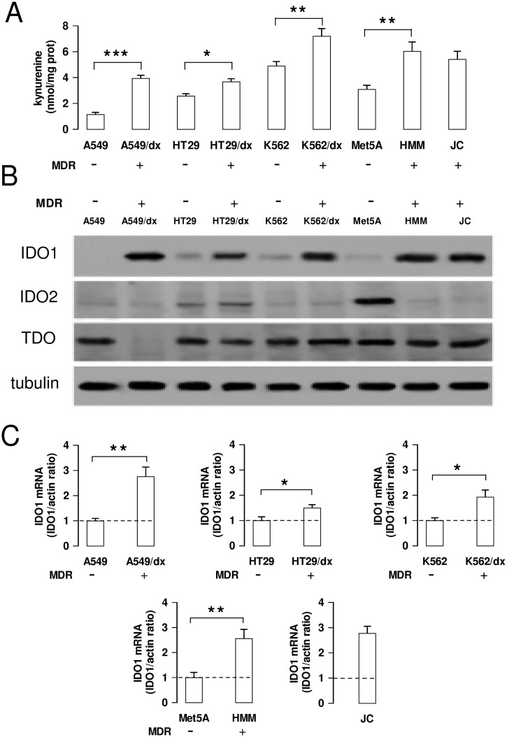
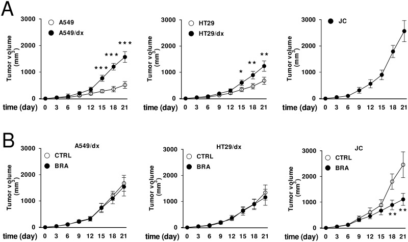
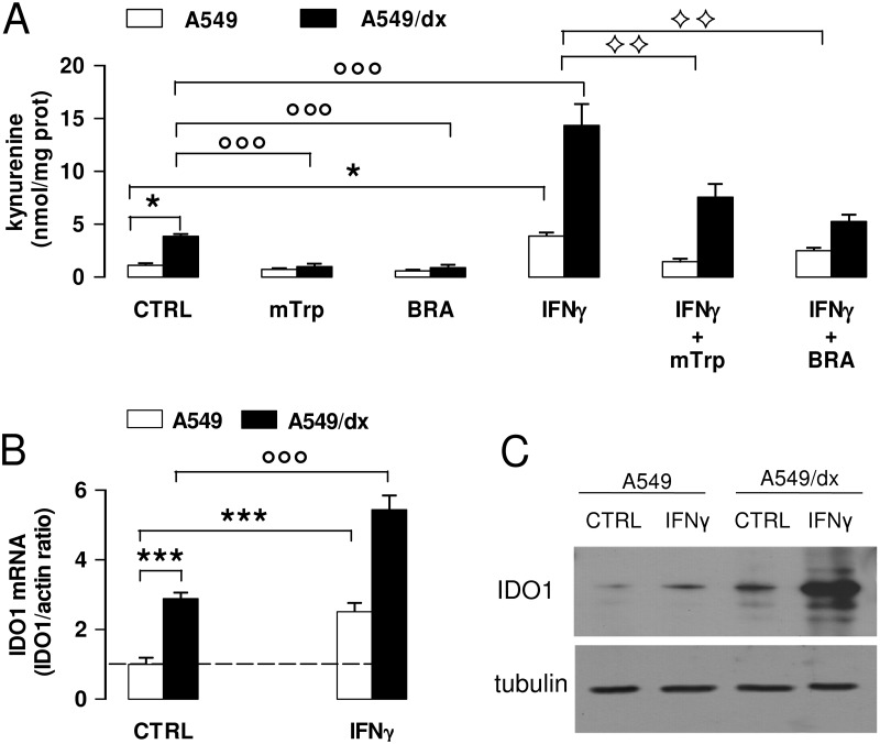
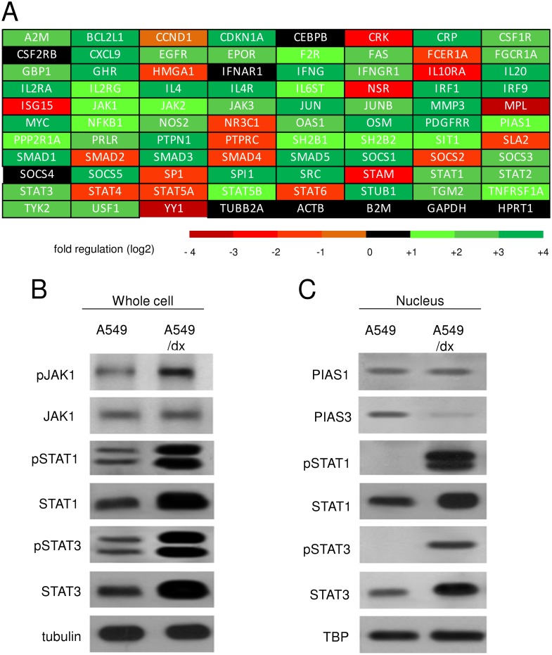
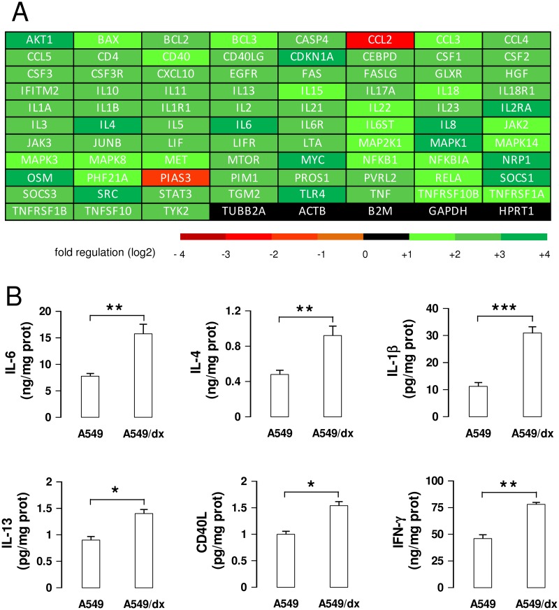
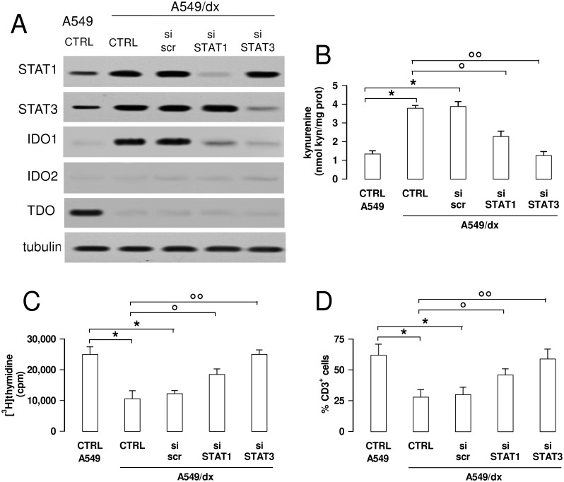
Results: We found that the activity and expression of indoleamine 2,3-dioxygenase 1 (IDO1), which catalyzes the conversion of tryptophan into the immunosuppressive metabolite kynurenine, was higher in all the multidrug resistant cells analyzed and that IDO1 inhibition reduced the growth of drug-resistant tumors in immunocompetent animals. In chemoresistant cells the basal activity of JAK1/STAT1 and JAK1/STAT3 signaling was higher, the STAT3 inhibitor PIAS3 was down-regulated, and the autocrine production of STAT3-target and IDO1-inducers cytokines IL-6, IL-4, IL-1β, IL-13, TNF-α and CD40L, was increased. The disruption of the JAK/STAT signaling lowered the IDO1 activity and reversed the kynurenine-induced pro-immunosuppressive effects, as revealed by the restored proliferation of T-lymphocytes in STAT-silenced chemoresistant cells.

Conclusions: Our work shows that multidrug resistant cells have a stronger immunosuppressive attitude than chemosensitive cells, due to the constitutive activation of the JAK/STAT/IDO1 axis, thus resulting chemo- and immune-evasive. Disrupting this axis may significantly improve the efficacy of chemo-immunotherapy protocols against resistant tumors.


Conflict of interest statement

Competing Interests: The authors have declared that no competing interests exist.

Figures

Fig 1
Fig 1. Kynurenine production and IDO1 expression in chemosensitive and multidrug resistant cells.
Human chemosensitive lung cancer A549 cells and chemoresistant A549/dx cells, human chemosensitive colon cancer HT29 cells and chemoresistant HT29/dx cells, human chemosensitive chronic myelogenous leukemia K562 cells and chemoresistant K562/dx cells, human chemosensitive mesothelial Met5A cells and human chemoresistant malignant mesothelioma HMM cells, murine chemoresistant mammary JC cells were subjected to the following investigations. A. The kynurenine levels in the cell culture supernatants were measured spectrophotometrically. Data are presented as means ± SD (n = 4). * p < 0.05, ** p < 0.01, *** p < 0.001: chemoresistant cells (MDR-positive) versus the corresponding chemosensitive (MDR-negative) cells. B. Western blot analysis of IDO1, IDO2 and TDO expression. The β-tubulin expression was used as control of equal protein loading. The figure is representative of 3 experiments with similar results. C. The expression level of IDO1 mRNA was measured by qRT-PCR. Data are presented as means ± SD (n = 4). * p < 0.01, ** p < 0.002: chemoresistant cells (MDR-positive) versus the corresponding chemosensitive (MDR-negative) cells.
Fig 2
Fig 2. Effects of IDO1 inhibition on the growth of multidrug resistant tumors.
A. 1 x 10⁵ human A549, A549/dx, HT29, HT29/dx cells were implanted subcutaneously in 6–8 weeks old female nude BALB/c mice, 1 x 10⁵ murine JC cells were implanted in immunocompetent BALB/c mice. Tumor growth was monitored daily by caliper measurement. Data are presented as means ± SD of 10 mice/group. * p < 0.02, ** p < 0.005, *** p < 0.001: A549/dx or HT29/dx cells versus A549 or HT29 cells, at the corresponding time points. B. Animals bearing A549/dx-, HT29/dx-, JC-tumors were randomized into two groups when tumors reached the volume of 100 mm3: “Control” group (treated with 100 μL of saline solution per os, 5 days/week for three weeks; CTRL); “Brassinin” group (treated with 400 mg/kg of the IDO1 inhibitor 5-Br-brassinin per os, 5 days/week for three weeks; BRA). Tumor growth was monitored daily by caliper measurement. Data are presented as means ± SD of 6 mice/group. ** p < 0.005: BRA-group versus CTRL-group, at the corresponding time points.
Fig 3
Fig 3. Effects of IFN-γ on kynurenine synthesis and IDO1 expression in chemosensitive and multidrug resistant cells.
A549 and A549/dx cells were incubated for 48 h in fresh medium (CTRL) or in medium containing the IDO1 inhibitors methyl-DL-tryptophan (1 mmol/L, mTrp) or 5-Br-brassinin (100 μmol/L, BRA), and the IDO1 inducer IFN-γ (100 ng/mL, IFNγ), alone or in combination. A. The kynurenine levels in the cell culture supernatants were measured spectrophotometrically. Data are presented as means ± SD (n = 4). * p < 0.01: versus A549 CTRL cells; °°° p < 0.001: versus A549/dx CTRL; ◊◊ p < 0.005: IFN-γ + mTrp-treated, IFN-γ + BRA-treated A549 and A549/dx cells versus the corresponding cells treated with IFN-γ alone. B. The expression level of IDO1 mRNA was measured by qRT-PCR. Data are presented as means ± SD (n = 4). *** p < 0.001: versus A549 CTRL cells; °°° p < 0.001: versus A549/dx CTRL cells. C. Western blot analysis of IDO1 expression. The β-tubulin expression was used as control of equal protein loading. The figure is representative of 3 experiments with similar results.
Fig 4
Fig 4. Multidrug resistant cells have a higher activity of JAK/STAT signaling than chemosensitive cells.
A. The cDNA from A549 and A549/dx cells was analyzed by a PCR array specific for JAK/STAT signaling, as reported under Materials and methods. The fold regulation of the 83 genes analyzed, expressed in logarithmic scale, is represented in a colorimetric scale. The figure is the mean of 4 experiments. B. The cells were lysed and subjected to the Western blot analysis for phospho(Tyr 1022/1023)-JAK1, JAK1, phospho(Tyr701)-STAT1, STAT1, phospho(Tyr705)-STAT3, STAT3. The β-tubulin expression was used as control of equal protein loading. The figure is representative of 3 experiments with similar results. C. The expression of PIAS1, PIAS3, phospho(Tyr701)-STAT1, STAT1, phospho(Tyr705)-STAT3, STAT3 in nuclear extracts was measured by Western blotting. The TBP expression was used as control of equal protein loading. The figure is representative of 3 experiments with similar results.
Fig 5
Fig 5. Multidrug resistant cells have a higher activity of IL-6/STAT3 signaling than chemosensitive cells.
A. The cDNA from A549 and A549/dx cells was analyzed by a PCR array specific for IL-6/STAT3 signaling, as reported under Materials and methods. The fold regulation of the 83 genes analyzed, expressed in logarithmic scale, was represented in a colorimetric scale. The figure is the mean of 4 experiments. B. The levels of IL-6, IL-4, IL-1β, IL-13, CD40L, IFN-γ were measured in the cell culture supernatants by specific ELISAs. Data are presented as means ± SD (n = 3). * p < 0.02, ** p < 0.005, *** p < 0.001: A549/dx cells versus A549 cells.
Fig 6
Fig 6. The inhibition of the STAT1/STAT3 signaling reverses the kynurenine-dependent immunosuppression in multidrug resistant cells.
A549/dx cells were grown for 48 h in fresh medium (CTRL), treated with a non-targeting scrambled siRNA (scr) or with a specific siRNAs pool targeting STAT1 or STAT3, respectively (si STAT1, si STAT3). Untreated chemosensitive A549 cells were used as control. A. The expression of STAT1, STAT3, IDO1, IDO2 and TDO was measured in whole cell lysates by Western blotting, 48 h after the transfection. The β-tubulin expression was used as control of equal protein loading. The figure is representative of 3 experiments with similar results. B. The kynurenine levels in the cell culture supernatants were measured spectrophotometrically. Data are presented as means ± SD (n = 4). * p < 0.01: versus A549 CTRL; ° p < 0.005, °° p < 0.001: versus A549/dx CTRL. C. The proliferation of activated T-lymphocytes collected from PBMC after a 72 h co-incubation with A549 and A549/dx cells was measured with the [3H]thymidine assay. In the presence of anti-CD3 and anti-CD28 stimulatory antibodies without tumor cells (positive control), the [3H]thymidine incorporation was 28,926 ± 1,426 cpm; in the presence of RPMI medium alone (negative control), the [3H]thymidine incorporation was 4,312 ± 529 cpm. Data are presented as means ± SD (n = 6). * p < 0.05: versus A549 CTRL; ° p < 0.01, °° p < 0.005: versus A549/dx CTRL. D. The percentage of CD3+ T-lymphocytes collected from PBMC, co-incubated with tumor cells as reported in C, was measured by flow cytometry. Data are presented as means ± SD (n = 6). * p < 0.01: versus A549 CTRL; ° p < 0.01, °° p < 0.002: versus A549/dx CTRL.

Similar articles

Cited by

References

    1. Gottesman MM, Fojo T, Bates SE (2002). Multidrug resistance in cancer: role of ATP-dependent transporters. Nat Rev Cancer 2(1): 48–58. - PubMed
    1. Agarwal S, Sane R, Oberoi R, Ohlfest JR, Elmquist WF (2011) Delivery of molecularly targeted therapy to malignant glioma, a disease of the whole brain. Expert Rev Mol Med 13: e17 10.1017/S1462399411001888 - DOI - PMC - PubMed
    1. Obeid M, Tesniere A, Ghiringhelli F, Fimia GM, Apetoh L, Perfettini JL, et al. (2007) Calreticulin exposure dictates the immunogenicity of cancer cell death. Nat Med 13(1): 54–61. - PubMed
    1. De Boo S, Kopecka J, Brusa D, Gazzano E, Matera L, Ghigo D, et al. (2009) iNOS activity is necessary for the cytotoxic and immunogenic effects of doxorubicin in human colon cancer cells. Mol Cancer 8: e108. - PMC - PubMed
    1. Kopecka J, Campia I, Brusa D, Doublier S, Matera L, Ghigo D, et al. (2011) Nitric oxide and P-glycoprotein modulate the phagocytosis of colon cancer cells. J Cell Mol Med 15(7): 1492–1504. 10.1111/j.1582-4934.2010.01137.x - DOI - PMC - PubMed

Publication types

MeSH terms

Grants and funding

This work was supported by grants from the Italian Association for Cancer Research (AIRC; grant MFAG 11475; www.airc.it), the Italian Ministry of University and Research (“Future in Research program” FIRB 2012, grant RBFR12SOQ1; www.istruzione.it). JK is fellow of the Italian Foundation for Cancer Research (FIRC; www.fondazionefirc.it). The funders had no role in study design, data collection and analysis, decision to publish, or preparation of the manuscript.