

Altered volume, morphology and composition of the pancreas in type 2 diabetes
- PMID: 25950180
- PMCID: PMC4423920
- DOI: 10.1371/journal.pone.0126825
Altered volume, morphology and composition of the pancreas in type 2 diabetes
Abstract
Objective: Although impairment in pancreatic insulin secretion is known to precede the clinical diagnosis of type 2 diabetes by up to a decade, fasting blood glucose concentration only rises abnormally once the impairment reaches a critical threshold. Despite its centrality to the pathogenesis of type 2 diabetes, the pancreas is the least studied organ due to its inaccessible anatomical position. Previous ultrasound and CT studies have suggested a possible decrease in pancreatic volume in type 2 diabetes. However, ultrasound techniques are relatively insensitive while CT uses ionizing radiation, making these modalities unsuitable for precise, longitudinal studies designed to explore the underlying mechanisms of type 2 diabetes. Hence there is a need to develop a non-invasive, safe and precise method to quantitate pancreas volume.
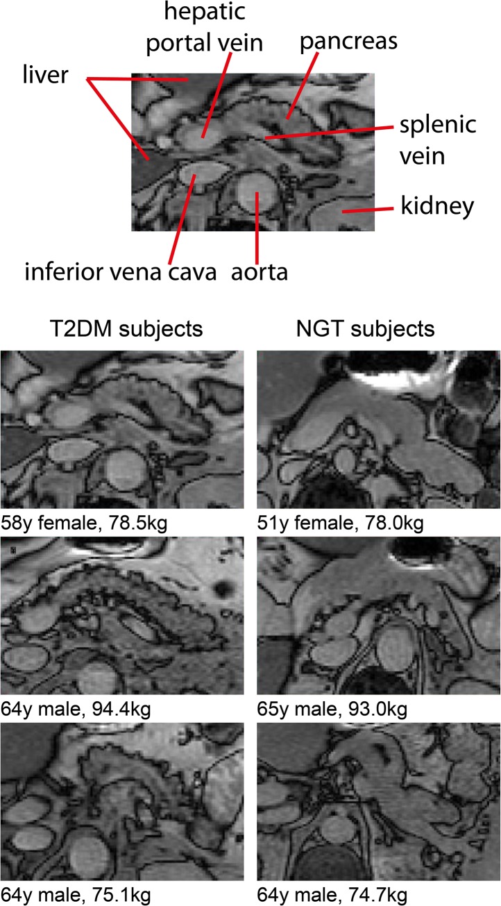
Methods: We developed and applied magnetic resonance imaging at 3.0T to obtain balanced turbo field echo (BTFE) structural images of the pancreas, together with 3-point Dixon images to quantify pancreatic triglyceride content. Pancreas volume, morphology and triglyceride content was quantified in a group of 41 subjects with well-controlled type 2 diabetes (HbA1c ≤ 7.6%) taking only metformin (duration of T2DM 5.7 ± 0.7 years), and a control group of 14 normal glucose tolerance subjects matched for age, weight and sex.
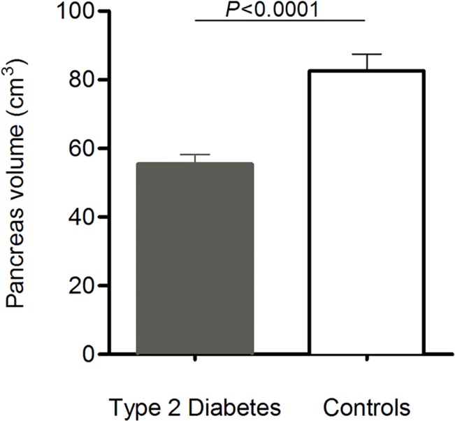
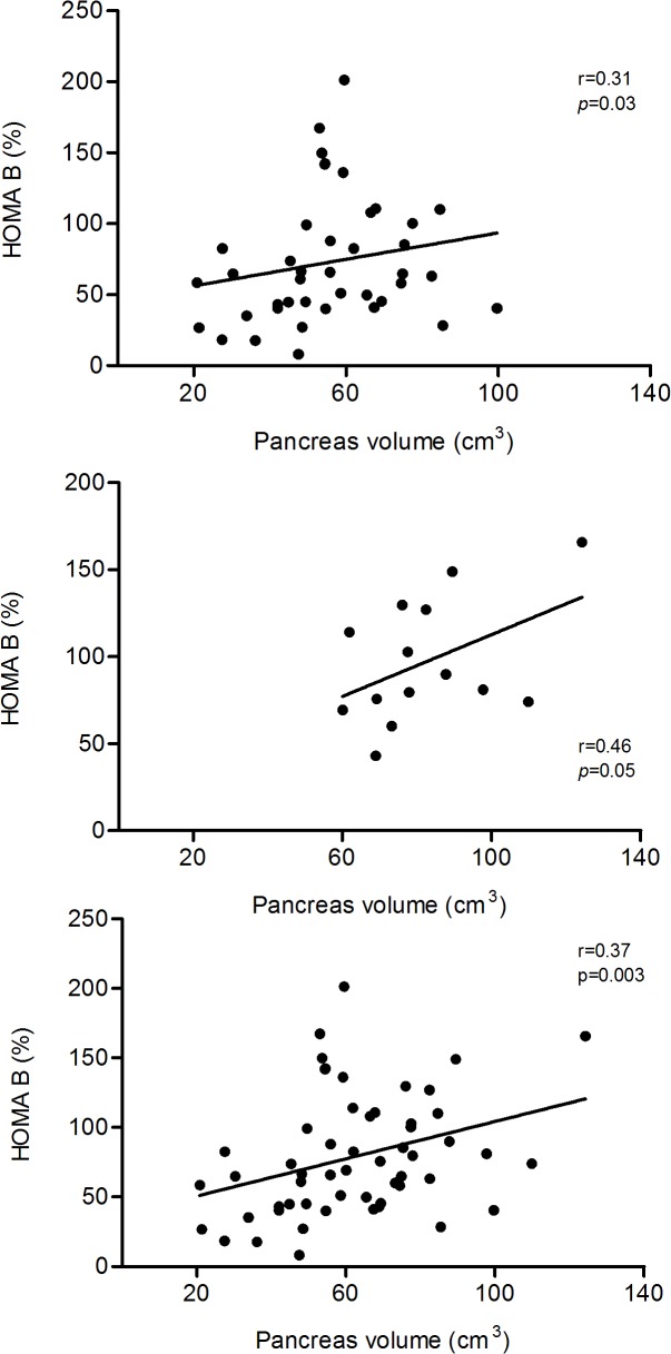
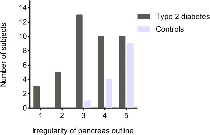
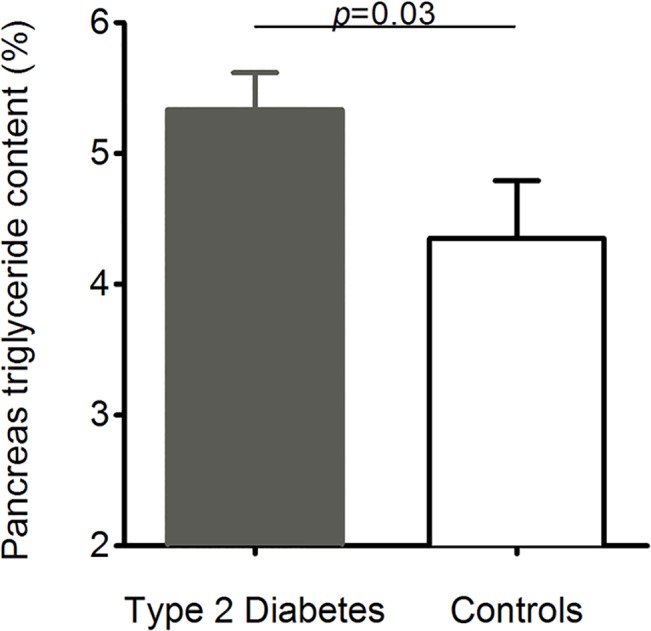
Results: The mean pancreatic volume was found to be 33% less in type 2 diabetes than in normal glucose tolerant subjects (55.5 ± 2.8 vs. 82.6 ± 4.8 cm3; p < 0.0001). Pancreas volume was positively correlated with HOMA-β in the type 2 diabetes subjects (r = 0.31; p = 0.03) and controls (r = 0.46; p = 0.05) considered separately; and in the whole population studied (r = 0.37; p = 0.003). In type 2 diabetes, the pancreas was typically involuted with a serrated border. Pancreatic triglyceride content was 23% greater (5.4 ± 0.3 vs. 4.4 ± 0.4%; p = 0.02) in the type 2 diabetes group.
Conclusion: This study describes for the first time gross abnormalities of the pancreas in early type 2 diabetes and quantifies the decrease in pancreas size, the irregular morphology and increase in fat content.
Conflict of interest statement
Figures
Similar articles
-
Quantification of ectopic fat storage in the liver and pancreas using six-point Dixon MRI and its association with insulin sensitivity and β-cell function in patients with central obesity.Eur Radiol. 2023 Dec;33(12):9213-9222. doi: 10.1007/s00330-023-09856-x. Epub 2023 Jul 6. Eur Radiol. 2023. PMID: 37410109
-
2-year remission of type 2 diabetes and pancreas morphology: a post-hoc analysis of the DiRECT open-label, cluster-randomised trial.Lancet Diabetes Endocrinol. 2020 Dec;8(12):939-948. doi: 10.1016/S2213-8587(20)30303-X. Epub 2020 Oct 5. Lancet Diabetes Endocrinol. 2020. PMID: 33031736 Clinical Trial.
-
Morphology of the pancreas in type 2 diabetes: effect of weight loss with or without normalisation of insulin secretory capacity.Diabetologia. 2016 Aug;59(8):1753-9. doi: 10.1007/s00125-016-3984-6. Epub 2016 May 14. Diabetologia. 2016. PMID: 27179658 Free PMC article.
-
Pancreatic size and fat content in diabetes: A systematic review and meta-analysis of imaging studies.PLoS One. 2017 Jul 24;12(7):e0180911. doi: 10.1371/journal.pone.0180911. eCollection 2017. PLoS One. 2017. PMID: 28742102 Free PMC article. Review.
-
Cross-sectional imaging of the pancreas in diabetes.Abdom Radiol (NY). 2024 Jun;49(6):2116-2124. doi: 10.1007/s00261-024-04310-y. Epub 2024 Apr 1. Abdom Radiol (NY). 2024. PMID: 38557767 Review.
Cited by
-
Fatty Pancreas and Cardiometabolic Risk: Response of Ectopic Fat to Lifestyle and Surgical Interventions.Nutrients. 2022 Nov 17;14(22):4873. doi: 10.3390/nu14224873. Nutrients. 2022. PMID: 36432559 Free PMC article. Review.
-
Intra-pancreatic fat deposition: bringing hidden fat to the fore.Nat Rev Gastroenterol Hepatol. 2022 Mar;19(3):153-168. doi: 10.1038/s41575-021-00551-0. Epub 2021 Dec 8. Nat Rev Gastroenterol Hepatol. 2022. PMID: 34880411 Review.
-
Radiomics-based machine learning (ML) classifier for detection of type 2 diabetes on standard-of-care abdomen CTs: a proof-of-concept study.Abdom Radiol (NY). 2022 Nov;47(11):3806-3816. doi: 10.1007/s00261-022-03668-1. Epub 2022 Sep 10. Abdom Radiol (NY). 2022. PMID: 36085379
-
Prediction of type 2 diabetes mellitus using noninvasive MRI quantitation of visceral abdominal adiposity tissue volume.Quant Imaging Med Surg. 2019 Jun;9(6):1076-1086. doi: 10.21037/qims.2019.06.01. Quant Imaging Med Surg. 2019. PMID: 31367561 Free PMC article.
-
Age-Related Changes in Pediatric Physiology: Quantitative Analysis of Organ Weights and Blood Flows : Age-Related Changes in Pediatric Physiology.AAPS J. 2021 Mar 31;23(3):50. doi: 10.1208/s12248-021-00581-1. AAPS J. 2021. PMID: 33791883
References
-
- Reaven GM. Banting lecture 1988. Role of insulin resistance in human disease. Diabetes. 1988;37(12):1595–607. Epub 1988/12/01. . - PubMed
-
- Rudenski AS, Hadden DR, Atkinson AB, Kennedy L, Matthew DR, Merrett JD, et al. Natural history of pancreatic islet B-cell function in type 2 diabetes mellitus studied over six years by homeostasis model assessment. Diabet Med. 1988;5:36–41. - PubMed
-
- Ferrannini E, Nannipieri M, Williams K, Gonzales C, Haffner SM, Stern MP. Mode of onset of type 2 diabetes from normal or impaired glucose tolerance. Diabetes. 2004;53(1):160–5. . - PubMed
-
- Cali AM, Man CD, Cobelli C, Dziura J, Seyal A, Shaw M, et al. Primary defects in beta-cell function further exacerbated by worsening of insulin resistance mark the development of impaired glucose tolerance in obese adolescents. Diabetes Care. 2009;32(3):456–61. Epub 2008/12/25. doi: dc08-1274 [pii] 10.2337/dc08-1274 - DOI - PMC - PubMed
Publication types
MeSH terms
Substances
Grants and funding
LinkOut - more resources
Full Text Sources
Other Literature Sources
Medical
