. 2015 Jun 26;290(26):15996-6020.
doi: 10.1074/jbc.M114.621730. Epub 2015 May 4.

La-related Protein 1 (LARP1) Represses Terminal Oligopyrimidine (TOP) mRNA Translation Downstream of mTOR Complex 1 (mTORC1)

Affiliations

La-related Protein 1 (LARP1) Represses Terminal Oligopyrimidine (TOP) mRNA Translation Downstream of mTOR Complex 1 (mTORC1)

Bruno D Fonseca et al. J Biol Chem. .

Abstract

The mammalian target of rapamycin complex 1 (mTORC1) is a critical regulator of protein synthesis. The best studied targets of mTORC1 in translation are the eukaryotic initiation factor-binding protein 1 (4E-BP1) and ribosomal protein S6 kinase 1 (S6K1). In this study, we identify the La-related protein 1 (LARP1) as a key novel target of mTORC1 with a fundamental role in terminal oligopyrimidine (TOP) mRNA translation. Recent genome-wide studies indicate that TOP and TOP-like mRNAs compose a large portion of the mTORC1 translatome, but the mechanism by which mTORC1 controls TOP mRNA translation is incompletely understood. Here, we report that LARP1 functions as a key repressor of TOP mRNA translation downstream of mTORC1. Our data show the following: (i) LARP1 associates with mTORC1 via RAPTOR; (ii) LARP1 interacts with TOP mRNAs in an mTORC1-dependent manner; (iii) LARP1 binds the 5'TOP motif to repress TOP mRNA translation; and (iv) LARP1 competes with the eukaryotic initiation factor (eIF) 4G for TOP mRNA binding. Importantly, from a drug resistance standpoint, our data also show that reducing LARP1 protein levels by RNA interference attenuates the inhibitory effect of rapamycin, Torin1, and amino acid deprivation on TOP mRNA translation. Collectively, our findings demonstrate that LARP1 functions as an important repressor of TOP mRNA translation downstream of mTORC1.

Keywords: 5'-terminal oligopyrimidine (5'TOP) motif; La-related protein 1 (LARP1); TOP mRNA translation; gene expression; mTOR complex 1 (mTORC1); mammalian target of rapamycin (mTOR); protein synthesis; repressor protein; ribosome biogenesis; translation.


Figures

FIGURE 1.
FIGURE 1.
Identification of LARP1 as a novel RAPTOR-binding partner. A, optimization of lysis conditions for preservation of an intact endogenous mTORC1. HEK293T cells were lysed in extraction buffer containing either 0.3% (w/v) CHAPS or 0.3% (v/v) Triton X-100 and low salt (120 mm NaCl) or high salt (500 mm NaCl). See under “Experimental Procedures” for additional buffer components and extraction conditions. Samples of lysates and immunoprecipitates (IP) were probed with antibodies against known mTORC1 components and substrates by SDS-PAGE/Western blot (WB). B, validation of lysis conditions for preservation of an intact exogenous mTORC1 complex. HEK293T cells were transfected with Myc-RAPTOR and lysed in extraction buffer containing 0.3% (w/v) CHAPS and low (120 mm) salt (NaCl). The Myc-RAPTOR immunoprecipitates were probed with the indicated antibodies. C, flow chart for affinity purification/mass spectrometry identification of LARP1 as a RAPTOR-binding partner. D, SYPRO Ruby-stained SDS-PAGE for RAPTOR-associated proteins and respective spectral count. HEK293T cells were transfected with 5 μg of Myc-tagged human RAPTOR and lysed in 0.3% (w/v) CHAPS, 120 mm NaCl buffer containing 1 μg/ml RNase A (see “Experimental Procedures” for complete buffer composition). 2 mg of lysate were used for immunoprecipitation with anti-Myc antibody. Immunoprecipitates were loaded onto a 4–12% gradient pre-cast SDS-polyacrylamide gel, resolved by electrophoresis, and visualized by SYPRO Ruby staining. E, list of previously validated RAPTOR-binding proteins identified in our Myc-RAPTOR immunoprecipitation LC-MS/MS screen and respective references (2–4, 6–13, 86–100). (Refer to Table 1 for complete list of mTORC1 targets identified by mass spectrometry.) F, exogenous RAPTOR interacts with endogenous LARP1. HEK293T cells were transfected with Myc-RAPTOR, and samples of lysates were subjected to immunoprecipitation with anti-Myc antibody. Lysates and immunoprecipitates were probed by SDS-PAGE/Western blot with the indicated antibodies. G, exogenous LARP1 interacts with endogenous RAPTOR. HEK293T cells were transfected with Myc/FLAG LARP1, and samples of lysates were subjected to immunoprecipitation with anti-Myc and analyzed as described in F. H, endogenous LARP1 interacts with endogenous mTORC1 via RAPTOR. HEK293T cells were stably depleted of RAPTOR using lentiviral shRNA. Samples of lysates were used for immunoprecipitation of endogenous LARP1. Lysates and immunoprecipitates were analyzed by SDS-PAGE/Western blot with the indicated antibodies.
FIGURE 2.
FIGURE 2.
LARP1 interacts and co-localizes with RAPTOR and PABP. A, lysate from HEK293T cells was used for immunoprecipitation (IP) of endogenous LARP1 and endogenous RAPTOR with commercial antibodies. LARP1 and RAPTOR immunoprecipitates were probed for LARP1, PABP, RAPTOR, and various mTORC1 components by SDS-PAGE/Western blot (WB). B, endogenous LARP1 co-immunoprecipitates with endogenous RAPTOR, using homemade RAPTOR antisera. HEK293T cell lysate was used for immunoprecipitation of endogenous RAPTOR using in-house anti-RAPTOR bleeds. Immunoprecipitates were resolved by SDS-PAGE/Western blot and probed with anti-LARP1 and anti-RAPTOR antibodies. C, GFP-LARP1 co-localizes with endogenous RAPTOR and PABP to stress granule-like structures.
FIGURE 3.
FIGURE 3.
LARP1 binds to RAPTOR via WD40 repeats and the RNC domains and to PABP via a PAM2 motif. A, schematic illustration of various RAPTOR domains. B, HEK293T cells were transfected with 2 μg of Myc-RAPTOR WT, 1.5 μg of Myc-RAPTOR(1–904), 4 μg of Myc-RAPTOR(526–904), 4 μg of Myc-RAPTOR(526–1335), and 6 μg of Myc/FLAG LARP1 WT. Twenty four hours following transfection, cells were stimulated with 10% (v/v) fetal bovine serum for 3 h and then lysed in CHAPS buffer in the presence of 1 μg/ml RNase A, and lysates were subjected to immunoprecipitation (IP) with FLAG antibody. Immunoprecipitates and inputs were analyzed by SDS-PAGE/Western blot (WB) with anti-Myc antibody. Asterisks denote Myc-RAPTOR fragments. C, schematic illustration of various LARP1 domains and the predicted PAM2-like and TOS-like motifs. LARP1 contains an N-terminal eIF4G-like motif spanning residues 60–191 (numbering refers to human sequence) as predicted by BLASTp search. It possesses an RG repeat region of unknown function and an La motif (LAM) spanning residues 305–429 (human sequence numbering). The La motif encompasses a PAM2-like motif that possesses the residues required for PABP binding. At the C-terminal region, LARP1 has a domain of unknown function (DM15) spanning residues 808–887. D and E, PAM2 motif in LARP1 is required for its interaction with PABP. HEK293T cells overexpressing wild type or the indicated mutants of LARP1 were stimulated with 10% (v/v) fetal bovine serum and subsequently lysed in low salt CHAPS buffer in the presence of 1 μg/ml RNase A, as described in B. LARP1 immunoprecipitates and lysates were analyzed by SDS-PAGE/Western blot using the antibodies indicated.
FIGURE 4.
FIGURE 4.
LARP1 association with mTORC1 is regulated by mTOR inhibitors and mRNA. A, rapamycin and Torin1 reduce binding of endogenous LARP1 to endogenous RAPTOR and endogenous mTOR. HEK293T cells were incubated with 10% (v/v) fetal bovine serum for 30 min in the presence of 0.1% (v/v) DMSO, 100 nm rapamycin, or 300 nm Torin1 and subsequently lysed as described under “Experimental Procedures.” Samples of lysates were used for immunoprecipitation (IP) with anti-LARP1 antibody and then probed with the indicated antibodies by SDS-PAGE/Western blot (WB). B, interaction between endogenous LARP1 and endogenous PABP is insensitive to mTORC1 inhibition (even after prolonged incubation with rapamycin or Torin1). HEK293T cells were stimulated with 10% (v/v) fetal bovine serum in the presence of 0.1% (v/v) DMSO (vehicle), 100 nm rapamycin, or 300 nm Torin1 for the times indicated and subsequently lysed in extraction buffer containing 0.3% (w/v) CHAPS, 120 mm NaCl, and 1 μg/ml RNase A. Lysates were subjected to immunoprecipitation with LARP1 antibody. Inputs and immunoprecipitates were analyzed by SDS-PAGE/Western blot with the indicated antibodies. C, amino acid deprivation reduces binding of endogenous LARP1 to endogenous RAPTOR and endogenous mTOR. HEK293T cells were starved of amino acids and/or dialyzed serum for 1 h in DMEM without amino acids in the presence or absence of 10% (v/v) dialyzed fetal bovine serum. D, mRNA impairs the interaction between endogenous LARP1 and mTORC1 but not between endogenous LARP1 and PABP. HEK293T cells were stimulated with 10% (v/v) fetal bovine serum and subsequently lysed in the absence or presence of various concentrations of RNase A. Lysates were subjected to immunoprecipitation with anti-LARP1 antibody. Lysates and immunoprecipitates were analyzed by SDS-PAGE/Western blot with the indicated antibodies. RNA degradation was monitored by agarose gel electrophoresis.
FIGURE 5.
FIGURE 5.
LARP1 associates with TOP mRNAs, and this interaction is enhanced upon mTORC1 inactivation. A, HEK293T cells were incubated with 100 nm rapamycin, 300 nm Torin1 or vehicle (DMSO) for 3 h in the presence of 10% (v/v) fetal bovine serum and lysed, and samples of lysates were subjected to RNA-immunoprecipitation (RIP) with anti-LARP1 or anti-eIF4G1 antibody conjugated to protein G-coated magnetic beads. RNA was extracted from immunoprecipitates, and cDNA was generated using oligo(dT) priming. TOP mRNA abundance for each immunoprecipitate was determined by RT-qPCR. Statistical significance was determined as detailed under the “Experimental Procedures” and the supplemental Workbook. B, samples of lysates were analyzed for mTORC1 and mTORC2 activation by SDS-PAGE/Western blot with the indicated antibodies. C, exogenous LARP1 binds RPL29 and PABP mRNAs in an mTORC1-dependent manner. HeLa cells stably overexpressing FLAG-LARP1 were starved for serum overnight, incubated with 100 nm rapamycin (where indicated), and subsequently stimulated with 10% (v/v) fetal bovine serum. Lysates were subjected to RNA immunoprecipitation and probed by Northern blot as detailed under “Experimental Procedures.”
FIGURE 6.
FIGURE 6.
LARP1 and PABP, but not mTORC1, co-sediment with polyribosomes. A, polysome profile traces of HEK293T cells lysed in the presence or absence of EDTA. HEK293T cells were stimulated with 10% (v/v) serum for 6 h and subsequently lysed in hypotonic buffer containing or lacking EDTA. Lysates were fractionated by sucrose density gradient ultracentrifugation as detailed under the “Experimental Procedures.” Polysome profile fractions were subjected to SDS-PAGE/Western blot (WB) analysis with antibodies against LARP1, PABP, mTORC1 components, and ribosomal protein markers. B, polysome profile traces of HEK293T cells in the presence or absence of DMSO/Torin1. HEK293T cells were stimulated with 10% (v/v) serum for 6 h in the presence or absence of 300 nm Torin1. Lysates were prepared and analyzed as described in A.
FIGURE 7.
FIGURE 7.
Endogenous LARP1 represses TOP mRNA translation under basal conditions. A, HEK293T cells were stably depleted of LARP1 by transduction of lentiviral particles encoding various shRNAs against the listed human LARP1. Lysates were analyzed by SDS-PAGE/Western blot (WB) for knockdown efficiency. B, stably knocked down LARP1 cells were propagated to 70% confluence and subsequently stimulated with 10% (v/v) fetal bovine serum for 6 h, following which they were lysed in hypotonic buffer and lysates fractionated by sucrose density gradient ultracentrifugation. Polysome profile traces for shCtrl, shLARP1_1, shLARP1_2, shLARP1_3, and shLARP1_4 are shown. C, RNA was extracted from lysates, and RPS6, RPS20, RPL32, and PABP mRNA levels were quantitated by RT-qPCR. Statistical analyses were performed as described under “Experimental Procedures” (see also supplemental Workbook for details). D, subpolysomal (Sub) and polysomal (Pol) fractions were pooled and subsequently analyzed for RPS6, RPS20, RPL32, and PABP mRNA abundance by RT-qPCR. Statistical analyses were performed as described under “Experimental Procedures” (see also supplemental Workbook for details).
FIGURE 8.
FIGURE 8.
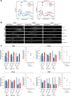
Endogenous LARP1 is required for repression of TOP mRNA translation by mTORC1 inhibitors. A, shLARP1_2 HEK293T cells described in Fig. 7 were further used to investigate effect of LARP1 knockdown on the ability of rapamycin and Torin1 to inhibit TOP mRNA translation by polysome profile analysis. Cells were stimulated with 10% (v/v) fetal bovine serum for 6 h in the presence of 0.1% (v/v) DMSO (vehicle), 100 nm rapamycin, or 300 nm Torin1 prior to lysis in hypotonic buffer. Polysome profiling was performed by loading an identical number of A260 nm units (a crude estimate of total RNA amounts) onto each gradient. Polysome profile traces are shown. RT-sqPCR analysis for RPS6, RPS20, RPL32, and PABP mRNA was performed for each fraction. B, RT-qPCR analysis of subpolysomal (S) and polysomal (P) mRNA abundance for RPS6, RPS20, RPL32, and PABP was performed as follows: RNA from fractions 1 to 6 and 7 to 14 (shown in A) were pooled and designated S and P, respectively. cDNA was synthesized from each pool, and qPCR was performed as detailed under “Experimental Procedures.” C, RT-qPCR analysis of subpolysomal (S) and polysomal (P) mRNA abundance for RPS6, RPS20, RPL32, and PABP and statistical analyses was performed as described under “Experimental Procedures” and the supplemental Workbook.
FIGURE 9.
FIGURE 9.
Endogenous LARP1 is required for repression of TOP mRNA translation upon amino acid starvation. A, shLARP1_2 HEK293T cells described in Fig. 7 were further used to investigate the effect of LARP1 knockdown on the ability of amino acid deprivation to inhibit TOP mRNA translation by polysome profile analysis. Cells were incubated for 3 h in DMEM containing or lacking amino acids in the presence of 10% (v/v) dialyzed serum. Polysome profiling was performed by an identical number of A260 nm units (a crude estimate of total RNA amounts) onto each gradient. Polysome profile traces are shown. B, Western blot analysis of LARP1 and GAPDH protein levels. C, RT-qPCR analysis of subpolysomal (S) and polysomal (P) mRNA abundance for RPS6, RPS20, RPL32, and PABP, and statistical analyses were performed as described under “Experimental Procedures” and the supplemental Workbook.
FIGURE 10.
FIGURE 10.
Ectopic LARP1 represses TOP mRNA translation. A, HEK293T cells were transiently transfected with 6 μg of Myc/FLAG LARP1 wild type into 2 × 10-cm plates (6 μg each). Thirty six hours after transfections, cells were stimulated with 10% (v/v) fetal bovine serum for 3 h and then lysed in hypotonic buffer as described under “Experimental Procedures.” Polysome profiling was performed by loading identical amounts of total RNA onto each gradient. Polysome profile traces are shown. B, inputs were analyzed for ectopic LARP1 expression by SDS-PAGE/Western blot. C, inputs were analyzed for specific mRNA levels by RT-qPCR, following normalization by total RNA concentration. D, RT-qPCR analysis of subpolysomal (S) and polysomal (P) mRNA abundance for RPS6, RPS20, RPL32 and PABP, and statistical analyses were performed as detailed under “Experimental Procedures” and the supplemental Workbook.
FIGURE 11.
FIGURE 11.
LARP1 binds the 5′TOP motif. A, schematic representation of RNA-EMSA. Synthetic RNA oligonucleotides were radiolabeled with [γ-32P]ATP at the 5′end using polynucleotide kinase. Radiolabeled oligonucleotides were incubated with recombinant commercial (Abnova) human LARP1 protein purified from wheat germ extracts. Binding of LARP1 to RNA probes was monitored by a nondenaturing electrophoretic mobility shift assay. The table lists synthetic RNA oligonucleotides spanning the 5′UTRs of RPL32 and RPS6 used in the RNA-EMSA. B and C, recombinant purified LARP1 binds to 5′UTR of RPL32 and RPS6 via their 5′TOP motif.
FIGURE 12.
FIGURE 12.
Translational repression by LARP1 is dependent on the 5′TOP motif. A, polysome profile traces of HeLa Tet-Off cells co-transfected with Tet-inducible β-globin reporters (expressing either their natural 5′UTR or the RPL32 5′UTR) and E.V. or Myc/FLAG-LARP1. B, distribution of β-globin reporter mRNAs in the sucrose gradients was monitored by Northern blot. C, Western blot (WB) analysis of steady-state protein levels of endogenous PABP, DDX6, and RPL32-β-globin fusion protein. D, Northern blot quantitation of steady-state protein levels of β-globin reporter mRNAs. E, effect of stable LARP1 overexpression on TOP mRNA stability.
FIGURE 13.
FIGURE 13.
LARP1 competes with eIF4G for TOP mRNA binding. A, HEK293T cells were cultured to near-confluency (70–80%) and then stimulated with 10% (v/v) fetal bovine serum for 3 h in the presence of 0.1% (v/v) DMSO (vehicle), 100 nm rapamycin, or 300 nm Torin1, followed by lysis in CHAPS lysis buffer in the presence of 1 μg/ml RNase A. Lysates were subjected to m7GTP chromatography and eIF4E-associated proteins probed by SDS-PAGE/Western blot (WB) with the antibodies indicated. B, HEK293T cells were propagated to near-confluency (70–80%) and then stimulated with 10% (v/v) fetal bovine serum for 3 h by media change. Cells were then lysed in CHAPS lysis buffer in the presence or absence of RNase A. Lysates were subjected to immunoprecipitation (IP) with LARP1 or eIF4G antibody, and immunoprecipitates were analyzed by SDS-PAGE/Western blot with the indicated antibodies. C, HEK293T cells were transduced with shCtrl or shLARP1_2 lentivirus and selected with puromycin was described under “Experimental Procedures.” Following selection, pools of cells were seeded in 15-cm plates in complete growth media without puromycin. Cells were cultured for ∼48 h until they reached 70–80% confluency at which point cells were stimulated with 10% (v/v) fetal bovine serum by media change for 3 h. Cells were then lysed in CHAPS lysis buffer, and lysates (2.5 mg of total protein) were subjected to immunoprecipitation with anti-eIF4G antibody (refer to “Experimental Procedures” for details). Immunoprecipitates were used for mRNA analysis by RT-qPCR and lysates for protein analysis by SDS-PAGE/Western blot. D, HEK293T cells were transiently transfected with 6 μg of wild type LARP1 in a 10 -cm plate. Twenty one hours after transfection, cells were stimulated with 10% (v/v) fetal bovine serum for 3 h by media change. Cells were then lysed in CHAPS lysis buffer, and lysates were subjected to immunoprecipitation with anti-eIF4G antibody (1 mg of lysate was used per immunoprecipitation). Immunoprecipitates were used for mRNA analysis by RT-qPCR and lysates for protein analysis by SDS-PAGE/Western blot.
FIGURE 14.
FIGURE 14.
Proposed model for LARP1-mediated repression of TOP mRNA translation downstream of mTORC1. mTORC1 phosphorylates LARP1 at multiple residues (34), effectively releasing LARP1 from the TOP motif. mTORC1 also controls the phosphorylation of 4E-BP1, releasing it from eIF4E thus allowing for eIF4G to bind eIF4E and recruit the pre-initiation complex to the mRNA, and translation ensues.

Similar articles

Cited by

References

    1. Laplante M., Sabatini D. M. (2009) mTOR signaling at a glance. J. Cell Sci. 122, 3589–3594 - PMC - PubMed
    1. Kim D. H., Sarbassov D. D., Ali S. M., King J. E., Latek R. R., Erdjument-Bromage H., Tempst P., Sabatini D. M. (2002) mTOR interacts with raptor to form a nutrient-sensitive complex that signals to the cell growth machinery. Cell 110, 163–175 - PubMed
    1. Hara K., Maruki Y., Long X., Yoshino K., Oshiro N., Hidayat S., Tokunaga C., Avruch J., Yonezawa K. (2002) Raptor, a binding partner of target of rapamycin (TOR), mediates TOR action. Cell 110, 177–189 - PubMed
    1. Loewith R., Jacinto E., Wullschleger S., Lorberg A., Crespo J. L., Bonenfant D., Oppliger W., Jenoe P., Hall M. N. (2002) Two TOR complexes, only one of which is rapamycin sensitive, have distinct roles in cell growth control. Mol. Cell 10, 457–468 - PubMed
    1. Coffman K., Yang B., Lu J., Tetlow A. L., Pelliccio E., Lu S., Guo D. C., Tang C., Dong M. Q., Tamanoi F. (2014) Characterization of the raptor/4E-BP1 interaction by chemical cross-linking coupled with mass spectrometry analysis. J. Biol. Chem. 289, 4723–4734 - PMC - PubMed

MeSH terms