Peptidylarginine deiminase IV promotes the development of chemoresistance through inducing autophagy in hepatocellular carcinoma
- PMID: 25922661
- PMCID: PMC4412294
- DOI: 10.1186/2045-3701-4-49
Peptidylarginine deiminase IV promotes the development of chemoresistance through inducing autophagy in hepatocellular carcinoma
Abstract
Background: Peptidylarginine deiminase IV (PADI4) is widely distributed in several tissues and the expression is correlated with many pathological processes. Chemotherapy remains a major treatment alternatively to surgery for a large number of patients at the advanced stage of hepatocellular carcinoma (HCC). However, the role of PADI4 in the chemoresistance of HCC has not been identified.
Methods: MTT and PI/Annexin V assay were employed to examine the proliferation and apoptosis of HCC cell lines. The expression of MDR1 is detected by Realtime PCR. GFP tagged LC3 expression vector and electron microscopy are utilized to demonstrate the occurrence of autophagy.
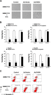
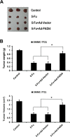
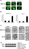
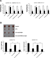
Results: We observed that the elevated PADI4 expression is associated with chemoresistance in HCC patients with TACE after surgery. In addition, we found that overexpression of PADI4 in HCC cell lines lead to the resistance to chemotherapeutic agents in vitro and in vivo. Interestingly, the HCC cells that overexpressed PADI4 were observed to undergo autophagy which was known as a protective mechanism for cells to resist the cell tosicity from chemotherapy. Autophagy inhibitor could effectively restore the sensitivity of HCC cells to chemotherapy in vitro and in vivo.
Conclusions: These results indicate that PADI4 may induce chemoresistance in HCC cells by leading autophagy.
Keywords: Autophagy; Chemoresistance; Hepatocellular Carcinoma; PADI4.
Figures


Similar articles
-
Mesenchymal stem cells contribute to the chemoresistance of hepatocellular carcinoma cells in inflammatory environment by inducing autophagy.Cell Biosci. 2014 Apr 28;4:22. doi: 10.1186/2045-3701-4-22. eCollection 2014. Cell Biosci. 2014. PMID: 24872873 Free PMC article.
-
Decreased PADI4 mRNA association with global hypomethylation in hepatocellular carcinoma during HBV exposure.Cell Biochem Biophys. 2013 Mar;65(2):187-95. doi: 10.1007/s12013-012-9417-3. Cell Biochem Biophys. 2013. PMID: 22907585
-
MiR-26 enhances chemosensitivity and promotes apoptosis of hepatocellular carcinoma cells through inhibiting autophagy.Cell Death Dis. 2017 Jan 12;8(1):e2540. doi: 10.1038/cddis.2016.461. Cell Death Dis. 2017. PMID: 28079894 Free PMC article.
-
Role of citrullination modification catalyzed by peptidylarginine deiminase 4 in gene transcriptional regulation.Acta Biochim Biophys Sin (Shanghai). 2017 Jul 1;49(7):567-572. doi: 10.1093/abbs/gmx042. Acta Biochim Biophys Sin (Shanghai). 2017. PMID: 28472221 Review.
-
MicroRNAs contribute to ATP-binding cassette transporter- and autophagy-mediated chemoresistance in hepatocellular carcinoma.World J Hepatol. 2019 Apr 27;11(4):344-358. doi: 10.4254/wjh.v11.i4.344. World J Hepatol. 2019. PMID: 31114639 Free PMC article. Review.
Cited by
-
The virtues and vices of protein citrullination.R Soc Open Sci. 2022 Jun 8;9(6):220125. doi: 10.1098/rsos.220125. eCollection 2022 Jun. R Soc Open Sci. 2022. PMID: 35706669 Free PMC article. Review.
-
Chloroquine sensitizes hepatocellular carcinoma cells to chemotherapy via blocking autophagy and promoting mitochondrial dysfunction.Int J Clin Exp Pathol. 2017 Sep 1;10(9):10056-10065. eCollection 2017. Int J Clin Exp Pathol. 2017. PMID: 31966896 Free PMC article.
-
Targeting epigenetic regulators to overcome drug resistance in cancers.Signal Transduct Target Ther. 2023 Feb 17;8(1):69. doi: 10.1038/s41392-023-01341-7. Signal Transduct Target Ther. 2023. PMID: 36797239 Free PMC article. Review.
-
Autophagy and liver cancer.Turk J Gastroenterol. 2018 May;29(3):270-282. doi: 10.5152/tjg.2018.150318. Turk J Gastroenterol. 2018. PMID: 29755011 Free PMC article. Review.
-
Targeting cell death mechanisms: the potential of autophagy and ferroptosis in hepatocellular carcinoma therapy.Front Immunol. 2024 Sep 9;15:1450487. doi: 10.3389/fimmu.2024.1450487. eCollection 2024. Front Immunol. 2024. PMID: 39315094 Free PMC article. Review.
References
LinkOut - more resources
Full Text Sources
Other Literature Sources
Research Materials
Miscellaneous

