

Hydration water mobility is enhanced around tau amyloid fibers
- PMID: 25918405
- PMCID: PMC4443308
- DOI: 10.1073/pnas.1422824112
Hydration water mobility is enhanced around tau amyloid fibers
Abstract
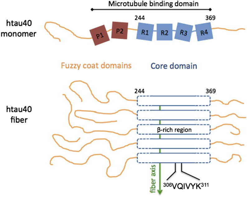
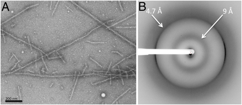
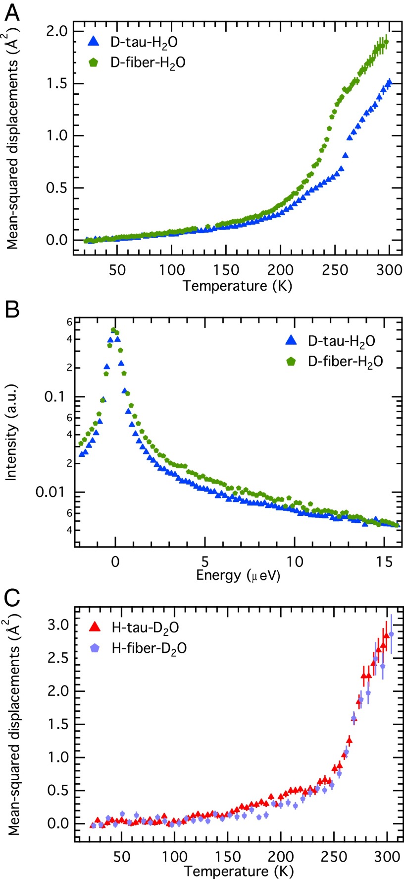
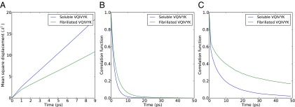
The paired helical filaments (PHF) formed by the intrinsically disordered human protein tau are one of the pathological hallmarks of Alzheimer disease. PHF are fibers of amyloid nature that are composed of a rigid core and an unstructured fuzzy coat. The mechanisms of fiber formation, in particular the role that hydration water might play, remain poorly understood. We combined protein deuteration, neutron scattering, and all-atom molecular dynamics simulations to study the dynamics of hydration water at the surface of fibers formed by the full-length human protein htau40. In comparison with monomeric tau, hydration water on the surface of tau fibers is more mobile, as evidenced by an increased fraction of translationally diffusing water molecules, a higher diffusion coefficient, and increased mean-squared displacements in neutron scattering experiments. Fibers formed by the hexapeptide (306)VQIVYK(311) were taken as a model for the tau fiber core and studied by molecular dynamics simulations, revealing that hydration water dynamics around the core domain is significantly reduced after fiber formation. Thus, an increase in water dynamics around the fuzzy coat is proposed to be at the origin of the experimentally observed increase in hydration water dynamics around the entire tau fiber. The observed increase in hydration water dynamics is suggested to promote fiber formation through entropic effects. Detection of the enhanced hydration water mobility around tau fibers is conjectured to potentially contribute to the early diagnosis of Alzheimer patients by diffusion MRI.
Keywords: amyloid fibers; hydration water; intrinsically disordered proteins; neutron scattering; tau protein.
Conflict of interest statement
The authors declare no conflict of interest.
Figures
Similar articles
-
Translational diffusion of hydration water correlates with functional motions in folded and intrinsically disordered proteins.Nat Commun. 2015 Mar 16;6:6490. doi: 10.1038/ncomms7490. Nat Commun. 2015. PMID: 25774711 Free PMC article.
-
Diffusion of Hydration Water around Intrinsically Disordered Proteins.J Phys Chem B. 2015 Oct 22;119(42):13262-70. doi: 10.1021/acs.jpcb.5b07248. Epub 2015 Oct 8. J Phys Chem B. 2015. PMID: 26418258
-
Molecular Dynamics Simulations of a Powder Model of the Intrinsically Disordered Protein Tau.J Phys Chem B. 2015 Oct 1;119(39):12580-9. doi: 10.1021/acs.jpcb.5b05849. Epub 2015 Sep 18. J Phys Chem B. 2015. PMID: 26351734
-
Role of water in protein folding, oligomerization, amyloidosis and miniprotein.J Pept Sci. 2014 Oct;20(10):747-59. doi: 10.1002/psc.2671. Epub 2014 Aug 6. J Pept Sci. 2014. PMID: 25098401 Review.
-
Computational studies of protein aggregation: methods and applications.Annu Rev Phys Chem. 2015 Apr;66:643-66. doi: 10.1146/annurev-physchem-040513-103738. Epub 2015 Feb 2. Annu Rev Phys Chem. 2015. PMID: 25648485 Review.
Cited by
-
Femtosecond Hydration Map of Intrinsically Disordered α-Synuclein.Biophys J. 2018 Jun 5;114(11):2540-2551. doi: 10.1016/j.bpj.2018.04.028. Biophys J. 2018. PMID: 29874605 Free PMC article.
-
In Vivo Detection of Gray Matter Neuropathology in the 3xTg Mouse Model of Alzheimer's Disease with Diffusion Tensor Imaging.J Alzheimers Dis. 2017;58(3):841-853. doi: 10.3233/JAD-170136. J Alzheimers Dis. 2017. PMID: 28505976 Free PMC article.
-
High protein flexibility and reduced hydration water dynamics are key pressure adaptive strategies in prokaryotes.Sci Rep. 2016 Sep 6;6:32816. doi: 10.1038/srep32816. Sci Rep. 2016. PMID: 27595789 Free PMC article.
-
Dynamical Behavior of Human α-Synuclein Studied by Quasielastic Neutron Scattering.PLoS One. 2016 Apr 20;11(4):e0151447. doi: 10.1371/journal.pone.0151447. eCollection 2016. PLoS One. 2016. PMID: 27097022 Free PMC article.
-
Molecular Mechanism of Tau Misfolding and Aggregation: Insights from Molecular Dynamics Simulation.Curr Med Chem. 2024;31(20):2855-2871. doi: 10.2174/0929867330666230409145247. Curr Med Chem. 2024. PMID: 37031392 Review.
References
-
- Chiti F, Dobson CM. Protein misfolding, functional amyloid, and human disease. Annu Rev Biochem. 2006;75(1):333–366. - PubMed
-
- Uversky VN, Oldfield CJ, Dunker AK. Intrinsically disordered proteins in human diseases: Introducing the D2 concept. Annu Rev Biophys. 2008;37(1):215–246. - PubMed
-
- Brion JP, Couck AM, Passareiro E, Flament-Durand J. Neurofibrillary tangles of Alzheimer’s disease: An immunohistochemical study. J Submicrosc Cytol. 1985;17(1):89–96. - PubMed
Publication types
MeSH terms
Substances
LinkOut - more resources
Full Text Sources
Other Literature Sources
Medical
