. 2015 Jun 1;194(11):5294-304.
doi: 10.4049/jimmunol.1402648. Epub 2015 Apr 27.

Mechanistic Assessment of PD-1H Coinhibitory Receptor-Induced T Cell Tolerance to Allogeneic Antigens

Affiliations

Mechanistic Assessment of PD-1H Coinhibitory Receptor-Induced T Cell Tolerance to Allogeneic Antigens

Dallas B Flies et al. J Immunol. .

Abstract

PD-1H is a recently identified cell surface coinhibitory molecule of the B7/CD28 immune modulatory gene family. We showed previously that single injection of a PD-1H agonistic mAb protected mice from graft-versus-host disease (GVHD). In this study, we report two distinct mechanisms operate in PD-1H-induced T cell tolerance. First, signaling via PD-1H coinhibitory receptor potently arrests alloreactive donor T cells from activation and expansion in the initiation phase. Second, donor regulatory T cells are subsequently expanded to maintain long-term tolerance and GVHD suppression. Our study reveals the crucial function of PD-1H as a coinhibitory receptor on alloreactive T cells and its function in the regulation of T cell tolerance. Therefore, PD-1H may be a target for the modulation of alloreactive T cells in GVHD and transplantation.


Conflict of interest statement

Conflict of interest: The authors declare no conflict of interest.

Figures

Figure 1
Figure 1
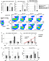
PD-1H mAb inhibits allo-reactive T cell expansion and cytotoxicity. 2.5 × 10⁷ total splenocytes from B6 wt mice were adoptively transferred to non-irradiated BDF1 mice with MH5A or control Ab to monitor donor expansion and cytotoxic killing of recipient cells. Splenocytes were counted and analyzed by flow cytometry at indicated time points for determination of cell numbers of (A) donor CD4+ T cells, (B) donor CD8+ T cells, (C) recipient CD19+ T cells, (D) recipient CD4+ T cells, and (E) recipient CD8+ T cells.
Figure 2
Figure 2
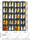
MH5A inhibits systemic expansion of allo-reactive donor T cells. B6.luc Tg donor T cells and WT B6 TCD-BM were adoptively transferred to lethally irradiated BALB/c recipients with (A and C) control Ab, or (B and D) MH5A. Mice were imaged for bioluminescence at 2, 24, 48, 96, and 120 hours for T cell accumulation and expansion measured as radiance for relative comparison between groups. Ventral (A and B) and right lateral (C and D) imaging of 2 mice per treatment group are shown as representatives of 5 mice/group. Minimum and maximum radiance levels and range are indicated at each time point for optimal view. Gating of (E) whole body, (F) cervical LN region and (G) abdominal region in the ventral position was performed for calculation of total flux.
Figure 3
Figure 3
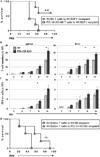
Adoptive transfer of PD-1H deficient donor T cells exacerbates GVHD. (A) PD-1H KO donor T cells + TCD-BM or wt littermate donor T cells + TCD-BM from B6 background were adoptively transferred to lethally irradiated wt BDF1 recipients and monitored for survival or (B) splenocytes and liver lymphocytes were isolated at indicated time points and absolute number of T cells was determined. (C) Total splenocytes or liver lymphocytes were restimulated ex vivo for 5 hours with PMA, ionomycin, and golgistop and intracellularly stained for IFN-γ production. (D) Wt BALB/c LN cells and BM were adoptively transferred to lethally irradiated B6 wt or PD-1H KO recipients and monitored for survival. 5 mice per group were used for survival experiments and 3 mice per group were used for time point analyses. Experiments were repeated 2–3 times.
Figure 4
Figure 4
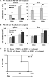
MH5A-directed inhibition of allo-reactive T cells requires PD-1H expression on T cells. (A) BALB/c wt T cells were adoptively transferred to lethally irradiated PD-1H KO recipients on a B6 background with MH5A or control Ab. Splenocytes and liver lymphocytes were isolated 6 days later for determination of absolute number of donor CD4+ and CD8+ T cells. (B) B6 PD-1H KO T cells were adoptively transferred to lethally irradiated BDF1 recipients with MH5A or control Ab. Splenocytes and liver lymphocytes were isolated on day 7 for determination of absolute number of donor CD4+ and CD8+ T cells. (C) PD-1H KO T cells + TCD-BM or wt littermate T cells + TCD-BM from B6 mice were adoptively transferred to lethally irradiated BDF1 recipients with MH5A or control Ab and mice were monitored for survival. 3 mice per group were analyzed for T cell numbers and 5 mice/group were used for survival studies. Experiments were repeated 2–3 times.
Figure 5
Figure 5
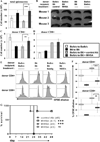
Donor T cell tolerance is induced by prophylactic MH5A treatment. BALB/c T cells + TCD-BM were adoptively transferred to lethally irradiated B6 recipients with MH5A or control Ab. Syngeneic adoptive transfers of BALB/c to lethally irradiated BALB/c and B6 to lethally irradiated B6 were also performed for controls in T cell tolerance studies. (A) Spleens were isolated on day 14 and analyzed for total splenocytes. (B) Pictures of spleens isolated from indicated mice on day 14. (C and D) Donor CD4+ and CD8+ T cell numbers on day 14. (E) Day 14 total splenocytes were labeled with CFSE and restimulated ex vivo with irradiated B6 splenocytes for 72 hours. Donor CD4+ and CD8+ T cells were analyzed by flow cytometry for cell division as determined by CFSE dilution. Syngeneic BALB/c to BALB/c splenocytes were used as positive controls while syngeneic B6 to B6 splenocytes were used as negative controls. (F) Average percentage of CFSE dilution was determined for donor CD4+ and CD8+ T cells. (G) Using the same BALB/c to lethally irradiated B6 GVHD model as above, mice were treated with MH5A on day -1, on day 0 at the time of adoptive T cell transfer, or 3 days after adoptive T cell transfer and monitored for survival. Day 14 tolerance experiments used 3 mice per group and survival experiments used 5 mice/group. Experiments repeated 2–3 times.
Figure 6
Figure 6
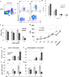
Regulatory T cells are transiently increased and promote tolerance in MH5A treated mice. BALB/c T cells + TCD-BM was adoptively transferred to lethally irradiated B6 mice with MH5A or control Ab. (A) Day 14 splenocytes were stained for surface CD4 and CD25, and for intracellular FoxP3 expression. (B) Representative dot plots of splenocyte Treg percentages in spleen. Plots are gated on CD4. On the far left is cells adoptively transferred and the right is analysis of FoxP3 and CD25 at indicated timepoints. (C and D) The ratio of the absolute number of donor CD8+ T cells to CD4+CD25+FoxP3+ T cells (C) and CD4+CD25+FoxP3− T cells to CD4+CD25+FoxP3+ T cells (D) was calculated on day 14. (E) Treg absolute cell numbers in the spleen on days 5, 10, 15 and 20. (F) The ratio of the absolute number donor CD8+ T cells to CD4+CD25+FoxP3+ T cells was calculated at the indicated time points. Red lines show trends over time. (G and H) Total splenocytes harvested on days 15 and 20 were restimulated ex vivo with PMA, ionomycin and golgistop for 5 hours and stained for intracellular levels of IFN-γ. Cells were gated on CD4+ or CD8+ T cells as indicated. (H) Serum from GVHD mice was isolated at the indicated time points and analyzed by ELISA for TNF-α levels.
Figure 7
Figure 7
Selective expansion of regulatory T cells in vivo with MH5A treatment. (A) Peripheral lymph nodes were isolated from untreated wt B6 mice and analyzed by flow cytometry. Surface staining was performed for CD4, CD25, and control Ab or PD-1H, followed by intracellular staining for FoxP3. Top panel is gated on CD4+CD25+ T cells. Bottom panel is gated on CD4+CD25(−) T cells. (B) Naïve CD4+CD25 negative T cells were isolated by MACS bead negative selection of CD4+ T cells followed by depletion of CD25+ cells with biotin labeled CD25 followed by anti-biotin MACS bead magnetic depletion. Cells were then cultured with anti-CD3, anti-CD28, recombinant IL-2 and titrated concentrations of TGF-β in the presence of soluble MH5A or control Ab for 5 days and analyzed for percentages of CD4+FoxP3+. (C) Treg cells were induced as in (B) but without control Ig or MH5A treatment. These cells were then added to 96-well plates with 5 ng/ml IL-2 and pre-coated with anti-CD3. MH5A or hamIg was added to wells at a final concentration of 10 ug/ml. Treg cell proliferation was analyzed 72 hours later. Experiment was repeated three times. (D) Treg cells were induced as in (B) and used in a mixed lymphocyte response (MLR) assay. Treg cells were pre-incubated with control IgG or MH5A, then washed and added at various ratios to an MLR of B6 responder T cells and irradiated BDF1 splenocytes as targets. Responder proliferation was analyzed 5 days later. (E–H) Total T cells (CD25 replete) (E,G) or CD25-depleted T cells (F,H) and TCD-BM from B6 mice were adoptively transferred to lethally irradiated BDF1 mice with MH5A or control Ab. Splenocytes were isolated at the indicated time points and absolute numbers of CD4+FoxP3+ T cells (E,F) and the ratio of absolute numbers of donor CD8+ to donor CD4+FoxP3+ T cells (G,H) were calculated. 3–5 mice per group repeated a minimum of three times.

Similar articles

Cited by

References

    1. Socie G, Blazar BR. Acute graft-versus-host disease: from the bench to the bedside. Blood. 2009;114:4327–4336. - PMC - PubMed
    1. Chen L, Flies DB. Molecular mechanisms of T cell co-stimulation and co-inhibition. Nat. Rev. Immunol. 2013;13:227–242. - PMC - PubMed
    1. Sharpe AH. Mechanisms of costimulation. Immunol. Rev. 2009;229:5–11. - PMC - PubMed
    1. Blazar BR, Carreno BM, Panoskaltsis-Mortari A, Carter L, Iwai Y, Yagita H, Nishimura H, Taylor PA. Blockade of programmed death-1 engagement accelerates graft-versus-host disease lethality by an IFN-gamma-dependent mechanism. J. Immunol. 2003;171:1272–1277. - PubMed
    1. Blazar BR, Taylor PA, Linsley PS, Vallera DA. In vivo blockade of CD28/CTLA4: B7/BB1 interaction with CTLA4-Ig reduces lethal murine graft-versus-host disease across the major histocompatibility complex barrier in mice. Blood. 1994;83:3815–3825. - PubMed

Publication types

MeSH terms