

Role of epidermal growth factor receptor and endoplasmic reticulum stress in vascular remodeling induced by angiotensin II
- PMID: 25916723
- PMCID: PMC4433406
- DOI: 10.1161/HYPERTENSIONAHA.115.05344
Role of epidermal growth factor receptor and endoplasmic reticulum stress in vascular remodeling induced by angiotensin II
Abstract
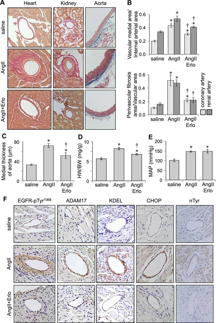
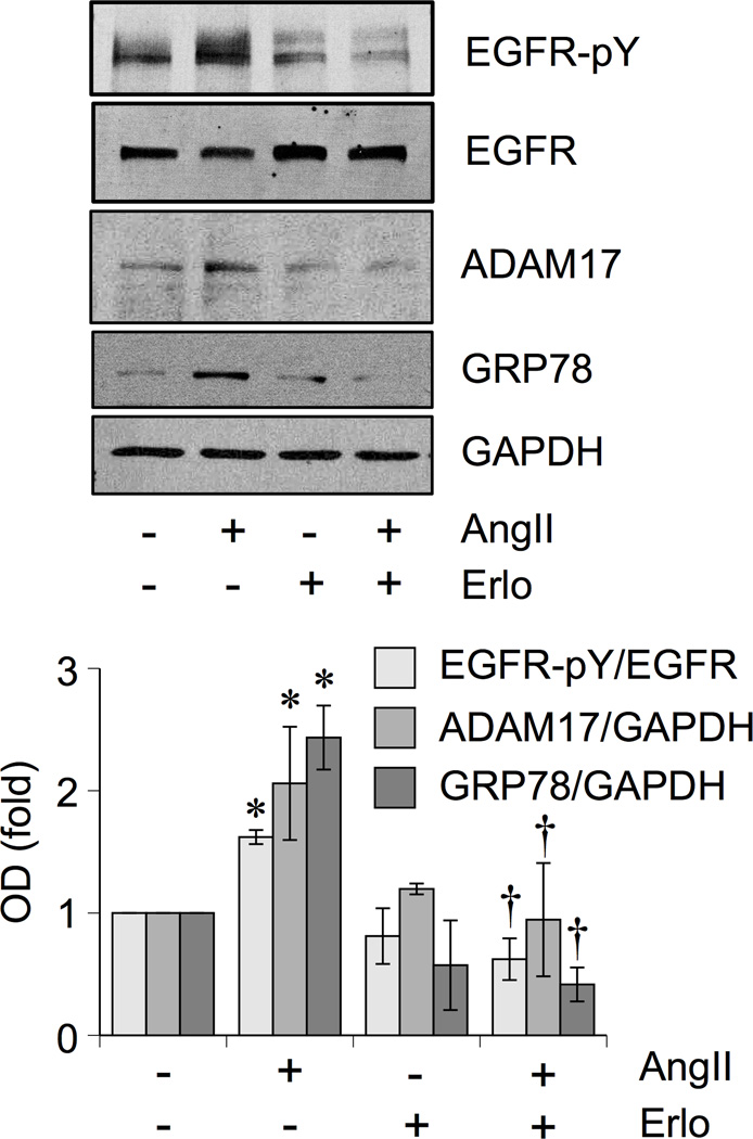
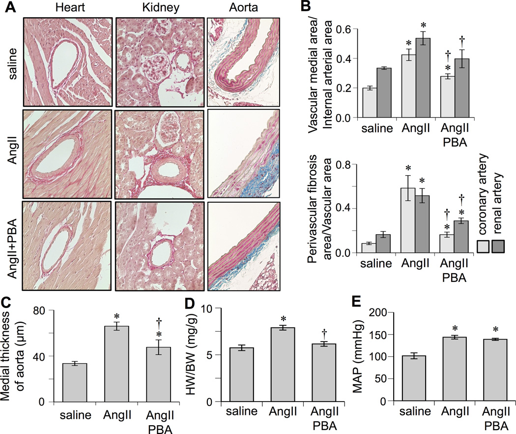
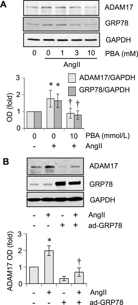
The mechanisms by which angiotensin II (AngII) elevates blood pressure and enhances end-organ damage seem to be distinct. However, the signal transduction cascade by which AngII specifically mediates vascular remodeling such as medial hypertrophy and perivascular fibrosis remains incomplete. We have previously shown that AngII-induced epidermal growth factor receptor (EGFR) transactivation is mediated by disintegrin and metalloproteinase domain 17 (ADAM17), and that this signaling is required for vascular smooth muscle cell hypertrophy but not for contractile signaling in response to AngII. Recent studies have implicated endoplasmic reticulum (ER) stress in hypertension. Interestingly, EGFR is capable of inducing ER stress. The aim of this study was to test the hypothesis that activation of EGFR and ER stress are critical components required for vascular remodeling but not hypertension induced by AngII. Mice were infused with AngII for 2 weeks with or without treatment of EGFR inhibitor, erlotinib, or ER chaperone, 4-phenylbutyrate. AngII infusion induced vascular medial hypertrophy in the heart, kidney and aorta, and perivascular fibrosis in heart and kidney, cardiac hypertrophy, and hypertension. Treatment with erlotinib as well as 4-phenylbutyrate attenuated vascular remodeling and cardiac hypertrophy but not hypertension. In addition, AngII infusion enhanced ADAM17 expression, EGFR activation, and ER/oxidative stress in the vasculature, which were diminished in both erlotinib-treated and 4-phenylbutyrate-treated mice. ADAM17 induction and EGFR activation by AngII in vascular cells were also prevented by inhibition of EGFR or ER stress. In conclusion, AngII induces vascular remodeling by EGFR activation and ER stress via a signaling mechanism involving ADAM17 induction independent of hypertension.
Keywords: angiotensin II; fibrosis; hypertension; hypertrophy; muscle, smooth, vascular; signal transduction.
© 2015 American Heart Association, Inc.
Figures
Similar articles
-
Vascular ADAM17 as a Novel Therapeutic Target in Mediating Cardiovascular Hypertrophy and Perivascular Fibrosis Induced by Angiotensin II.Hypertension. 2016 Oct;68(4):949-955. doi: 10.1161/HYPERTENSIONAHA.116.07620. Epub 2016 Aug 1. Hypertension. 2016. PMID: 27480833 Free PMC article.
-
Caveolin-1 Deletion Prevents Hypertensive Vascular Remodeling Induced by Angiotensin II.Hypertension. 2017 Jan;69(1):79-86. doi: 10.1161/HYPERTENSIONAHA.116.08278. Epub 2016 Nov 28. Hypertension. 2017. PMID: 27895190 Free PMC article.
-
Vascular ADAM17 (a Disintegrin and Metalloproteinase Domain 17) Is Required for Angiotensin II/β-Aminopropionitrile-Induced Abdominal Aortic Aneurysm.Hypertension. 2017 Nov;70(5):959-963. doi: 10.1161/HYPERTENSIONAHA.117.09822. Epub 2017 Sep 25. Hypertension. 2017. PMID: 28947615 Free PMC article.
-
Angiotensin II regulates vascular and endothelial dysfunction: recent topics of Angiotensin II type-1 receptor signaling in the vasculature.Curr Vasc Pharmacol. 2006 Jan;4(1):67-78. doi: 10.2174/157016106775203126. Curr Vasc Pharmacol. 2006. PMID: 16472178 Review.
-
Angiotensin II signal transduction through the AT1 receptor: novel insights into mechanisms and pathophysiology.Clin Sci (Lond). 2007 Apr;112(8):417-28. doi: 10.1042/CS20060342. Clin Sci (Lond). 2007. PMID: 17346243 Review.
Cited by
-
Direct GPCR-EGFR interaction enables synergistic membrane-to-nucleus information transfer.Cell Mol Life Sci. 2024 Jun 20;81(1):272. doi: 10.1007/s00018-024-05281-5. Cell Mol Life Sci. 2024. PMID: 38900158 Free PMC article.
-
Glucose consumption of vascular cell types in culture: toward optimization of experimental conditions.Am J Physiol Cell Physiol. 2022 Jan 1;322(1):C73-C85. doi: 10.1152/ajpcell.00257.2021. Epub 2021 Nov 24. Am J Physiol Cell Physiol. 2022. PMID: 34817269 Free PMC article.
-
EGFR inhibition attenuates diabetic nephropathy through decreasing ROS and endoplasmic reticulum stress.Oncotarget. 2017 May 16;8(20):32655-32667. doi: 10.18632/oncotarget.15948. Oncotarget. 2017. PMID: 28427241 Free PMC article.
-
Silencing Epidermal Growth Factor Receptor in Hypothalamic Paraventricular Nucleus Reduces Extracellular Signal-regulated Kinase 1 and 2 Signaling and Sympathetic Excitation in Heart Failure Rats.Neuroscience. 2021 May 21;463:227-237. doi: 10.1016/j.neuroscience.2021.01.025. Epub 2021 Feb 2. Neuroscience. 2021. PMID: 33540053 Free PMC article.
-
Milestone Papers on Signal Transduction Mechanisms of Hypertension and Its Complications.Hypertension. 2024 May;81(5):977-990. doi: 10.1161/HYPERTENSIONAHA.123.21365. Epub 2024 Feb 19. Hypertension. 2024. PMID: 38372140 Review.
References
-
- Kang N, Walther T, Tian XL, Bohlender J, Fukamizu A, Ganten D, Bader M. Reduced hypertension-induced end-organ damage in mice lacking cardiac and renal angiotensinogen synthesis. J Mol Med (Berl) 2002;80:359–366. - PubMed
-
- Gibbons GH. Angiotensin-converting enzyme inhibition and vascular structure in hypertension. J Cardiovasc Pharmacol. 1991;18(Suppl 7):S19–S24. - PubMed
-
- Briet M, Schiffrin EL. Treatment of arterial remodeling in essential hypertension. Curr Hypertens Rep. 2013;15:3–9. - PubMed
-
- Kanaide H, Ichiki T, Nishimura J, Hirano K. Cellular mechanism of vasoconstriction induced by angiotensin II: it remains to be determined. Circ Res. 2003;93:1015–1017. - PubMed
-
- Ohtsu H, Dempsey PJ, Frank GD, Brailoiu E, Higuchi S, Suzuki H, Nakashima H, Eguchi K, Eguchi S. ADAM17 mediates epidermal growth factor receptor transactivation and vascular smooth muscle cell hypertrophy induced by angiotensin II. Arterioscler Thromb Vasc Biol. 2006;26:e133–e137. - PubMed
Publication types
MeSH terms
Substances
Grants and funding
LinkOut - more resources
Full Text Sources
Other Literature Sources
Research Materials
Miscellaneous
