. 2015 Nov;22(11):1877-89.
doi: 10.1038/cdd.2015.49. Epub 2015 Apr 24.

Astrocyte NMDA receptors' activity sustains neuronal survival through a Cdk5-Nrf2 pathway

Affiliations

Astrocyte NMDA receptors' activity sustains neuronal survival through a Cdk5-Nrf2 pathway

D Jimenez-Blasco et al. Cell Death Differ. 2015 Nov.

Abstract

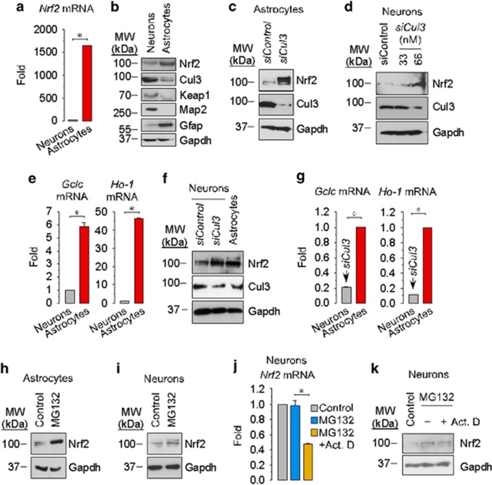
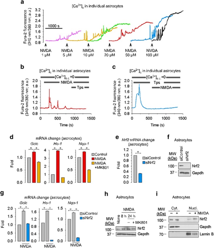
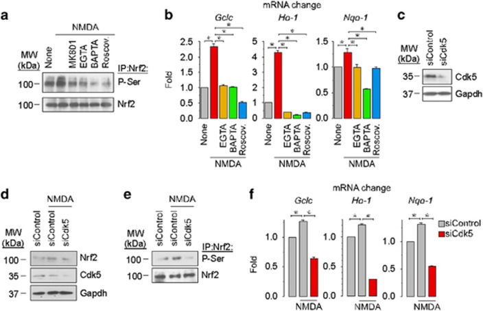
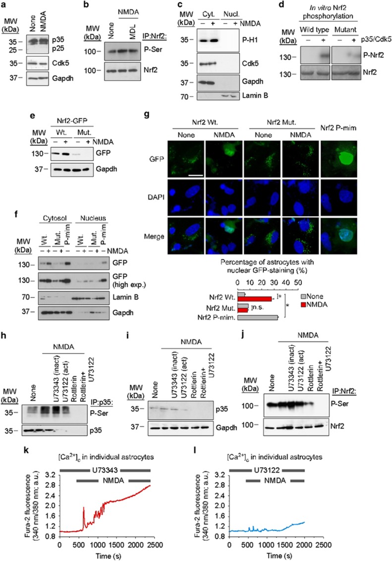
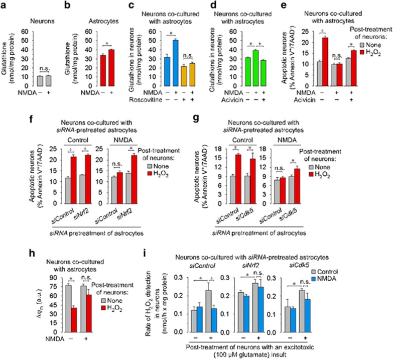
Neurotransmission unavoidably increases mitochondrial reactive oxygen species. However, the intrinsic antioxidant defense of neurons is weak and hence the mechanism whereby these cells are physiologically protected against oxidative damage is unknown. Here we found that the antioxidant defense of neurons is repressed owing to the continuous protein destabilization of the master antioxidant transcriptional activator, nuclear factor-erythroid 2-related factor-2 (Nrf2). By contrast, Nrf2 is highly stable in neighbor astrocytes explaining their robust antioxidant defense and resistance against oxidative stress. We also show that subtle and persistent stimulation of N-methyl-d-aspartate receptors (NMDAR) in astrocytes, through a mechanism not requiring extracellular Ca²⁺ influx, upregulates a signal transduction pathway involving phospholipase C-mediated endoplasmic reticulum release of Ca²⁺ and protein kinase Cδ activation. Active protein kinase Cδ promotes, by phosphorylation, the stabilization of p35, a cyclin-dependent kinase-5 (Cdk5) cofactor. Active p35/Cdk5 complex in the cytosol phosphorylates Nrf2 at Thr(395), Ser(433) and Thr(439) that is sufficient to promote Nrf2 translocation to the nucleus and induce the expression of antioxidant genes. Furthermore, this Cdk5-Nrf2 transduction pathway boosts glutathione metabolism in astrocytes efficiently protecting closely spaced neurons against oxidative damage. Thus, intercellular communication through NMDAR couples neurotransmission with neuronal survival.


Figures

Figure 1
Figure 1
Higher Nrf2 protein destabilization rate in neurons when compared with astrocytes explains low Nrf2 functional abundance in neurons. (a) Quantitative real-time PCR (RT-qPCR) analysis of the relative mRNA abundance of Nrf2 in rat cortical primary neurons and rat cortical primary astrocytes; Gapdh mRNA was used for normalization, and the astrocytes data are expressed as the change relative to neurons. Results reveals a ~1600-fold higher Nrf2 mRNA abundance in astrocytes than in neurons. (b) Western blot analysis indicates lower Nrf2 and higher Cul3 and Keap1 protein abundances in neurons when compared with astrocytes. Map2 and Gfap protein levels indicate the purity of neurons and astrocytes cultures, respectively. Gapdh was used a loading marker. (c) siRNA against Cul3 (siCul3, 100 nM) leading to decreased Cul3 protein increases Nrf2 abundance in astrocytes; siControl (100 nM) was a siRNA against luciferase. (d) siCul3 treatment dose-dependently (33-66 nM) increases Nrf2 in neurons. (e) RT-qPCR analysis of Nrf2-target genes glutamate–cysteine ligase catalytic subunit (Gclc) and heme-oxygenase-1 (Ho-1) reveals 6- and 40-fold lower mRNA abundances, respectively, in neurons than in astrocytes. (f) siCul3 (66 nM) in neurons increases Nrf2 protein to levels similar to those of the astrocytes. (g) Gclc and Ho-1 mRNA levels in neurons after siCul3 (66 nM) are ~20 and 10%, respectively, of those in control astrocytes. (h) The proteasome inhibitor MG132 (20 μM, 2 h) increased Nrf2 protein abundance in astrocytes. (i) MG132 increased Nrf2 protein abundance in neurons. (j) MG132 did not increase Nrf2 mRNA abundance in neurons, and the transcriptional inhibitor actinomycine D (Act. D; 100 ng/ml) decreased Nrf2 mRNA levels. (k) The increase in Nrf2 protein levels in neurons by MG132 was unaffected by actinomycine D. Data are expressed as mean±S.E.M. *P<0.05 (Student's t-test; n=3–4 independent experiments)
Figure 2
Figure 2
NMDA causes sustained cytosolic Ca2+ rise and Nrf2 functional activation in astrocytes. (a) NMDA dose-dependently (1–100 μM) induced a delayed and sustained increase in cytosolic free Ca2+ concentrations ([Ca2+]c) in individual astrocytes. (b) Astrocytes were incubated with NMDA (20 μM) in the absence of extracellular Ca2+ ([Ca2+]e.c.=0), which revealed increased [Ca2+]c during a limited period, after which the sarco-ER Ca2+-ATPase inhibitor, thapsigargin (Tps, 1 μM), had no effect. (c) Incubation of astrocytes under [Ca2+]e.c.=0 with Tps (1 μM) in the absence of NMDA robustly increased [Ca2+]c transiently, after which addition of NMDA had no effect on [Ca2+]c. (d) Incubation of astrocytes with NMDA (20 μM) for 8 h increased Gclc, Ho-1 and NADPH quinone oxidorreductase-1 (Nqo1) mRNA levels, an effect that was abolished by the NMDA antagonist MK801 (10 μM). (e) siNrf2 (100 nM, 72 h) decreased Nrf2 mRNA levels in astrocytes; siControl (100 nM) was an siRNA against luciferase. (f) siNrf2 decreased Nrf2 protein levels in astrocytes. (g) siNrf2 abolished the NMDA (20 μM, 8 h)-induced increase in Gclc, Ho-1 and Nqo1 mRNA levels in astrocytes. (h) NMDA (20 μM, 8 or 24 h) increased Nrf2 protein abundance in astrocytes, and MK801 abolished this effect. (i) NMDA (20 μM, 8 or 24 h) increased endogenous Nrf2 levels in the nucleus of astrocytes. Data are expressed as mean±S.E.M. *P<0.05 (Student's t-test for e; ANOVA followed by Bonferroni test for d and g; n=3–4 independent experiments)
Figure 3
Figure 3
Stimulation of NMDA receptors in astrocytes promotes Cdk5-mediated Nrf2 phosphorylation and activation. (a) Nrf2 immunoprecipitation in astrocytes followed by western blotting against an anti-serine antibody revealed increased Nrf2 phosphorylation by NMDA treatment (20 μM for 8 h), an effect that was prevented by MK801 (10 μM), by extracellular Ca2+ chelation with EGTA (100 μM in Ca2+-free medium), by intracellular Ca2+ chelation with BAPTA (10 μM) and by the general cyclin-dependent kinase (Cdk) inhibitor roscovitine (10 μM). (b) EGTA, BAPTA and roscovitine abolishes NMDA (20 μM, 8 h)-induced increase in the mRNA levels of the Nrf2-target genes Gclc, Ho-1 and Nqo1 in astrocytes. (c) siRNA against Cdk5 (siCdk5, 100 nM; 72 h) efficiently knocks down Cdk5 in astrocytes; siControl was a siRNA against luciferase. (d) siCdk5 prevents NMDA (20 μM, 8 h)-dependent increase in Nrf2 protein abundance in astrocytes. (e) NMDA (20 μM, 8 h)-induced Nrf2 phosphorylation is prevented by siCdk5 in astrocytes. (f) siCdk5 abolishes NMDA (20 μM, 8 h)-induced increase in the mRNA levels of the Nrf2-target genes Gclc, Ho-1 and Nqo1 in astrocytes. Data are expressed as mean±S.E.M. *P<0.05 (ANOVA followed by Bonferroni test; n=3–4 independent experiments)
Figure 4
Figure 4
NMDAR activation in astrocytes promotes a phospholipase C (PLC)-protein kinase Cδ (PKCδ)-mediated p35 phosphorylation leading to Cdk5-triggered Nrf2 phosphorylation at residues Thr-395, Ser-433 and Thr-439 and Nrf2 nuclear localization. (a) NMDA (20 μM, 8 h) did not alter the p35 protein levels and did not result in the appearance of p25 in astrocytes. (b) The calpain inhibitor MDL (100 μM) did not prevent NMDA (20 μM, 8 h)-mediated increase in Nrf2 phosphorylation in astrocytes. (c) NMDA (20 μM, 8 h) increased histone H1 phosphorylation (P-H1) in cytosolic, but not nuclear protein extracts from astrocytes previously immunoprecipitated with an antibody against Cdk5; Gapdh was used as a cytosolic marker, and lamin B was used as a nuclear marker. (d) In vitro Nrf2 phosphorylation assay reveals p35/Cdk5-mediated phosphorylation of wild-type Nrf2 and, at a lower degree, of T395A/S433A/T439A mutant form of Nrf2. (e) Transfection of astrocytes with either wild type (Wt) or T395A/S433A/T439A mutant (Mut) form of Nrf2 fused with GFP, followed by NMDA (20 μM, 8 h) treatment, shows that NMDA induces the accumulation of wild type, but not T395A/S433A/T439A mutant Nrf2-GFP. (f) Transfection of astrocytes with either Wt or T395A/S433A/T439A Mut of Nrf2 fused with GFP, followed by NMDA (20 μM, 8 h) treatment, shows that NMDA induces cytosolic and nuclear accumulation of the wild type, but not of the T395A/S433A/T439A mutant form; a higher exposition time of the film (high exp.) shows Nrf2 in the nucleus; transfection of astrocytes with the phosphomimetic T395D/S433D/T439D Nrf2 mutant (P-mim) shows Nrf2 accumulation in the cytosol and in the nucleus in the absence of NMDA treatment. (g) Transfection of astrocytes with either wild type or T395A/S433A/T439A mutant form of Nrf2 fused with GFP, followed by NMDA (20 μM, 8 h) treatment and GFP immunofluorescence analysis shows that NMDA induces nuclear accumulation of the wild type, but not of the T395A/S433A/T439A mutant Nrf2 form; transfection of astrocytes with the phosphomimetic T395D/S433D/T439D Nrf2 mutant (Nrf2 P-mim) shows Nrf2 accumulation in the nucleus in the absence of NMDA treatment. Quantification of the proportion of astrocytes with GFP-DAPI co-localization was performed in ~150 cells, and the results are shown. (h) NMDA (20 μM, 8 h) increases p35 protein phosphorylation in astrocytes, as revealed by anti-phospho-Ser immunoblotting in p35-immunoprecipitated protein extracts; the PLC inactive antagonist, U73433 (10 nM), did not prevent p35 phosphorylation; however, the active PLC antagonist, U73122 (10 nM), prevented p35 phosphorylation; the PKCδ inhibitor, rottlerin (25 μM), either alone or in combination with U73122, abolished the abundance of phospho-p35. (i) PLC inactive antagonist, U73433 (10 nM), did not decrease p35 levels in NMDA (20 μM, 8 h)-treated astrocytes; however, the active PLC antagonist, U73122 (10 nM), decreased p35 abundance; and the PKCδ inhibitor, rottlerin (25 μM), either alone or in combination with U73122, induced a loss of p35 protein. (j) The increase in Nrf2 protein phosphorylation, as revealed by anti-phospho-Ser immunoblotting in Nrf2-immunoprecipitated protein extracts of astrocytes treated with NMDA (20 μM, 8 h), was prevented by U73122 (but not U73433), and by rottlerin, either alone or in combination with U73122. (k) U73343 is unable to alter the NMDA (20 μM)-mediated increase in [Ca2+]c in individual astrocytes. (l) U73122 largely prevented NMDA (20 μM)-induced increase in [Ca2+]c. Data are expressed as mean±S.E.M. *P<0.05 (ANOVA followed by Bonferroni test; n=3–4 independent experiments)
Figure 5
Figure 5
NMDA-mediated activation of the Cdk5–Nrf2 pathway in astrocytes sustains the antioxidant protection and survival of co-cultured neurons. (a) NMDA (20 μM, 8 h) did not change GSH concentration in neurons. (b) NMDA (20 μM, 8 h) increased GSH concentration in astrocytes. (c) Incubation of neurons with astrocytes in co-culture for 8 h increased GSH concentrations in neurons by approximately threefold when compared with neurons cultured alone (a); incubation of the astrocyte–neuronal co-culture with NMDA (20 μM, 8 h) increased, by ~1.7-fold, GSH concentration in neurons, an effect that prevented by roscovitine (10 μM). (d) Incubation of the astrocyte–neuronal co-culture with acivicin (100 μM) prevented NMDA (20 μM, 8 h)-mediated increase in neuronal GSH concentration. (e) Incubation of neurons, after the 8 h co-incubation period with astrocytes, with H2O2 (400 μM) increased neuronal apoptotic death, determined after 1 h; the presence of NMDA (20 μM) during co-incubation yielded neurons resistant to the H2O2 insult; however, this resistance was lost by acivicin. (f) Incubation of neurons, after the 8 h co-incubation period with astrocytes, with H2O2 (400 μM) increased neuronal apoptotic death (after 1 h) if astrocytes in co-culture were transfected with a siControl, but the presence of NMDA (20 μM) during the co-culture prevented this effect; however, Nrf2 knockdown (siNrf2) in astrocytes before the co-culture was unable to prevent H2O2-induced neuronal apoptotic death, regardless the presence of NMDA during co-culture. (g) Incubation of neurons, after the 8 h co-incubation period with astrocytes, with H2O2 (400 μM) increased neuronal apoptotic death (after 1 h) if astrocytes in co-culture were transfected with a siControl, but the presence of NMDA (20 μM) during the co-culture prevented this effect; however, Cdk5 knockdown (siCdk5) in astrocytes before the co-culture was unable to prevent H2O2-induced neuronal apoptotic death, regardless of the presence of NMDA during co-culture. (h) Incubation of neurons, after the 8-h co-incubation period with astrocytes, with H2O2 (400 μM) decreased neuronal mitochondrial inner membrane potential (Δψm), determined after 1 h, an effect that was abolished if NMDA (20 μM) was present during the astrocyte–neuronal co-culture incubation period. (i) After 8 h of neuronal co-culture with astrocytes (either in the absence or in the presence of NMDA 20 μM), astrocytes were removed, and neurons alone were further incubated (or not) with glutamate (100 μM/15 min, followed by 1 h in DMEM; post treatment with an excitotoxic insult); this excitotoxic insult increased H2O2, an effect that was prevented if NMDA was present during the co-culture period; however, NMDA was unable to prevent excitotoxic-induced H2O2 increase if Nrf2 or Cdk5 were knocked down (siNrf2 or siCdk5) in astrocytes before the co-culture. Data are expressed as mean±S.E.M. *P<0.05 (Student's t-test for ac, eh; ANOVA followed by Bonferroni test for d and i; n=3–4 independent experiments)

Similar articles

Cited by

References

    1. 1Bossy-Wetzel E, Schwarzenbacher R, Lipton SA. Molecular pathways to neurodegeneration. Nat Med 2004; 10: S2–S9. - PubMed
    1. 2Hardingham GE, Bading H. Synaptic versus extrasynaptic NMDA receptor signalling: implications for neurodegenerative disorders. Nat Rev Neurosci 2010; 11: 682–696. - PMC - PubMed
    1. 3Vaughn AE, Deshmukh M. Glucose metabolism inhibits apoptosis in neurons and cancer cells by redox inactivation of cytochrome c. Nat Cell Biol 2008; 10: 1477–1483. - PMC - PubMed
    1. 4Herrero-Mendez A, Almeida A, Fernandez E, Maestre C, Moncada S, Bolanos JP. The bioenergetic and antioxidant status of neurons is controlled by continuous degradation of a key glycolytic enzyme by APC/C-Cdh1. Nat Cell Biol 2009; 11: 747–752. - PubMed
    1. 5Papadia S, Soriano FX, Leveille F, Martel MA, Dakin KA, Hansen HH et al. Synaptic NMDA receptor activity boosts intrinsic antioxidant defenses. Nat Neurosci 2008; 11: 476–487. - PMC - PubMed

Publication types

MeSH terms