Review
. 2015 May;25(3):350-64.
doi: 10.1111/bpa.12248.

The neuropathology of chronic traumatic encephalopathy

Affiliations
Review

The neuropathology of chronic traumatic encephalopathy

Ann C McKee et al. Brain Pathol. 2015 May.

Abstract

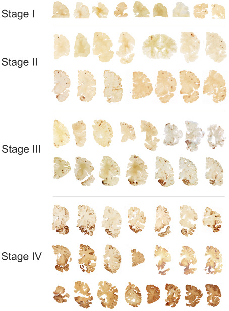
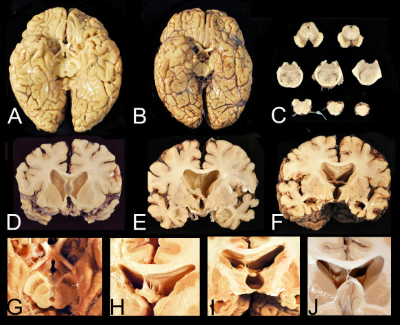
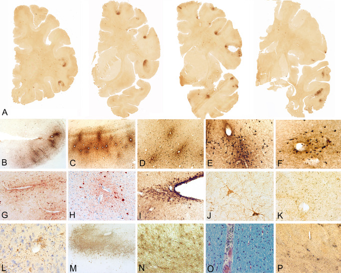
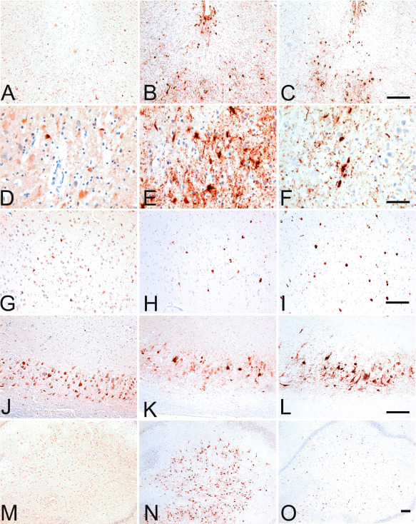
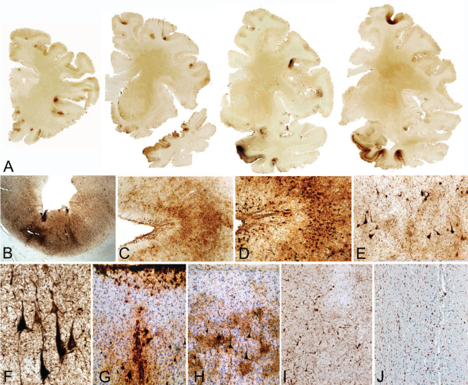
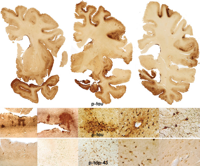
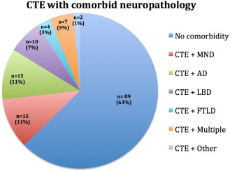
Repetitive brain trauma is associated with a progressive neurological deterioration, now termed as chronic traumatic encephalopathy (CTE). Most instances of CTE occur in association with the play of sports, but CTE has also been reported in association with blast injuries and other neurotrauma. Symptoms of CTE include behavioral and mood changes, memory loss, cognitive impairment and dementia. Like many other neurodegenerative diseases, CTE is diagnosed with certainty only by neuropathological examination of brain tissue. CTE is a tauopathy characterized by the deposition of hyperphosphorylated tau (p-tau) protein as neurofibrillary tangles, astrocytic tangles and neurites in striking clusters around small blood vessels of the cortex, typically at the sulcal depths. Severely affected cases show p-tau pathology throughout the brain. Abnormalities in phosphorylated 43 kDa TAR DNA-binding protein are found in most cases of CTE; beta-amyloid is identified in 43%, associated with age. Given the importance of sports participation and physical exercise to physical and psychological health as well as disease resilience, it is critical to identify the genetic risk factors for CTE as well as to understand how other variables, such as stress, age at exposure, gender, substance abuse and other exposures, contribute to the development of CTE.

Keywords: Alzheimer's disease; TDP-43; chronic traumatic encephalopathy; tauopathy; traumatic brain injury.


Figures

Figure 1
Figure 1
Stages of chronic traumatic encephalopathy (CTE) [adapted from McKee et al 59]. In stage I CTE, p‐tau pathology is found in discrete foci in the cerebral cortex, most commonly in the superior or lateral frontal cortices, typically around small vessels at the depths of sulci. In stage II CTE, there are multiple foci of p‐tau at the depths of the cerebral sulci and there is localized spread of neurofibrillary pathology from these epicenters to the superficial layers of adjacent cortex. The medial temporal lobe is spared neurofibrillary p‐tau pathology. In stage III CTE, p‐tau pathology is widespread; the frontal, insular, temporal and parietal cortices show widespread neurofibrillary degeneration with greatest severity in the frontal and temporal lobes, concentrated at the depths of the sulci. Also in stage III CTE, the amygdala, hippocampus and entorhinal cortex show substantial neurofibrillary pathology that is found in earlier CTE stages. In stage IV CTE, there is widespread severe p‐tau pathology affecting most regions of the cerebral cortex and the medial temporal lobe, sparing calcarine cortex in all but the most severe cases. All images, CP‐13 immunostained 50 μ tissue sections.
Figure 2
Figure 2
Gross neuropathological features of advanced chronic traumatic encephalopathy (CTE). A. CTE stage IV in a former professional football player. Despite the relatively large size of the brain, there is generalized, symmetrical brain atrophy that is most severe in the frontal and temporal lobes. There is also flattening of the hypothalamic floor, atrophy of the mammillary bodies and pallor of the substantia nigra. B. The brain of a subject with Alzheimer's disease, by contrast, also shows generalized, symmetrical brain atrophy but the hypothalamic floor, mammillary bodies and substantia nigra are normal. C. There is depigmentation of the substantia nigra and locus ceruleus and degeneration of the frontopontine tracts in the cerebral peduncle. D, E, F. Coronal sections show pronounced frontal and temporal lobe atrophy, marked ventricular dilatation, as well as cavum septum (A) and absence of the posterior septum (B) and (C). G. The floor of the hypothalamus is severely thinned and the mammillary bodies are markedly atrophic. H. Multiple septal perforations may be present. I. The posterior septum may be absent with only small blood vessels remaining. J. A large single septal perforation.
Figure 3
Figure 3
Patterns of 3R and 4R tau in chronic traumatic encephalopathy (CTE) [adapted from McKee et al 59]. A, D. 3R immunostaining shows scattered RD3 immunoreactive neurons in middle frontal cortex. B, E. 4R immunostaining shows many ET3 immunoreactive neurons and astrocytic tangles in the subpial region of the middle frontal cortex and at the depth of the sulcus. C, F. AT8 immunostaining shows 3R and 4R immunopositive neurons and astrocytic tangles in middle frontal cortex. G, J, M. 3R immunostaining shows scattered RD3 immunoreactive neurons in CA1 (G), CA2 (J) and CA4 hippocampus (M). H, K, N. 4R immunostaining shows many ET3 immunoreactive neurons in CA1 ( H ), CA2 (K) and CA4 hippocampus ( N ). I, L, O . I. AT8 immunostaining shows 3R and 4R immunopositive neurons in CA1 ( I ), CA2 (L) and CA4 hippocampus (O). All 10 μ paraffin embedded sections, magnification bars 50 μm.
Figure 4
Figure 4
Microscopic findings in stage II chronic traumatic encephalopathy (CTE). A. Whole mount coronal sections in stage II CTE show multiple foci of p‐tau pathology primarily located at the depths of the cortical sulci of the frontal and temporal lobes (free floating 50 μ sections, AT8 immunostain). B–H. The p‐tau pathology consists of neurofibrillary tangles and dot‐like and thread‐like dystrophic neurites and is characteristically found around small blood vessels (B–F, free floating 50 μ sections, AT8 immunostain; G, H, 10 μ paraffin embedded sections, AT8 immunostain). (I). Subpial astrocytic tangles are also found at the cortical depths (free floating 50 μ sections, AT8 immunostain). Other pathologies include pretangles (J), dystrophic neurites in the white matter (K) and occasional p‐tau astrocytes (L) (free floating 50 μ sections, AT8 immunostain). There may be marked astrocytosis of the white matter (M, N) (free floating 50 μ sections, Glial fibrillary acidic protein immunostain). Hemosiderin‐laden macrophages (O) (10 μ paraffin section, Luxol fast blue hematoxylin and eosin stain) and multiple perivascular foci of reactive microglia (P) are found around small vessels in the cerebral white matter (free floating 50 μ sections, LN3 immunostain).
Figure 5
Figure 5
Microscopic changes in stage III chronic traumatic encephalopathy (CTE). (A) Whole mount coronal sections in stage III CTE show multiple cortical foci of p‐tau pathology throughout the frontal and temporal cortices. The cortical epicenters and depths of the sulci often consist of confluent masses of neurofibrillary tangles (NFT) and astrocytic tangles (ATs). (B–D) The p‐tau pathology consists of NFTs, ATs and dot‐like and thread‐like dystrophic neurites clustered around the penetrating cortical vessels. (E) Cortex adjacent to the cortical epicenters shows scattered NFT. (F ) The hippocampus shows dense neurofibrillary pathology. Subpial ATs (G) and p‐tau immunoreactive astrocytes may be prominent (H). There may be dystrophic neurites in the white matter (I). SMI‐34 immunostaining shows reduction in axonal staining and numerous large, irregular axonal varicosities (J). (A–I. free floating 50 μ sections, AT8 immunostain; J. SMI‐34 immunostain, 10‐μ paraffin section).
Figure 6
Figure 6
Microscopic changes in stage IV chronic traumatic encephalopathy (CTE). Whole mount coronal sections in stage IV CTE show widespread p‐tau pathology affecting most regions of the cerebral cortex and medial temporal lobe. Astrocytic tangles are prominent and there is marked neuronal loss in the cortex, amygdala and hippocampus. There are also widespread pTDP‐43 abnormalities. All images: 50 μ tissue sections, CP‐13 or pTDP‐43 immunostain.
Figure 7
Figure 7
Chronic traumatic encephalopathy (CTE) with comorbid neuropathology. Of 142 neuropathologically confirmed cases of CTE at the VABUSLI CTE Brain Bank, 15 (10.56%) had coexistent motor neuron disease (MND), 15 (10.56%) had Alzheimer's disease (AD), 10 (7.04%) had Lewy Body disease, 4 (2.%) had frontotemporal lobar degeneration (FTLD) and 7 (4.51%) had two or more of these comorbidities; 89 (63.16%) cases were diagnosed only with CTE.
Figure 8
Figure 8
Coronal T1‐weighted Magnetic resonance imaging shows a cavum septum pellucidum, cortical and white matter atrophy in former professional football player with neuropathologically confirmed chronic traumatic encephalopathy stage IV (arrows indicate the location of the cavum septum).

Similar articles

Cited by

References

    1. Amadoro G, Ciotti MT, Costanzi M, Cestari V, Calissano P, Canu N (2006) NMDA receptor mediates tau‐induced neurotoxicity by calpain and ERK/MAPK activation. Proc Natl Acad Sci U S A 103:2892–2897. - PMC - PubMed
    1. Anthony IC, Norrby KE, Dingwall T, Carnie FW, Millar T, Arango JC et al (2010) Predisposition to accelerated Alzheimer‐related changes in the brains of human immunodeficiency virus negative opiate abusers. Brain 133(Pt 12):3685–3698. - PubMed
    1. Baugh CM, Robbins CA, Stern RA, McKee AC (2014) Current Understanding of Chronic Traumatic Encephalopathy. Curr Treat Options Neurol 16:1–13. - PMC - PubMed
    1. Bazarian JJ, Zhu T, Zhong J, Janigro D, Rozen E, Roberts A et al (2014) Persistent, long‐term cerebral white matter changes after sports‐related repetitive head impacts. PLoS ONE 9:e94734. - PMC - PubMed
    1. Bieniek KF, Stein TD, Alvarez VE, Fry BT, Dickson DW, McKee AC (2014) Poster abstracts. Brain Pathol 24:39–103.

Publication types