. 2015 May 5;112(18):E2376-84.
doi: 10.1073/pnas.1424241112. Epub 2015 Apr 21.

NK cells require IL-28R for optimal in vivo activity

Affiliations

NK cells require IL-28R for optimal in vivo activity

Fernando Souza-Fonseca-Guimaraes et al. Proc Natl Acad Sci U S A. .

Expression of concern in

Abstract

Natural killer (NK) cells are naturally circulating innate lymphoid cells that protect against tumor initiation and metastasis and contribute to immunopathology during inflammation. The signals that prime NK cells are not completely understood, and, although the importance of IFN type I is well recognized, the role of type III IFN is comparatively very poorly studied. IL-28R-deficient mice were resistant to LPS and cecal ligation puncture-induced septic shock, and hallmark cytokines in these disease models were dysregulated in the absence of IL-28R. IL-28R-deficient mice were more sensitive to experimental tumor metastasis and carcinogen-induced tumor formation than WT mice, and additional blockade of interferon alpha/beta receptor 1 (IFNAR1), but not IFN-γ, further enhanced metastasis and tumor development. IL-28R-deficient mice were also more susceptible to growth of the NK cell-sensitive lymphoma, RMAs. Specific loss of IL-28R in NK cells transferred into lymphocyte-deficient mice resulted in reduced LPS-induced IFN-γ levels and enhanced tumor metastasis. Therefore, by using IL-28R-deficient mice, which are unable to signal type III IFN-λ, we demonstrate for the first time, to our knowledge, the ability of IFN-λ to directly regulate NK cell effector functions in vivo, alone and in the context of IFN-αβ.

Keywords: IL-28R; LPS; NK cells; anti-tumor; interferon.


Conflict of interest statement

The authors declare no conflict of interest.

Figures

Fig. 1.
Fig. 1.
LPS stimulation in vivo. (A) IL-28R−/− mice display resistance to endotoxicosis compared with WT mice. Groups of WT and various gene-targeted mice (IL-28R−/−, IFN-γ−/−, and IFNAR1−/−) were inoculated i.p. with 1.25 mg LPS/30 g mouse. Groups received either 100 μg i.p. of cIg or anti-asialoGM1 (to deplete NK cells) on day −1 and 0 (where day 0 is LPS challenge). Statistical analysis was performed using Mantel–Cox Log-rank test, ***P < 0.001, n = 10 in two independent experiments. (B) Spleen NK cells from IL-28R−/− mice, but not IFNAR1−/− mice, display normal expression of CD69 in vivo, in response to LPS (i.p. injection of 0.1 mg/20 g – 6 h). Representative FACS plots are shown and mean ± SEM. CD69 mean fluorescence intensity (MFI) depicted in bar graphs, with n = 10 from two independent experiments. Statistical analysis was performed using Mann–Whitney test; *P < 0.05, **P < 0.01, ***P < 0.001. (C) IL-28R−/− and IFNAR1−/− spleen NK cells demonstrated defective IFN-γ production compared with WT mice at various time points (0–12 h) post-LPS. Representative FACS plots are shown and mean ± SEM. CD69 MFI depicted in bar graphs, with n = 9 from two independent experiments. Statistical analysis was performed using Mann–Whitney test; *P < 0.05, **P < 0.01. (D) Serum cytokines at various time points (0–12 h) post-LPS reveal decreased IFN-γ production in the IFNAR1−/− and IL-28R−/− mice. Results are expressed in mean ± SEM. Statistical analysis was performed using Mann–Whitney test; *P < 0.05, with n = 5 mice per time point.
Fig. 2.
Fig. 2.
(A) Sorted IL-28R−/− or WT spleen NK cells display normal peripheral blood reconstitution in RAG2−/−γc−/− recipient mice 5 d post cell transfer (2 × 10⁵ NK cells injected i.v.). Representative FACS plots are shown, and all data from individual mice are depicted by symbols in bar graphs (Lower). (B) IL-28R−/− NK reconstituted RAG2−/−γc−/− mice display decreased NK cell IFN-γ expression in spleen 6 h after LPS (0.1 mg/30 g) challenge. Representative FACS plots are shown, and all data from individual mice are depicted by symbols in bar graphs (Lower). Results are expressed as mean ± SEM; n = 15 pooled from three independent experiments. Statistical analysis was performed using Mann–Whitney test; **P < 0.01. (C) From the same mice as B, cytokines were assessed from serum after 6 h post LPS challenge. IL-28R−/− NK cell reconstituted RAG2−/−γc−/− mice displayed decreased levels of IFN-γ. Results are expressed as mean ± SEM; n = 8 pooled from two independent experiments, and all data from individual mice are depicted by symbols in bar graphs. Statistical analysis was performed using Mann–Whitney test; *P < 0.05.
Fig. 3.
Fig. 3.
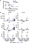
(A) IL-28R−/− mice display enhanced survival after CLP-induced septic shock. Statistical analysis was performed using Mantel–Cox Log-rank test; **P < 0.01, n = 18 pooled from three independent experiments. (B) Cytokine assessment from serum after 3 and 12 h post-CLP. IL-28R−/− mice have increased levels of G-CSF and KC, and decreased levels of IL-17A, MIP1α, MIP1β, and TNF-α. Results are expressed as mean ± SEM, with all data from individual mice depicted by symbols in bar graphs. Statistical analysis was performed using Mann–Whitney test; *P < 0.05, **P < 0.01, and ***P < 0.001.
Fig. 4.
Fig. 4.
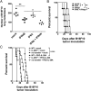
IL-28R−/− and IFNAR1−/− mice have decreased control of B16F10 and RM-1 experimental lung metastases. (A) Groups of five WT or gene-targeted mice were injected i.v. with B16F10 melanoma cells (dose as shown). (B) Groups of seven to eight RAG2−/−γc−/− recipients reconstituted for 5 d with 2 × 10⁵ IL-28R−/− or WT NKs (sorted by TCRβneg, NKp46+, NK1.1+) and injected i.v. with B16F10 melanoma cells (5 × 10⁴). (C) Groups of five WT or gene-targeted mice were injected i.v. with B16F10 melanoma cells (5 × 10⁴). Some mice received cIg (250 μg), anti-IFNAR1 (250 μg), anti-IFN-γ (250 μg), or anti-asGM1 (100 μg) i.p. on days −1, 0, and 7 relative to tumor inoculation as indicated. (D) Groups of 5–10 WT or gene-targeted mice were injected i.v. with RM-1 prostate carcinoma cells (5 × 10³). Some mice received cIg (250 μg), anti-IFNAR1 (250 μg), or anti-IFN-γ (250 μg) i.p. on days −1, 0, and 7 relative to tumor inoculation as indicated. In AD, 14 d after tumor inoculation, the lungs of these mice were harvested and fixed, and the number of B16F10 or RM-1 colonies was counted under a dissection microscope. Symbols in scatter plots represent the number of B16F10 or RM-1 tumor colonies in the lung from individual mice (with mean and SEM shown by cross-bar and errors). Mann–Whitney test was used to compare differences between the groups of mice as indicated (**P < 0.01; ***P < 0.001; ns, not significant).
Fig. 5.
Fig. 5.
IFN-λ (PEG-IL-28A) suppresses B16F10 metastases in an NK cell-dependent fashion. (A and B) Groups of five WT mice in each panel were injected i.v. with B16F10 melanoma cells (2 × 10⁵). Mice received PEG-IL-28A or mIFN-αβ (25 μg i.p.) daily (days 0–5). (C) Groups of 4–12 RAG2−/−γc−/− recipient mice were reconstituted for 5 d with 2 × 10⁵ IL-28R−/− or WT NKs (sorted by TCRβneg, NKp46+, NK1.1+) and injected i.v. with B16F10 melanoma cells (2 × 10⁵). Some mice received no NK cell transfer. Mice then received mock or PEG-IL-28A (25 μg i.p.) daily (days 0–5). In A, 14 d after tumor inoculation, the lungs of these mice were harvested and fixed, and the number of B16F10 colonies was counted under a dissection microscope. In B and C, survival of mice was plotted, and statistical analysis was performed by Mantel–Cox Log-rank test; *P < 0.05, **P < 0.01, ****P < 0.0001.
Fig. 6.
Fig. 6.
IL-28R is necessary for effective control of NK cell-sensitive lymphoma and MCA-induced sarcoma in vivo. (A) Enhanced RMAs (5 × 10⁴ Luc+ RMAs cells) lymphoma growth in vivo in IL-28R−/− mice after i.p. inoculation is depicted by images taken every 5 d from one of these independent experiments in SI Appendix, Fig. S10A; ND, not determined. (B) IL-28R−/− mice display decreased survival after i.p. challenge with 5 × 10² NK cell-sensitive RMAs lymphoma cells. Statistical analysis was performed using a Mantel–Cox test; ***P < 0.001, n = 10–13 per group. (C and D). Groups of 20 male C57BL/6 WT or gene-targeted mice as indicated were inoculated s.c. with 5 μg of MCA in corn oil and subsequently monitored for tumor development over 250 d. WT, IL-28R−/−, or IFNAR1−/− mice were treated with (C and D) control Ig (cIg) or (D) neutralized for IFNAR1 or IFN-γ (250 μg i.p. at day −1 and 0 and then weekly until week 8). Results are shown as survival curves defined as the percentage of tumor-free mice at each time point. Statistical differences in tumor incidence were determined by Mantel-Cox Log-rank test (*P < 0.05; **P < 0.01).

Similar articles

Cited by

References

    1. Kotenko SV, et al. IFN-lambdas mediate antiviral protection through a distinct class II cytokine receptor complex. Nat Immunol. 2003;4(1):69–77. - PubMed
    1. Sheppard P, et al. IL-28, IL-29 and their class II cytokine receptor IL-28R. Nat Immunol. 2003;4(1):63–68. - PubMed
    1. Jewell NA, et al. Lambda interferon is the predominant interferon induced by influenza A virus infection in vivo. J Virol. 2010;84(21):11515–11522. - PMC - PubMed
    1. Prokunina-Olsson L, et al. A variant upstream of IFNL3 (IL28B) creating a new interferon gene IFNL4 is associated with impaired clearance of hepatitis C virus. Nat Genet. 2013;45(2):164–171. - PMC - PubMed
    1. Donnelly RP, Kotenko SV. Interferon-lambda: A new addition to an old family. J Interferon Cytokine Res. 2010;30(8):555–564. - PMC - PubMed

Publication types

MeSH terms