

Protocadherins branch out: Multiple roles in dendrite development
- PMID: 25869446
- PMCID: PMC4594470
- DOI: 10.1080/19336918.2014.1000069
Protocadherins branch out: Multiple roles in dendrite development
Abstract
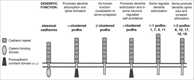
The proper formation of dendritic arbors is a critical step in neural circuit formation, and as such defects in arborization are associated with a variety of neurodevelopmental disorders. Among the best gene candidates are those encoding cell adhesion molecules, including members of the diverse cadherin superfamily characterized by distinctive, repeated adhesive domains in their extracellular regions. Protocadherins (Pcdhs) make up the largest group within this superfamily, encompassing over 80 genes, including the ∼60 genes of the α-, β-, and γ-Pcdh gene clusters and the non-clustered δ-Pcdh genes. An additional group includes the atypical cadherin genes encoding the giant Fat and Dachsous proteins and the 7-transmembrane cadherins. In this review we highlight the many roles that Pcdhs and atypical cadherins have been demonstrated to play in dendritogenesis, dendrite arborization, and dendritic spine regulation. Together, the published studies we discuss implicate these members of the cadherin superfamily as key regulators of dendrite development and function, and as potential therapeutic targets for future interventions in neurodevelopmental disorders.
Keywords: CNR, Cadherin related neuronal receptor; CTCF, CCCTC-binding factor; CaMKII, Ca2+/calmodulin-dependent protein kinase II.; Celsr, Cadherin EGF LAG 7-pass G-type receptor 1; DSCAM, Down syndrome cell adhesion molecule; Dnmt3b, DNA (cytosine-5-)-methyltransferase 3 β; Ds, Dachsous; EC, extracellular cadherin; EGF, Epidermal growth factor; FAK, Focal adhesion kinase; FMRP, Fragile X mental retardation protein; Fj, Four jointed; Fjx1, Four jointed box 1; GPCR, G-protein-coupled receptor; Gogo, Golden Goal; LIM domain, Lin11, Isl-1 & Mec-3 domain; MARCKS, Myristoylated alanine-rich C-kinase substrate; MEF2, Myocyte enhancer factor 2; MEK3, Mitogen-activated protein kinase kinase 3; PCP, planar cell polarity; PKC, Protein kinase C; PSD, Post-synaptic density; PYK2, Protein tyrosine kinase 2; Pcdh; Pcdh, Protocadherin; RGC, Retinal ganglion cell; RNAi, RNA interference; Rac1, Ras-related C3 botulinum toxin substrate 1; S2 cells, Schneider 2 cells; SAC, starburst amacrine cell; TAF1, Template-activating factor 1; TAO2β, Thousand and one amino acid protein kinase 2 β; TM, transmembrane; arborization; atypical cadherin; branching; cadherin superfamily; cell adhesion; da neuron, dendritic arborization neuron; dendritic; dendritic spine; dendritogenesis; fmi, Flamingo; md neuron, multiple dendrite neuron; neural circuit formation; p38 MAPK, p38 mitogen-activated protein kinase; self avoidance; synaptogenesis.
Figures

Similar articles
-
A Unique Role for Protocadherin γC3 in Promoting Dendrite Arborization through an Axin1-Dependent Mechanism.J Neurosci. 2023 Feb 8;43(6):918-935. doi: 10.1523/JNEUROSCI.0729-22.2022. Epub 2023 Jan 5. J Neurosci. 2023. PMID: 36604170 Free PMC article.
-
Protein Kinase C Phosphorylation of a γ-Protocadherin C-terminal Lipid Binding Domain Regulates Focal Adhesion Kinase Inhibition and Dendrite Arborization.J Biol Chem. 2015 Aug 21;290(34):20674-20686. doi: 10.1074/jbc.M115.642306. Epub 2015 Jul 2. J Biol Chem. 2015. PMID: 26139604 Free PMC article.
-
Combinatorial Effects of Alpha- and Gamma-Protocadherins on Neuronal Survival and Dendritic Self-Avoidance.J Neurosci. 2018 Mar 14;38(11):2713-2729. doi: 10.1523/JNEUROSCI.3035-17.2018. Epub 2018 Feb 8. J Neurosci. 2018. PMID: 29439167 Free PMC article.
-
Cadherins and catenins in dendrite and synapse morphogenesis.Cell Adh Migr. 2015;9(3):202-13. doi: 10.4161/19336918.2014.994919. Cell Adh Migr. 2015. PMID: 25914083 Free PMC article. Review.
-
Regulation of neural circuit formation by protocadherins.Cell Mol Life Sci. 2017 Nov;74(22):4133-4157. doi: 10.1007/s00018-017-2572-3. Epub 2017 Jun 19. Cell Mol Life Sci. 2017. PMID: 28631008 Free PMC article. Review.
Cited by
-
The γ-Protocadherin-C3 isoform inhibits canonical Wnt signalling by binding to and stabilizing Axin1 at the membrane.Sci Rep. 2016 Aug 17;6:31665. doi: 10.1038/srep31665. Sci Rep. 2016. PMID: 27530555 Free PMC article.
-
Expression of protocadherin-γC4 protein in the rat brain.J Comp Neurol. 2020 Apr 1;528(5):840-864. doi: 10.1002/cne.24783. Epub 2019 Nov 6. J Comp Neurol. 2020. PMID: 31609469 Free PMC article.
-
Collective mechanical responses of cadherin-based adhesive junctions as predicted by simulations.Biophys J. 2022 Mar 15;121(6):991-1012. doi: 10.1016/j.bpj.2022.02.008. Epub 2022 Feb 10. Biophys J. 2022. PMID: 35150618 Free PMC article.
-
A Unique Role for Protocadherin γC3 in Promoting Dendrite Arborization through an Axin1-Dependent Mechanism.J Neurosci. 2023 Feb 8;43(6):918-935. doi: 10.1523/JNEUROSCI.0729-22.2022. Epub 2023 Jan 5. J Neurosci. 2023. PMID: 36604170 Free PMC article.
-
Genomic Programming of Human Neonatal Dendritic Cells in Congenital Systemic and In Vitro Cytomegalovirus Infection Reveal Plastic and Robust Immune Pathway Biology Responses.Front Immunol. 2017 Sep 25;8:1146. doi: 10.3389/fimmu.2017.01146. eCollection 2017. Front Immunol. 2017. PMID: 28993767 Free PMC article.
References
-
- Vaughn JE, Barber RP, Sims TJ. Dendritic development and preferential growth into synaptogenic fields: A quantitative study of Golgi-impregnated spinal motor neurons. Synapse 1988; 2:69-78; PMID:2458630; http://dx.doi.org/10.1002/syn.890020110 - DOI - PubMed
-
- Vaughn J, Anonymous . Fine structure of synaptogenesis in the vertebrate central nervous system. Synapse 1989; 3:255; PMID:2655146; http://dx.doi.org/10.1002/syn.890030312 - DOI - PubMed
-
- Cline H, Haas K. The regulation of dendritic arbor development and plasticity by glutamatergic synaptic input: a review of the synaptotrophic hypothesis. J Physiol 2008; 586:1509; PMID:18202093; http://dx.doi.org/10.1113/jphysiol.2007.150029 - DOI - PMC - PubMed
-
- Jan YN, Jan LY. Branching out: mechanisms of dendritic arborization. Nat Rev Neurosci 2010; 11:316; PMID:20404840; http://dx.doi.org/10.1038/nrn2836 - DOI - PMC - PubMed
-
- Dierssen M, Ramakers GJ. Dendritic pathology in mental retardation: from molecular genetics to neurobiology. Genes, Brain, and Behav 2006; 5 Suppl 2):48; PMID:16681800; http://dx.doi.org/10.1111/j.1601-183X.2006.00224.x - DOI - PubMed
Publication types
MeSH terms
Substances
Grants and funding
LinkOut - more resources
Full Text Sources
Other Literature Sources
Research Materials
Miscellaneous