. 2015 May 22;290(21):13372-85.
doi: 10.1074/jbc.M115.643767. Epub 2015 Apr 10.

TRAF Family Member-associated NF-κB Activator (TANK) Inhibits Genotoxic Nuclear Factor κB Activation by Facilitating Deubiquitinase USP10-dependent Deubiquitination of TRAF6 Ligase

Affiliations

TRAF Family Member-associated NF-κB Activator (TANK) Inhibits Genotoxic Nuclear Factor κB Activation by Facilitating Deubiquitinase USP10-dependent Deubiquitination of TRAF6 Ligase

Wei Wang et al. J Biol Chem. .

Abstract

DNA damage-induced NF-κB activation plays a critical role in regulating cellular response to genotoxic stress. However, the molecular mechanisms controlling the magnitude and duration of this genotoxic NF-κB signaling cascade are poorly understood. We recently demonstrated that genotoxic NF-κB activation is regulated by reversible ubiquitination of several essential mediators involved in this signaling pathway. Here we show that TRAF family member-associated NF-κB activator (TANK) negatively regulates NF-κB activation by DNA damage via inhibiting ubiquitination of TRAF6. Despite the lack of a deubiquitination enzyme domain, TANK has been shown to negatively regulate the ubiquitination of TRAF proteins. We found TANK formed a complex with MCPIP1 (also known as ZC3H12A) and a deubiquitinase, USP10, which was essential for the USP10-dependent deubiquitination of TRAF6 and the resolution of genotoxic NF-κB activation upon DNA damage. Clustered regularly interspaced short palindromic repeats (CRISPR)/Cas9-mediated deletion of TANK in human cells significantly enhanced NF-κB activation by genotoxic treatment, resulting in enhanced cell survival and increased inflammatory cytokine production. Furthermore, we found that the TANK-MCPIP1-USP10 complex also decreased TRAF6 ubiquitination in cells treated with IL-1β or LPS. In accordance, depletion of USP10 enhanced NF-κB activation induced by IL-1β or LPS. Collectively, our data demonstrate that TANK serves as an important negative regulator of NF-κB signaling cascades induced by genotoxic stress and IL-1R/Toll-like receptor stimulation in a manner dependent on MCPIP1/USP10-mediated TRAF6 deubiquitination.

Keywords: DNA damage; NF-κB; TANK; TNF receptor-associated factor (TRAF); ubiquitin-dependent protease; ubiquitylation (ubiquitination).


Figures

FIGURE 1.
FIGURE 1.
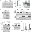
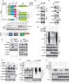
TANK negatively regulates genotoxic stress-induced NF-κB activation. A, control (Ctrl) or HA-TANK-transfected HEK293T cells were treated with TNF-α (10 ng/ml, 30 min), VP16 (10 μm, 2 h), CPT (10 μm, 2 h), or doxorubicin (Dox, 2 μg/ml, 2 h). Whole cell lysates were analyzed by EMSA (NF-κB and Oct1) and immunoblotting. For EMSA, signals were quantified with a PhosphorImager, and normalized activity (NF-κB/Oct1) is shown as -fold induction. B, HEK293T cells were transfected with the NF-κB-Fluc and Tk-Rluc reporter along with a control or HA-TANK. Cells were treated with VP16 (10 μm, 4 h), and luciferase activity was quantified. The histogram represents the normalized data (FLuc/RLuc) from three independent experiments, shown as mean ± S.D. **, p < 0.01. RLU, relative luciferase unit. C and D, control or HA-TANK-transfected HEK293T cells were treated with VP16 (10 μm, 1 h) or CPT (10 μm, 1 h). Whole lysates were immunoblotted with antibodies (C) or immunoprecipitated with IKKα/β antibody followed by immunoblotting (D). E and F, wild-type or TANK knockout HEK293T cells generated with two different sgRNAs were transfected with HA-TANK as indicated. Cells were then treated with VP16 (10 μm, 2 h) or CPT (10 μm, 2 h) (E) or ionizing radiation (IR, 10 gray, 2 h) (F) and analyzed as in A. Arrow, endogenous TANK; arrowhead, HA-TANK. G, HEK293T cells were transfected with control siRNA or TANK-targeting siRNA. After 48 h, cells were treated and analyzed as in A. H, wild-type or TANK KO HEK293T cells were transfected, treated, and analyzed as in B. *, p < 0.05; **, p < 0.01.
FIGURE 2.
FIGURE 2.
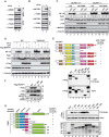
MCPIP1 is required for TANK repression of genotoxic NF-κB activation. A and B, HEK293T cells were treated with VP16 (10 μm, 2 h), and whole cell lysates were immunoprecipitated with the antibody against TANK. The precipitates were immunoblotted with the antibody against NEMO (A) or MCPIP1 (B). C, wild-type and MCPIP1-deficient MEFs were transfected with HA-TANK as indicated. Cells were treated with CPT (10 μm, 2 h) or doxorubicin (Dox, 2 μg/ml, 2 h). Whole cell lysates were analyzed by EMSA (NF-κB and Oct1) and immunoblotting. Normalized NF-κB activation (NF-κB/Oct1) is shown as -fold induction. Ctrl, control. D, wild-type and TANK-KO HEK293T cells were transfected with FLAG-MCPIP1 or a control plasmid. After 48 h, cells were treated with VP16 (10 μm, 2 h) or CPT (10 μm, 2 h) and analyzed as in C. E, cell lysates containing FLAG-MCPIP1 wild-type or the respective mutants were incubated with recombinant GST-TANK proteins. TANK-associated proteins were enriched by IP with glutathione beads and detected by immunoblotting. UBA, ubiquitin associated domain; NCR, N-terminal conserved region; proline rich domain; CCR, C-terminal conserved region. F, HEK293T cells transfected with HA-TANK alone or along with FLAG-MCPIP1 WT or mutant as indicated. Whole cell lysates were immunoprecipitated with anti-HA, followed by immunoblotting with the antibodies as shown. IR, ionizing radiation. G, wild-type or the indicated mutants of recombinant GST-TANK proteins were incubated with cell lysates containing FLAG-MCPIP1 wild-type proteins. TANK-associated FLAG-MCPIP1 was analyzed as in E. Input GST-TANK proteins were visualized by Coomassie Blue staining.
FIGURE 3.
FIGURE 3.
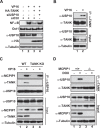
USP10 is required for TANK to inhibit NF-κB activation. A, HEK293T cells were transfected with HA-TANK along with control siRNA (siCtrl) or USP10-targeting siRNA as indicated. After 48 h, cells were treated with VP16 (10 μm, 2 h), and whole cell lysates were analyzed by EMSA (NF-κB and Oct1) and immunoblotting. B, HEK293T cells were treated with VP16 (10 μm, 2 h), and then whole cell lysates were immunoprecipitated with antibody against TANK. The precipitates were immunoblotted with antibodies as shown. C, wild-type and TANK KO HEK293T cells were treated with VP16 (10 μm, 2 h), and whole lysates were immunoprecipitated with antibody against USP10, followed by immunoblotting with the indicated antibodies. D, wild-type and MCPIP1-deficient MEFs were treated with doxorubicin (DOX, 2 μg/ml, 2 h) and analyzed as in B.
FIGURE 4.
FIGURE 4.
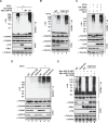
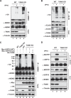
USP10 is essential for TANK-dependent deubiquitination of TRAF6 upon genotoxic stress. A, HEK293T cells were transfected with HA-TANK, wild-type USP10 or catalytically inactive (CA) USP10 (C424A) as indicated and treated with VP16 (10 μm, 2 h). TRAF6 ubiquitination was analyzed with TRAF6 immunoprecipitation under denatured conditions, followed by immunoblotting. B, wild-type or TANK KO HEK293T cells were treated with VP16. TRAF6 ubiquitination was analyzed as in A. C, HEK293T cells were transfected with HA-TANK along with control (siCtrl) or USP10-targeting siRNA. Cells were treated with VP16 (10 μm, 2 h), and TRAF6 ubiquitination was analyzed as in A. D, wild-type HEK293T cells, USP10 KO cells, or TANK KO cells were treated with VP16, and TRAF6 ubiquitination was analyzed as in A. E, wild-type and TANK KO HEK293T cells were transfected with HA-TANK, wild-type USP10 (WT) or catalytically inactive USP10 as indicated. TRAF6 ubiquitination following VP16 (10 μm, 2 h) treatment was analyzed as in A.
FIGURE 5.
FIGURE 5.
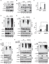
The TRAF-binding domain of TANK is required for USP10 to deubiquitinate TRAF6. A, recombinant full-length GST-TANK or varying mutants harboring different binding domains were incubated with lysates from HEK293 cells expressing FLAG-TRAF6. TANK-associated TRAF6 was determined by IP with glutathione beads, followed by immunoblotting with anti-FLAG antibody. A diagram of the TRAF6 function domain is shown at the bottom. RING, really interesting new gene domain; CC, coiled coil domain; FL, full length. B, HEK293T cells were transfected with wild-type HA-TANK or a TANK mutant with TRAF-binding domain depletion (Δ170–191). Whole cell lysates were subjected to IP with anti-HA and followed by immunoblotting as indicated. C, Similar analyses as in B were carried out in HEK293T cells transfected with HA-TRAF6 wild-type or a TRAF-C depletion mutant. D, wild-type and TANK KO HEK293T cells were treated with VP16 (10 μm, 2 h). Whole cell lysates were immunoprecipitated with antibody against TRAF6. The precipitates were immunoblotted as indicated. E, wild-type and MCPIP1−/− MEFs were treated with doxorubicin (DOX, 2 μg/ml, 2 h), and whole cell lysates were immunoprecipitated with anti-TANK antibody, followed by immunoblotting with the indicated antibodies. F, TANK KO cells were transfected with a vector control, TANK WT, or TANK (31–425) mutant. Cells were treated with VP16 (10 μm, 2 h), and TRAF6 ubiquitination was analyzed by TRAF6 IP, followed by immunoblotting with anti-ubiquitin (Ub). Input whole cell lysates were analyzed by immunoblotting using the indicated antibodies. G and H, HEK293T cells were transfected with HA-TANK or TRAF-binding domain-depleted TANK mutants as shown. Cells were then treated with VP16 (10 μm, 2 h) or CPT (10 μm, 2 h). Whole cell lysates were subjected to analyses of TRAF6 ubiquitination (G) or analyzed by EMSA (NF-κB and Oct1) and immunoblotting (H). Normalized NF-κB activation (NF-κB/Oct1) is shown as -fold induction.
FIGURE 6.
FIGURE 6.
TANK enhances NEMO deubiquitination by MCPIP1/USP10 upon DNA damage. A, wild-type and TANK-deficient HEK293T cells were treated with VP16 (10 μm) for 2 h. VP16-induced NEMO ubiquitination was analyzed with NEMO IP under denatured conditions. The immunoprecipitates were further analyzed by immunoblotting using the indicated antibodies. Ub, ubiquitin. B, VP16-induced ELKS ubiquitination was analyzed as in A. C, wild-type and TANK KO HEK293T cells were transfected with HA-TANK, wild-type USP10 (WT), or catalytically inactive USP10 (CA) as indicated. NEMO ubiquitination following VP16 (10 μm, 2 h) treatment was analyzed as in A. D, wild-type and TANK KO HEK293T cells were treated with VP16 (10 μm) for 2 h. Whole cell lysates were subjected to immunoprecipitation with anti-NEMO antibody and analyzed by immunoblotting using antibodies as shown.
FIGURE 7.
FIGURE 7.
The TANK-MCPIP1-USP10 complex inhibits IL-1R/TLR-mediated NF-κB activation by deubiquitinating TRAF6. A, HEK293T cells were transfected with control siRNA (siCtrl) or USP10-specific siRNA (CST#7747). Cells were treated with IL-1β (2 ng/ml, 30 min) and then analyzed by EMSA (NF-κB and Oct1) and immunoblotting with the indicated antibodies. Normalized NF-κB activation (NF-κB/Oct1) is shown as -fold induction. B, HEK293T cells were transfected with control siRNA or USP10-targeting siRNA alone or along with Myc-USP10 (siRNA-resistant). 48 h later, cells were treated with IL-1β (2 ng/ml, 30 min). Whole cell lysates were analyzed as in A. C, HEK293T cells were transfected as in A and treated with IL-1β (2 ng/ml) for 2 h. NF-κB luciferase reporter activity was measured with a Dual-Luciferase assay, and data from three independent experiments were pooled and are shown as mean ± S.D. *, p < 0.05. RLU, relative luciferase unit. D, HEK293T cells were transfected and treated as in A. TRAF6 ubiquitination in response to IL-1β treatment was analyzed with TRAF6 IP, followed by immunoblotting as indicated. Ub, ubiquitin. E, wild-type and TANK KO HEK293T cells were transfected with HA-TANK as indicated. Cells were treated with IL-1β (2 ng/ml) for 1 h, and TRAF6 ubiquitination was analyzed as in D. Arrow, endogenous TANK; arrowhead, HA-TANK. F, IL-1β-induced NF-κB luciferase reporter activity in wild-type and TANK KO HEK293T cells was determined as in C. *, p < 0.05. G, HEK293T cells were transfected with HA-TANK along with control siRNA or USP10-targeting siRNA as indicated. After 48 h, cells were treated with IL-1β (2 ng/ml, 30 min), and TRAF6 ubiquitination was analyzed as in D. H, wild-type and TANK KO HEK293T cells were transfected with Myc-USP10 (WT) or Myc-USP10 (CA) as shown. TRAF6 ubiquitination in response to IL-1β treatment was analyzed as in D. I, wild-type and MCPIP1-deficient MEFs were transfected with HA-TANK or a control. 48 h later, cells were treated with LPS (10 μg/ml, 30 min), and LPS-induced TRAF6 ubiquitination was analyzed as in D.
FIGURE 8.
FIGURE 8.
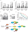
TANK overexpression promotes cell apoptosis upon DNA damage. A and B, wild type and TANK KO HEK293T cells were transfected with HA-TANK or a control vector. After 48 h, cells were treated with VP16 (10 μm, 4 h), and expression of the antiapoptosis genes cIAP1, cIAP2, and BCL-XL (A) as well as the cytokine genes TNF-α, IL-6, and IL-8 (B) was measured by quantitative RT-PCR. GAPDH expression was used as the internal control. Normalized -fold change data were from three independent experiments and are shown as mean ± S.D. C, wild-type and TANK KO HEK293T cells were transfected with HA-TANK as shown. Cells were treated with VP16 (1 μm) for 24 h and whole cell lysates were subjected to immunoblotting using the indicated antibodies. Arrow, endogenous TANK; arrowhead, HA-TANK. D, MDA-MB-231 cells were transfected with HA-TANK or a control vector. Cells were treated with doxorubicin (DOX, 0.5 μm) for 24 h and analyzed as in C. E, wild-type and TANK KO HEK293T cells were transfected with HA-TANK or a control for 24 h. Cells were then treated with 1 μm VP16 for the indicated times, and the survival fraction of the cells was determined by trypan blue staining. The cell survival fraction data are from three independent experiments and are shown as mean ± S.D. F, a model illustrating the TANK/MCPIP1/USP10-mediated negative feedback response to suppress NF-κB activation via deubiquitinating TRAF6 and NEMO. *, p < 0.05; **, p < 0.01.

Similar articles

Cited by

References

    1. Baldwin A. S. (2012) Regulation of cell death and autophagy by IKK and NF-κB: critical mechanisms in immune function and cancer. Immunol. Rev. 246, 327–345 - PubMed
    1. McCool K. W., Miyamoto S. (2012) DNA damage-dependent NF-κB activation: NEMO turns nuclear signaling inside out. Immunol. Rev. 246, 311–326 - PMC - PubMed
    1. Wang C. Y., Mayo M. W., Korneluk R. G., Goeddel D. V., Baldwin A. S., Jr. (1998) NF-κB antiapoptosis: induction of TRAF1 and TRAF2 and c-IAP1 and c-IAP2 to suppress caspase-8 activation. Science 281, 1680–1683 - PubMed
    1. Nakanishi C., Toi M. (2005) Nuclear factor-κB inhibitors as sensitizers to anticancer drugs. Nat Rev Cancer 5, 297–309 - PubMed
    1. Niu J., Shi Y., Tan G., Yang C. H., Fan M., Pfeffer L. M., Wu Z. H. (2012) DNA damage induces NF-κB-dependent microRNA-21 upregulation and promotes breast cancer cell invasion. J. Biol. Chem. 287, 21783–21795 - PMC - PubMed

Publication types

MeSH terms