Review
. 2015:33:291-353.
doi: 10.1146/annurev-immunol-032414-112212.

Ion channels in innate and adaptive immunity

Affiliations
Review

Ion channels in innate and adaptive immunity

Stefan Feske et al. Annu Rev Immunol. 2015.

Abstract

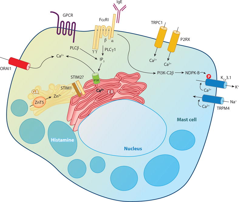
Ion channels and transporters mediate the transport of charged ions across hydrophobic lipid membranes. In immune cells, divalent cations such as calcium, magnesium, and zinc have important roles as second messengers to regulate intracellular signaling pathways. By contrast, monovalent cations such as sodium and potassium mainly regulate the membrane potential, which indirectly controls the influx of calcium and immune cell signaling. Studies investigating human patients with mutations in ion channels and transporters, analysis of gene-targeted mice, or pharmacological experiments with ion channel inhibitors have revealed important roles of ionic signals in lymphocyte development and in innate and adaptive immune responses. We here review the mechanisms underlying the function of ion channels and transporters in lymphocytes and innate immune cells and discuss their roles in lymphocyte development, adaptive and innate immune responses, and autoimmunity, as well as recent efforts to develop pharmacological inhibitors of ion channels for immunomodulatory therapy.

Keywords: CRAC; KCa3.1; KV1.3; ORAI; STIM; TRP, T cells, B cells, macrophages, mast cells, DC, disease, therapy; calcium; chloride; ion channels; magnesium; potassium; sodium; transporters; zinc.


Figures

Figure 1
Figure 1
Ion channels and regulation of divalent cation signaling in immune cells. A variety of ion channels and transporters regulate the function of immune cells. Rather than depicting a specific immune cell type or specific ion channels/transporters, this figure presents an overview of the classes of ion channels and transporters in the plasma membrane and the pathways and compartments involved in regulating intracellular ion concentrations. (a) Divalent cation channels and transporters and the concentrations of extra- and intracellular Ca2+, Mg2+, and Zn2+ ions (concentration gradients indicated by triangles). (b) Monovalent cation (K+, Na+) and anion (Cl) channels that control the Vm in immune cells and the concentrations of extra- and intracellular K+, Na+, and Cl ions. (See Figure 2 for details on Vm regulation.) (c) Mechanisms regulating Ca2+ homeostasis in immune cells. Antigen receptor or GPCR binding results in PLC activation, production of IP3 and Ca2+ release from ER Ca2+ stores via IP3R channels resulting in (1) a transient increase in [Ca2+]i and (2) activation of store-operated Ca2+ entry (SOCE; for details see Figures 3, 4, 5 and the sidebar, below). The resulting increase in [Ca2+]i is balanced by Ca2+ reuptake into the ER via SERCA pumps, Ca2+ export via PMCA pumps, and uptake into the mitochondrial matrix via the MCU. (Abbreviations: [Ca2+]i, intracellular Ca2+ concentration; GPCR, G protein–coupled receptor; IP3R, inositol 1,4,5-trisphosphate receptor; MCU, mitochondrial Ca2+ uniporter; PLC, phospholipase C; PMCA, plasma membrane Ca2+ pump; SERCA, sarco/endoplasmic reticulum Ca2+ ATPase; Vm, membrane potential. For additional abbreviations, see Table 1, below.)
Figure 2
Figure 2
Regulation of membrane potential (Vm) and Ca2+ influx. (a) In nonactivated lymphocytes, the resting Vm is approximately −59 mV, as determined by the equilibrium potentials of K+ (KV1.3, KCa3.1), Na+ (TRPM4), and Cl channels in the plasma membrane (PM). The equilibrium potentials (E) and Vm are shown; the intra- and extracellular ion concentrations are as indicated in Figure 1. (b) K+ channels hyperpolarize the PM and enhance Ca2+ influx. The opening of ORAI1 Ca2+ channels following T cell activation results in Ca2+ influx ❶ and depolarization of Vm, causing the subsequent opening of Ca2+-activated KCa3.1 channels and voltage-gated KV1.3 channels, respectively. ❷ Open KCa3.1 and KV1.3 channels mediate K+ efflux and hyperpolarization of the PM, thereby sustaining Ca2+ influx ❸. (c) TRPM4 depolarizes the PM and inhibits Ca2+ influx. Ca2+ influx through ORAI1 channels ❶ results in the opening of Ca2+-regulated TRPM4 channels ❷ that mediate Na+ influx and depolarization of the PM, thereby inhibiting Ca2+ influx via ORAI1 ❸. (d) Opening of Cl channels in lymphocytes results in efflux of Cl ions due to the relatively high [Cl]i of approximately 38 mM and an equilibrium potential (approximately −33 mV) that is positive relative to Vm ❶. Cl efflux depolarizes Vm and inhibits Ca2+ influx ❷.
Figure 3
Figure 3
Immunodeficiency due to mutations in ORAI1, STIM1, and MAGT1 genes. ORAI1 is the pore-forming subunit of the CRAC channel in the PM and mediates Ca2+ influx following TCR stimulation. It is activated by the Ca2+-sensing protein STIM1 localized in the ER. Mutations in ORAI1 and STIM1 genes (indicated by red circles) cause CRAC channelopathy, a primary immunodeficiency syndrome characterized by severe, life-threatening infections, autoimmunity, and nonimmunological symptoms. MAGT1 is a selective Mg2+ transporter in the PM activated by TCR stimulation. Mutations in the human MAGT1 gene (indicated by red circles) cause XMEN disease. For details, see sidebar. (Abbreviations: CAD, CRAC activation domain; CC, coiled-coil domain; CRAC, Ca2+ release–activated Ca2+ channel; EBV, Epstein-Barr virus; EFh; EF hand; ER, endoplasmic reticulum; IP3, inositol 1,4,5-trisphosphate; IP3R, IP3 receptor; MAGT1, Mg2+ transporter; NKG2D, natural killer group 2, member D; PLC, phospholipase C; PM, plasma membrane; SAM, sterile alpha motif; SCID, severe combined immunodeficiency; STIM1, stromal interaction molecule 1; TCR, T cell receptor; Treg, regulatory T cell; XMEN, X-linked immunodeficiency with Mg2+ defect, EBV infection, and neoplasia.)
Figure 4
Figure 4
CRAC channel structure. The CRAC channel is a multimer of ORAI1 subunits that form the pore of the channel. (a) Membrane topology of one ORAI1 subunit. Each subunit has four transmembrane domains (M1–M4), intracellular N and C termini, and two extracellular loops. (b) Ion pore of the Drosophila Orai channel, whose transmembrane domains are conserved in the human ORAI1 channel. M1 lines the conduction pathway for Ca2+. Two M1 alpha-helices from two separate ORAI1 subunits are shown, with amino acid side chains protruding into the pore indicated in yellow. Amino acid residue numbers refer to human ORAI1. Glutamate (E) 106 at the outer end of the pore is the Ca2+-binding site in the selectivity filter of the CRAC channel that determines its preference for conducting Ca2+ over other divalent or monovalent cations. Arginine (R) 91, together with lysine (K) 87 and R83, is part of a basic region of residues that form the narrowest part of the pore; R91 is mutated in patients with abolished CRAC channel function (see sidebar and Figure 3). (c) Crystal structure of the Drosophila Orai channel. The hexameric assembly of Orai subunits is shown in an orthogonal view from the extracellular side. The colors for each transmembrane helix in this panel are the same as in panel a (e.g., M1 helices are shown in blue, E106 is depicted in yellow, and a Ca2+ ion in magenta). Panels b and c have been reproduced with permission from Reference 381. A Ca2+ ion bound to E106 and the path of Ca2+ through the pore have been added to panel b.
Figure 5
Figure 5
Ion channels in T cells. Antigen binding to the TCR results in activation of tyrosine kinases and phosphorylation of ITAM motifs of the TCR and phosphorylation of the scaffold proteins SLP-76 and LAT (not shown) that facilitate recruitment and activation of PLCγ1, production of IP3, and release of Ca2+ from ER stores. Other second messengers such as NAADP and cADPR have also been reported to cause ER store depletion via RyR after TCR activation. Reduction of [Ca2+]ER activates STIM1 and STIM2, which then translocate to ER-PM junctions, where they bind to ORAI1. ORAI1 encodes the CRAC channel and mediates SOCE; the role of ORAI2 and ORAI3 isoforms for SOCE in T cells and T cell function is unknown. Other Ca2+ channels reported in T cells include P2X1, P2X4, and P2X7 receptors; TRPM2; and several L-type voltage-gated Ca2+ channels (CaV), although their roles in T cell function are less well defined. Ca2+ influx depends on the negative membrane potential (Vm) established by two K+ channels, KV1.3 and KCa3.1, which are counteracted by the Ca2+-activated TRPM4 channel that depolarizes Vm. KV1.3 activation is mediated by membrane depolarization, whereas KCa3.1 activation is mediated by two signals that include Ca2+ binding to the calmodulin-bound C terminus of KCa3.1 and the recruitment of PI3K-C2β to the immunological synapse, which results in histidine phosphorylation of KCa3.1 by NDPK-B. The Mg2+ transporter MAGT1 and the divalent cation channel TRPM7 mediate Mg2+ influx; their activation mechanisms following TCR stimulation are not understood, but that for TRPM7 is regulated by PIP2 and inhibited by intracellular Mg2+. Several Zn2+ transporters, including ZIP6, ZIP3, and ZIP8, reportedly mediate Zn2+ influx from the extracellular space and release from intracellular compartments, respectively. The Cl channels LRRC8A, CFTR, and GABAA regulate T cell function, likely by mediating Cl efflux and depolarization of Vm. (Abbreviations: cADPR, cyclic ADP ribose; CaV, voltage-gated Ca2+ channel; CFTR, cystic fibrosis transmembrane conductance regulator; CRAC, Ca2+ release–activated Ca2+ channel; ER, endoplasmic reticulum; GABA, γ-aminobutyric acid; GPCR, G protein–coupled receptor; IP3, inositol 1,4,5-trisphosphate; ITAM, immunoreceptor tyrosine-based activation motif; LAT, linker for activation of T cells; LRRC8A, leucine-rich repeat containing 8 family member A; NAADP, nicotinic acid adenine dinucleotide phosphate; NDPK-B, nucleoside diphosphate kinase B; PLC, phospholipase C; PM, plasma membrane; RyR, ryanodine receptor; SOCE, store-operated Ca2+ entry; STIM, stromal interaction molecule; TCR, T cell receptor; TRP, transient receptor potential; Vm, membrane potential; ZIP, Zir/Irt-like protein.)
Figure 6
Figure 6
Ion channels in T cell development and lineage differentiation. Studies in knockout mice and human patients have implicated a number of ion channels and transporters in T cell development. During T cell development, common lymphoid progenitors (CLPs) enter the thymus and differentiate into CD2+, CD4CD8 (double-negative, DN) T cells that have not yet rearranged their T cell receptor (TCR) genes. These DN cells further develop into CD4+ CD8+ (double-positive, DP) thymocytes that express low levels of a rearranged TCRαβ. This developmental step is regulated by the Mg2+-permeable channel TRPM7 and potentially also by the Zn2+ transporter ZIP3. The volume-regulated Cl channel LRRC8A also controls thymocyte development, and Lrrc8a/ mice have strongly reduced numbers of DN, DP, and SP thymocytes. DP thymocytes next undergo positive selection, during which cells whose TCR interacts weakly with self-peptide-MHC on thymic epithelial cells are rescued from death by neglect. The voltage-gated Na+ channel NaV1.5 and potentially also the voltage-gated Ca2+ channel CaV1.4 regulate positive selection. During negative selection, DP thymocytes with high-affinity TCR-self-peptide-MHC interactions and strong TCR signals are eliminated to delete potentially self-reactive T cells. A subset of DP thymocytes escapes negative selection despite expressing a high-affinity TCR, and instead they become agonist-selected T cells whose development requires strong and sustained TCR signals. These include Foxp3+ natural regulatory T (nTreg) cells, invariant natural killer T cells (iNKT), and CD8αα+ intestinal intraepithelial lymphocytes (IELs). The thymic development of nTreg, iNKT, and IEL cells, in contrast to the development of conventional αβ T cells (denoted in figure as Tcon), requires Ca2+ influx via CRAC channels, likely by controlling the expression of cytokines needed for their homeostasis. The development or survival of CD4+ T cells partially depends on Mg2+ influx through the Mg2+ transporter MAGT1. Once conventional CD4+ T cells leave the thymus, they further differentiate into distinct T helper lineages of which Th17 cells, but not Th1 or Th2 cells, have been suggested to depend on CRAC channels for differentiation. The proliferation and differentiation of CD8+ T cells into effector and memory cells (in figure as Tmem) is regulated by the Cl/HCO3 anion exchanger Ae2, whereas the maintenance of memory CD8+ T cells requires Ca2+ influx via CRAC channels.
Figure 7
Figure 7
Ion channels in B cells. Similar to TCR activation in T cells, cross-linking of the BCR results in tyrosine phosphorylation of ITAM motifs in the Igα/β chains of the BCR and the scaffold protein SLP-65 (not shown), which helps to recruit PLCγ2 to the plasma membrane of B cells and activate it. The subsequent generation of IP3 and the IP3R-mediated Ca2+ release from the ER result in the activation of STIM1 and STIM2 and in the opening of ORAI1/CRAC channels. The nonselective cation channels TRPC1 and TRPC7 have been implicated in store-dependent and -independent Ca2+ influx in chicken DT-40 B cells. Likewise, the nonselective cation channel TRPM7 regulates total cellular Mg2+ levels and, thereby, the proliferation and viability of DT-40 B cells. The Vm in B cells is established by the voltage-gated K+ channel KV1.3 and the Ca2+-activated KCa3.1 channel. The proton channel HV1 associates with the BCR complex and is required for activation of NOX and the generation of reactive oxygen species, and these events promote BCR signaling by inhibiting the tyrosine phosphatase SHP1, leading to enhanced activation of signaling pathways such as Syk, PI3-kinase, and Akt. (Abbreviations: BCR, B cell receptor; CRAC, Ca2+ release–activated Ca2+ channel; ER, endoplasmic reticulum; GPCR, G protein–coupled receptor; IP3, inositol 1,4,5-trisphosphate; ITAM, immunoreceptor tyrosine-based activation motif; NOX, NADPH oxidase; PLC, phospholipase C; PM, plasma membrane; STIM, stromal interaction molecule; TRP, transient receptor potential; Vm, membrane potential.)
Figure 8
Figure 8
Ion channels in macrophages. Macrophages express a variety of receptors that activate ion channels including FcγRI and FcγRIII, G protein–coupled chemokine receptors, and innate immunoreceptors such as Dectin-1/2 and Toll-like receptors (TLRs). FcγRI cross-linking and Dectin-1 binding result in activation of PLCγ1, and chemokine receptor engagement results in activation of PLCβ and in the opening of CRAC channels, as described in Figures 5 and 7 for T and B cells. The membrane potential (Vm) required for SOCE in macrophages is established by two K+ channels, KV1.3 and KCa3.1, and by the Na+ channel TRPM4 channel, as described in Figure 2. Other Ca2+ channels in macrophages are TRPM2, TRPV2, and P2RX7. TRPM2 is a nonselective Ca2+ channel located in both the plasma and ER membranes. TRPM2 is activated by the second messengers ADPR, cADPR, and NAAPD, resulting in Ca2+ influx and release from the ER. TRPM2 function in macrophages is further regulated by ROS levels, which increase after TLR stimulation. TRPV2 is a nonselective, Ca2+-permeable channel that is recruited to the early phagosome after FcγR or zymosan stimulation and is required for the early steps of phagocytosis, presumably by mediating Na+ but not Ca2+ influx, leading to membrane depolarization resulting in the generation of PIP2 and actin depolymerization. P2RX7 appears to play a dual role in macrophages by mediating the ATP-induced influx of Ca2+ and the efflux of K+ that is required for inflammasome activation (see Figure 9). The Zn2+ transporters ZIP6 and ZIP8 reportedly regulate Zn2+ levels in macrophages by mediating Zn2+ influx after TLR stimulation, thereby inhibiting NF-κB activation. The proton channel HV1 is closely associated with the NADPH oxidase complex (not shown) and is essential for the generation of ROS in macrophages (see Figure 10). (Abbreviations: CRAC, Ca2+ release–activated Ca2+ channel; ER, endoplasmic reticulum; GPCR, G protein–coupled receptor; NLRP3, NLR family, pyrin domain containing 3; PIP2, phosphatidylinositol 4,5-bisphosphate; ROS, reactive oxygen species; SOCE, store-operated Ca2+ entry; TLR, Toll-like receptor; TRP, transient receptor potential; ZIP, Zir/Irt-like protein.)
Figure 9
Figure 9
P2RX7 activation of the NLRP3 inflammasome in macrophages. High concentrations of extracellular ATP released from dying cells function as danger signals to initiate NLRP3 inflammasome activation, which requires two signals: ❶ During the priming phase, NLRP3 and pro-IL-1β are transcribed in response to signals such as TLR activation or IFN-γ, TNF-α, or other stimuli that activate NF-κB. ❷ A second signal then activates the NLRP3 inflammasome, resulting in the activation of caspase-1, cleavage of pro-IL-1β and pro-IL-18 into their mature forms, and ❸ secretion of IL-1β and IL-18. Central to NLRP3 activation during the second phase is K+ efflux from the cytoplasm of macrophages, which mediates assembly and activation of the NLRP3 inflammasome by an unknown mechanism(s). P2RX7 mediates only ATP-stimulated K+ efflux, whereas K+ efflux by other second signals can be mediated by plasma membrane insertion of bacterial pore-forming toxins or by yet-to-be-defined mechanisms, as is the case for particulate matter. (Abbreviations: ASC, apoptosis associated speck-like protein containing a CARD; ATP, adenosine trisphosphate; IFN-γ, interferon-γ; IL-1β, interleukin-1β; IRAK, interleukin-1 receptor associated kinase; MyD88, myeloid differentiation primary response gene 88; NF-κB, nuclear factor-κB; NLRP3, NLR family, pyrin domain containing 3; TLR, Toll-like receptor; TNF-α, tumor necrosis factor α.)
Figure 10
Figure 10
Ion channels in NADPH oxidase (NOX) activation and phagocytic respiratory burst. The multiprotein NOX complex is critical for the killing of microorganisms by macrophages and neutrophils by mediating the generation of reactive oxygen species (ROS). The NOX2 subunit (also known as gp91phox, not shown) located in the nascent phagosome membrane accepts electrons from cytosolic NADPH, which are then transported across the plasma membrane where they are transferred to O2, generating O2. The transfer of electrons results in the depolarization and acidification of the cytosol, either of which would inhibit NOX activity. This is prevented by the transport of H+ from the cytosol into the phagosome by the proton channel HV1. H+ reacts with O2 in the phagosome to generate H2O2, which undergoes further conversion to HOCl that is catalyzed by MPO. (Abbreviations: HOCl, hydrochlorus acid; MPO, myeloperoxidase; NADPH, nicotinamide adenine dinucleotide phosphate.)
Figure 11
Figure 11
Ion channels in mast cells. Antigen binding and cross-linking of IgE antibodies bound to the high-affinity FcɛRI are central to mast cell activation, Ca2+ influx, and the secretion of mediators of the allergic response. Ca2+ mediates both acute mast cell degranulation and the delayed transcriptional production of cytokines, leukotrienes, and other secondary mediators by mast cells. IgE cross-linking leads to phosphorylation of the β and γ FcɛRI subunits and to the activation of several signaling molecules including PLCγ, IP3 production, Ca2+ release from ER stores, and SOCE via STIM1 and ORAI1 activation. Ca2+ influx results in the activation of KCa3.1, which by mediating the efflux of K+ maintains a hyperpolarized membrane potential (Vm) and thereby enables Ca2+ influx via ORAI1. The function of KCa3.1 is counteracted by TRPM4, which mediates Na+ influx and Vm depolarization, thereby limiting the magnitude of Ca2+ influx in mast cells. FcɛRI activation also results in the activation of the class II PI3K-C2β, which is critical for KCa3.1 activation by stimulating the histidine phosphorylation at the C terminus of KCa3.1 by NDPK-B. Mast cells express several TRP channels and P2X receptors that potentially mediate Ca2+ influx, but their role in mast cell function is not well defined. The Zn2+ transporter ZnT5 is located in the Golgi apparatus (GA) and mediates Zn2+ uptake, thereby regulating FcɛRI-mediated PKC recruitment to the plasma membrane and NF-κB activation. (Abbreviations: ER, endoplasmic reticulum; IP3, inositol 1,4,5-trisphosphate; NDPK-B, nucleoside diphosphate kinase B; PKC, protein kinase C; PLC, phospholipase C; SOCE, store-operated Ca2+ entry; STIM, stromal interaction molecule; TRP, transient receptor potential; Vm, membrane potential.)

Similar articles

Cited by

References

    1. Cahalan MD, Chandy KG. The functional network of ion channels in T lymphocytes. Immunol Rev. 2009;231:59–87. - PMC - PubMed
    1. Clapham DE. Calcium signaling. Cell. 2007;131:1047–58. - PubMed
    1. Feske S. Calcium signalling in lymphocyte activation and disease. Nat Rev Immunol. 2007;7:690–702. - PubMed
    1. Feske S. ORAI1 and STIM1 deficiency in human and mice: roles of store-operated Ca2+ entry in the immune system and beyond. Immunol Rev. 2009;231:189–209. - PMC - PubMed
    1. Oh-hora M. Calcium signaling in the development and function of T-lineage cells. Immunol Rev. 2009;231:210–24. - PubMed

Publication types

MeSH terms