. 2015 Jun 12;290(24):14798-809.
doi: 10.1074/jbc.M115.637488. Epub 2015 Apr 2.

Soluble Urokinase Receptor Is Released Selectively by Glioblastoma Cells That Express Epidermal Growth Factor Receptor Variant III and Promotes Tumor Cell Migration and Invasion

Affiliations

Soluble Urokinase Receptor Is Released Selectively by Glioblastoma Cells That Express Epidermal Growth Factor Receptor Variant III and Promotes Tumor Cell Migration and Invasion

Andrew S Gilder et al. J Biol Chem. .

Abstract

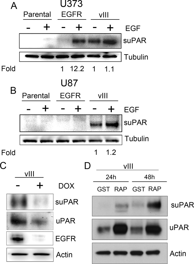
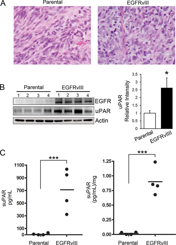
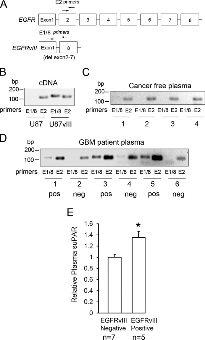
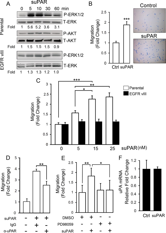
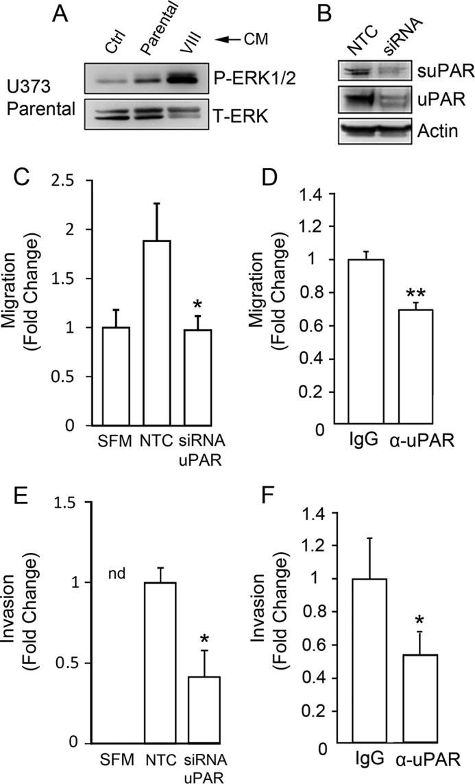
Genomic heterogeneity is characteristic of glioblastoma (GBM). In many GBMs, the EGF receptor gene (EGFR) is amplified and may be truncated to generate a constitutively active form of the receptor called EGFRvIII. EGFR gene amplification and EGFRvIII are associated with GBM progression, even when only a small fraction of the tumor cells express EGFRvIII. In this study, we show that EGFRvIII-positive GBM cells express significantly increased levels of cellular urokinase receptor (uPAR) and release increased amounts of soluble uPAR (suPAR). When mice were xenografted with human EGFRvIII-expressing GBM cells, tumor-derived suPAR was detected in the plasma, and the level was significantly increased compared with that detected in plasma samples from control mice xenografted with EGFRvIII-negative GBM cells. suPAR also was increased in plasma from patients with EGFRvIII-positive GBMs. Purified suPAR was biologically active when added to cultures of EGFRvIII-negative GBM cells, activating cell signaling and promoting cell migration and invasion. suPAR did not significantly stimulate cell signaling or migration of EGFRvIII-positive cells, probably because cell signaling was already substantially activated in these cells. The activities of suPAR were replicated by conditioned medium (CM) from EGFRvIII-positive GBM cells. When the CM was preincubated with uPAR-neutralizing antibody or when uPAR gene expression was silenced in cells used to prepare CM, the activity of the CM was significantly attenuated. These results suggest that suPAR may function as an important paracrine signaling factor in EGFRvIII-positive GBMs, inducing an aggressive phenotype in tumor cells that are EGFRvIII-negative.

Keywords: EGFRvIII; LRP1; cell invasion; cell migration; epidermal growth factor receptor (EGFR); glioblastoma; paracrine interaction; paracrine signaling; tumor microenvironment; urokinase receptor.


Figures

FIGURE 1.
FIGURE 1.
EGFR signaling increases expression of cellular uPAR in human GBM cells. A, immunoblot analysis comparing EGFR expression in U87MG and U373MG parental cells and in the same cells that express EGFRvIII. Activation of the EGFR (P-Y1068), ERK1/2, Akt, and STAT5 was assessed after serum-starving cells for 18 h. Mean relative signal intensities for P-ERK1/2 and P-Akt relative to T-ERK1/2 and T-Akt are shown below the blots (n = 3). B, immunoblot analysis comparing cellular uPAR in parental U87MG and U373MG cells and in the same cells that overexpress WT-EGFR or express EGFRvIII (vIII). uPAR immunoblots were performed using the identical samples but separate blots because the antibody requires nonreducing conditions. Blots were re-probed for tubulin as a control for load. C, U373MG cells that were serum-starved for 18 h were pretreated with 50 nm AG1478 for 2 h, as indicated (+), and then with 10 ng/ml EGF for 6 h, as indicated (+). Immunoblot analysis was performed to detect cellular uPAR, EGFR, and actin as a control for load. D, qPCR was performed to detect uPAR mRNA after completing the identical incubations as in C (mean ± S.D. relative to control, n = 3; **, p < 0.01). E, EGFRvIII-expressing U373MG cells were serum-starved for 18 h and subsequently treated (+) or untreated (−) for 18 h with 25 μm PD98059 in serum-free medium. Immunoblotting was performed to assess the indicated antigens. F, parental and WT-EGFR-expressing U87MG cells were treated with 10 ng/ml EGF (+) or with vehicle (−) in serum-free medium for 18 h. Immunoblot analysis was then performed.
FIGURE 2.
FIGURE 2.
EGFRvIII-expressing GBM cells generate increased levels of suPAR. A, parental U373MG cells and U373MG cells that overexpress WT-EGFR (EGFR) or express EGFRvIII (vIII) were serum-starved for 18 h. The cells were then treated with 10 ng/ml EGF (+) or with vehicle (−) for an additional 18 h. CM was recovered, concentrated 50×, and subjected to immunoblot analysis to detect suPAR. The cells from which CM was recovered also were subjected to immunoblot analysis to detect tubulin, as a control to ensure that CM was generated by an equivalent load of cells. Mean relative signal intensities are shown below the blots (n = 3). In each case, the signal intensity is standardized against that observed in the absence of EGF. B, U87MG cells were treated as described in A. CM was subjected to immunoblot analysis to detect suPAR. Cell extracts were analyzed to detect tubulin. C, U373MG cells that express EGFRvIII under the control of a doxycycline (DOX)-repressible promoter were treated (+) or untreated (−) with 1 μg/ml doxycycline for 3 days and subsequently serum-starved for 24 h in the presence or absence of doxycycline. Immunoblot analysis was performed to detect suPAR in CM and uPAR in cell extracts. EGFR and actin in cell extracts also were assessed. D, U87MG cells were cultured in serum-free medium with GST-RAP or GST (150 nm) for 24 or 48 h. CM and cell extracts were subjected to immunoblot analysis to detect uPAR and actin.
FIGURE 3.
FIGURE 3.
Detection of suPAR in the plasma of mice xenografted with GBM cells. A, representative sections of xenografts recovered from mice when the tumors were 2 cm in maximum diameter. The tissue was formalin-fixed and paraffin-embedded. Sections were stained with hematoxylin and eosin. B, tissue from each xenograft was extracted in RIPA buffer and subjected to SDS-PAGE and immunoblot analysis to detect uPAR and EGFR. Densitometry was performed to quantitate uPAR in each lane, standardized against actin (mean ± S.E.; n = 4; *, p < 0.05). C, plasma was recovered from mice with xenografts at the time of euthanasia. Equivalent samples of plasma were subjected to ELISA to detect human suPAR. Each value represents a different plasma sample tested in duplicate. Results are presented as uncorrected values (left) and relative to tumor weight (right) (n = 4; ***, p < 0.001).
FIGURE 4.
FIGURE 4.
GBM patients with circulating tumor DNA corresponding to EGFRvIII have increased levels of plasma suPAR. A, diagram showing the primers (arrows) used to amplify DNA derived from WT-EGFR for EGFRvIII in plasma samples. B, cDNA was generated from parental U87MG cells and EGFRvIII-expressing U87MG cells. The cDNA was amplified using the primers that detect EGFRvIII (E1/8) and WT-EGFR (E2). The resulting amplicons, which were 119 and 104 bp, respectively, were compared by agarose gel electrophoresis with SYBR Green staining. C, PCR amplification of circulating DNA within cancer-free control plasma samples using the indicated primers. D, PCR amplification of circulating tumor DNA in six representative GBM patient plasma samples. Samples that were judged to be positive for EGFRvIII-derived DNA are marked “pos.” Those judged to be negative are marked “neg.” E, ELISA analysis of suPAR levels in 12 GBM patient plasma samples. Cases were sorted as EGFRvIII-positive (n = 5) or negative (n = 7) based on E1/8 DNA amplification and analysis as shown in D (mean ± S.E.; *, p < 0.05).
FIGURE 5.
FIGURE 5.
Purified suPAR activates ERK1/2 and promotes migration of GBM cells. A, purified recombinant uPAR (10 nm) was incubated with parental U373MG cells (parental) or with EGFRvIII-expressing U373MG cells, which had been serum-starved for 18 h, for the indicated time points. Immunoblot analysis was performed to detect phospho-ERK1/2, total ERK1/2, phospho-Akt, and total Akt. Mean relative signal intensities for P-ERK1/2 and P-Akt relative to T-ERK1/2 and T-Akt are shown below the blots (n = 3). B, parental U373MG cells were serum-starved for 18 h, added to Transwells, and allowed to migrate toward 1% serum supplemented with 10 nm suPAR or vehicle (Ctrl) for 18 h. The number of cells that migrated to the underside of the membrane were counted and expressed relative to the control (mean ± S.E.; n = 3, ***, p < 0.001). C, cell migration was studied as in B with parental and EGFRvIII-expressing U373MG cells. The concentration of suPAR was varied as shown (mean ± S.E.; n = 3; *, p < 0.05; **, p < 0.01; ***, p < 0.001). D, parental U373MG cells were allowed to migrate in Transwells for 18 h. The bottom chamber contained serum-free medium and 15 nm suPAR (+) or vehicle (−), as indicated. Other additives to the lower chamber included uPAR-neutralizing antibody (α-uPAR, 50 μg/ml ATN-658 + 15 μg/ml MAB807) or control mouse IgG (65 μg/ml). The number of cells that migrated to the underside of the membranes are expressed relative to the control, which was not suPAR-treated (mean ± S.E.; n = 3; **, p < 0.01). E, parental U373MG cells were allowed to migrate toward 1% serum-containing medium and 15 nm suPAR for 18 h, as indicated (+). 20 μm PD98059 was added to both chambers, as indicated (+). The number of migrated cells were determined and expressed relative to the vehicle-treated control (mean ± S.E.; n = 3; **, p < 0.01; *, p < 0.05). F, qPCR was performed to detect uPA mRNA after treating parental U373MG cells with suPAR for 4 h (mean ± S.E.; n = 3).
FIGURE 6.
FIGURE 6.
suPAR in conditioned medium from EGFRvIII-expressing GBM cells promotes cell migration. A, serum-free CM was recovered from parental U373MG cells and EGFRvIII-expressing U373MG cells. Nonconditioned medium was used as a control (Ctrl). The CM samples were concentrated 50× and incubated with parental U373MG cells for 10 min. The parental cells were serum-starved for 4 h prior to adding CM or control medium. Immunoblot analysis was performed to detect phospho-ERK1/2 and total ERK1/2. B, uPAR gene expression was silenced in EGFRvIII-expressing cells used to generate CM. Extracts of these cells and cells transfected with NTC siRNA were subjected to immunoblot analysis to detect cellular uPAR. CM was processed to detect suPAR. C, parental U373MG cells were allowed to migrate in Transwells containing serum-free medium (SFM) and coated with 5 μg/ml vitronectin toward CM from EGFRvIII-expressing U373MG cells transfected with NTC or uPAR-specific siRNA (siRNA uPAR). Migration was determined relative to controls in which CM was not added (SFM) (mean ± S.E.; n = 3; *, p < 0.05). D, migration was performed as described in C. CM from EGFRvIII-expressing U373MG cells was added to the lower chamber together with uPAR-specific antibody (α-uPAR, 50 μg/ml ATN-658 + 15 μg/ml MAB807) or nonspecific mouse IgG (65 μg/ml) (mean ± S.E.; n = 3; **, p < 0.01). E, Matrigel invasion assays were performed with parental U373MG cells. The lower chamber contained CM obtained from EGFRvIII-expressing U373MG cells transfected with NTC siRNA (NTC) or uPAR-specific siRNA (siRNA uPAR). Invasion was allowed to proceed for 48 h. No cells invaded toward serum-free medium in the absence of CM (nd). The number of invading cells, detected on the lower membrane surface, was expressed relative to that observed when CM from cells transfected with NTC siRNA was added (mean ± S.E.; n = 3; *, p < 0.05). F, Matrigel invasion was examined as described in E. CM from EGFRvIII-expressing U373MG cells transfected with NTC siRNA was added to the lower chamber. uPAR-neutralizing antibody (α-uPAR) or control mouse IgG was added as specified (mean ± S.E.; n = 3; *, p < 0.05).

Similar articles

Cited by

References

    1. Smith H. W., Marshall C. J. (2010) Regulation of cell signalling by uPAR. Nat. Rev. Mol. Cell Biol. 11, 23–36 - PubMed
    1. Collen D. (1999) The plasminogen (fibrinolytic) system. Thromb. Haemost. 82, 259–270 - PubMed
    1. Liu D., Aguirre Ghiso J., Estrada Y., Ossowski L. (2002) EGFR is a transducer of the urokinase receptor initiated signal that is required for in vivo growth of a human carcinoma. Cancer Cell 1, 445–457 - PubMed
    1. Wei Y., Lukashev M., Simon D. I., Bodary S. C., Rosenberg S., Doyle M. V., Chapman H. A. (1996) Regulation of integrin function by the urokinase receptor. Science 273, 1551–1555 - PubMed
    1. Tarui T., Mazar A. P., Cines D. B., Takada Y. (2001) Urokinase-type plasminogen activator receptor (CD87) is a ligand for integrins and mediates cell-cell interaction. J. Biol. Chem. 276, 3983–3990 - PubMed

Publication types

Substances