. 2015 Jun;158(2):193-203.
doi: 10.1016/j.clim.2015.03.017. Epub 2015 Mar 28.

T cell PKCδ kinase inactivation induces lupus-like autoimmunity in mice

Affiliations

T cell PKCδ kinase inactivation induces lupus-like autoimmunity in mice

Gabriela Gorelik et al. Clin Immunol. 2015 Jun.

Abstract

Genetic and environmental factors contribute to the onset and progression of lupus. CD4+ T cells from patients with active lupus show a decreased ERK signaling pathway, which causes changes in gene expression. The defect points to its upstream regulator, PKCδ, which exhibits a deficient activity due to oxidative stress. Our aim was to investigate the effect of a defective PKCδ in the development of lupus. We generated a double transgenic C57BL6 × SJL mouse that expresses a doxycycline-induced dominant negative PKCδ (dnPKCδ) in T cells. The transgenic mice displayed decreased T cell ERK signaling, decreased DNMT1 expression and overexpression of methylation sensitive genes involved in the exaggerated immune response in the pathogenesis of lupus. The mice developed anti-dsDNA autoantibodies and glomerulonephritis with IgG deposition. The study indicates common pathogenic mechanisms with human lupus, suggesting that environmentally-mediated T cell PKCδ inactivation plays a causative role in lupus.

Keywords: Autoimmunity; Extracellular signal-regulated kinase (ERK); Lupus; PKCδ; T cells; Transgenic mouse model.


Figures

Fig. 1
Fig. 1
Schematic representation of the Tet-On transgenic mouse model. This double transgenic mouse (dnPKCδ+/CD2-rtTA+) results from the breeding of two different strains of mice. One strain expresses the reverse tetracycline-controlled transactivator (rtTA) which comprises the TetR repressor and the VP16 transactivation domain. This vector is driven by the CD2 promoter (CD2+ cell-specific, PCD2), which causes rtTA expression only in CD2+ cells. The other strain expresses the dnPKCδ (PKCδ cDNA) under the control of a tetracycline-responding element (TRE). As consequence, the double transgenic mouse responds with transcription of the transgene only in the presence of the tetracycline derivative, doxycycline (doxy). In the absence of doxycycline, the transcription cannot be completed.
Fig.2
Fig.2
Inducibility of dnPKCδ expression in different tissues and cell populations. A. DnPKCδ expression was determined by RT-PCR in different tissues as indicated in the figure from four double transgenic mice that were given doxycycline (2mg/ml) with 5% sucrose (doxy) in the drinking water for two weeks. Expression was compared with same tissues from four control animals that only received 5% sucrose (no doxy) in the drinking water. Values represent mean ± SEM. * p≤0.05 vs no doxy. B. Representative experiment showing the expression of dnPKCδ in splenic T and B cells isolated from dnPKCδ/CD2rtTA double transgenic (+/+), dnPKCδ single transgenic (+/−) or, wild type (−/−) mice. Cells were cultured and treated (+) or not (−) with 1 mg/ml doxy for 18 h, where indicated. mRNA was purified, reversed transcribed and PCR amplified by using dnPKCδ specific primers. PCR products were analyzed by electrophoresis on 0.8% agarose gel. Similar results were obtained from 4 additional mice per strain. GAPDH was used as housekeeping gene.
Fig. 3
Fig. 3
Decreased p-ERK levels in mouse T cells expressing dnPKCδ. A. Representative experiment showing the levels of ERK phosphorylation inCD4+ T cells isolated from wild type (−/−), dnPKCδ single transgenic (+/−) or dnPKCδ / CD2rtTA double transgenic (+/+) mice that were given doxy in the drinking water. Cells were isolated from the spleen and unstimulated (−) or stimulated (+) with 50 ng/ml PMA for 15 min. Lysates were subjected to SDS-electrophoresis followed by immunoblotting with an antibody against phospho-ERK. The blot was then stripped and reprobed with anti ERK antibody as loading control. Results are representative of 4 independent experiments. B. Bar graph shown is a quantitative densitometric analysis of p-ERK and ERK in the lysates of CD4+ T cells isolated from four double transgenic mice doxy-treated (+) or not (−) for two weeks, and PMA-stimulated (in vivo). The ratio p-ERK/ERK in CD4+ T cells isolated from spleens of double transgenic mice and treated (+) or not (−) with doxyduring 18h and then PMA-stimulated for 15 min (in vitro) it is also shown. Values are the mean ± SEM of 5 independent experiments. * p≤0.05 vs non-doxy (−).C. Splenic T cells from dnPKCδ/CD2rtTA double transgenic (+/+), dnPKCδ single transgenic (+/−) or wild type (−/−) mice were treated with doxy for 18h and stimulated (+) or not (−) with anti-CD3/anti-CD4 where indicated. After stripping, the blot was reprobed with anti-total ERK antibody. p-ERK/ERK ratio corresponding to each sample is shown below. Results are representative of three independent experiments D. Protein extract from splenic T cells from doxy treated wild type animals that were stimulated (+) or not (−) with PMA; dnPKC δ (+/−) and double (+/+) transgenic mice that were doxy treated (+) or not (−) and PMA- stimulated were resolved by electrophoresis. Blot was probed with anti pPKCδ antibody, stripped and reprobed with anti-total PKCδ antibody. Data shown are representative of three experiments. pPKC δ/PKCδ ratio is shown below each sample. E. Splenic T cells from dnPKCδ/CD2rtTA double transgenic (+/+), dnPKCδ single transgenic (+/−) or wild type (−/−) mice were untreated or treated with doxy for 18h where indicated, and PMA stimulated for 15 min. Phosphorylation of PKCθ and PKCα was analyzed by immunoblotting with anti PKCθ p-Thr538 and anti-PKCα p-Thr638/641 antibodies. Beta actin was used as loading control. Results are representative of 4-5 mice per strain.
Fig. 4
Fig. 4
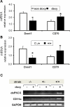
Decreased DNMT1 expression correlates with higher CD70 and CD11a mRNA levels in dnPKCδ transgenic T cells. A. Double transgenic animals were given doxy (doxy, n=8) or sucrose alone (no doxy, n=7) in the drinking water for two weeks. CD3+ T cells were then isolated from spleens and Dnmt1 and CD70 mRNA levels were measured relative to β-actin by RT-PCR. *p≤0.03 vs no doxy. Results represent mean ± SE of 4 independent experiments. B. CD3+ splenic T cells were isolated from four CD2rtTA single transgenic animals (−/+) or from five dnPKCδ/ CD2rtTA double transgenic animals (+/+) treated in vitro with doxy for 72 h. Dnmt1 and CD70 mRNA levels were then measured relative to β-actin by RT-PCR as in A. *p≤0.05 vs −/+. C. CD3+T cells isolated from wild type (−/−), single transgenic (+/−) or double transgenic (+/+) mice treated with doxy for two weeks were compared to non-treated animals. mRNA was purified and the expression of dnPKCδ and CD11a were analyzed by PCR amplification using specific primers. The PCR products were resolved by electrophoresis on 0.8% agarose gel. β actin was used as housekeeping gene. Data represent four independent experiments.
Fig 5
Fig 5
Increase in memory T cell population in mice expressing dnPKCδ. Flow cytometric analysis for CD44 and CD62L expression on CD3+ splenic cells from double transgenic mice treated or not with doxy. Percentages are shown in each quadrant. Data are representative of three independent experiments.
Fig.6
Fig.6
Serum ds-DNA autoantibody production when T cell PKCδ activity is decreased. Blood was obtained from C57BL/6 × SJL mice carrying the mutant dnPKCδ and CD2rtTA genes and given 2 mg/ml doxy/5% sucrose (doxy) or 5% sucrose alone (no doxy) in their drinking water during the indicated times. Serum concentrations of IgG anti ds-DNA were determined by ELISA. Values are the mean ± SEM of 6 independent experiments. Comparison between both groups by ANOVA regression. Results are the mean ± SEM of 4-6 mice per point; p=0.044.
Fig.7
Fig.7
Glomerulonephritis, pneumonitis and IgG deposition in mice lacking T cell PKCδ activity. Hematoxylin and eosin staining of sections of kidneys from A. dnPKCδ/CD2rtTA double transgenic mice (+/+) treated or B. no treated with doxy or; C. CD2+rtTA single transgenic mice (−/+) treated with doxy for 6 months. A glomerulonephritis with leukocyte infiltration is seen in A. Second row: D. ungs from the same doxy treated double transgenic animals show pneumonitis with leukocyte infiltration as well E. Absence of cell infiltration in animals non-doxy treated or, F. in single transgenic mice treated with doxy. Data are representative of 5 to 7 different animals. Magnification: 400X. G. Representative immunohistochemical staining of kidneys from six dnPKCδ/rtTA double transgenic mice receiving doxy. Intense IgG deposition in all the glomeruli along capillary walls and in the mesangial region is seen. H. Similar staining of representative kidney sections from double transgenic animals receiving only sucrose (n=5) and, I. from single CD2 rtTA transgenic animals receiving doxy (n=5) were used as controls. In both controls, the blank areas are glomeruli with no immune complex deposition. Magnification: 400X.

Similar articles

Cited by

References

    1. Sawalha AH, Wang L, Nadig A, Somers EC, McCune WJ, Hughes T, Merrill JT, Scofield RH, Strickland FM, Richardson B. Sex-specific differences in the relationship between genetic susceptibility, T cell DNA demethylation and lupus flare severity. J Autoimmun. 2012;38:J216–22. - PMC - PubMed
    1. Hewagama A, Richardson B. The genetics and epigenetics of autoimmune diseases. J Autoimmun. 2009;33:3–11. - PMC - PubMed
    1. Richardson B. Effect of an inhibitor of DNA methylation on T cells. II. 5-Azacytidine induces self-reactivity in antigen-specific T4+ cells. Hum Immunol. 1986;17:456–70. - PubMed
    1. Richardson B, Scheinbart L, Strahler J, Gross L, Hanash S, Johnson M. Evidence for impaired T cell DNA methylation in systemic lupus erythematosus and rheumatoid arthritis. Arthritis Rheum. 1990;33:1665–73. - PubMed
    1. Yung RL, Richardson BC. Drug-induced lupus. Rheum Dis Clin North Am. 1994;20:61–86. - PubMed

Publication types

MeSH terms