. 2015 Mar 24;108(6):1459-1469.
doi: 10.1016/j.bpj.2015.02.008.

Switch-like responses of two cholesterol sensors do not require protein oligomerization in membranes

Affiliations

Switch-like responses of two cholesterol sensors do not require protein oligomerization in membranes

Austin Gay et al. Biophys J. .

Abstract

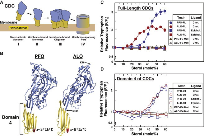
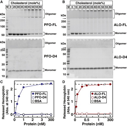
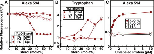
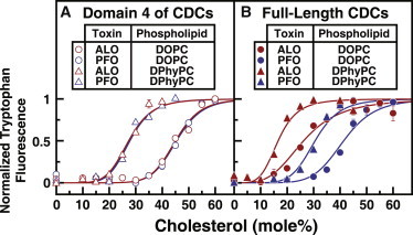
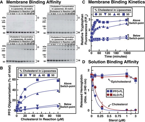
Many cellular processes are sensitive to levels of cholesterol in specific membranes and show a strongly sigmoidal dependence on membrane composition. The sigmoidal responses of the cholesterol sensors involved in these processes could arise from several mechanisms, including positive cooperativity (protein effects) and limited cholesterol accessibility (membrane effects). Here, we describe a sigmoidal response that arises primarily from membrane effects due to sharp changes in the chemical activity of cholesterol. Our models for eukaryotic membrane-bound cholesterol sensors are soluble bacterial toxins that show an identical switch-like specificity for endoplasmic reticulum membrane cholesterol. We show that truncated versions of these toxins fail to form oligomers but still show sigmoidal binding to cholesterol-containing membranes. The nonlinear response emerges because interactions between bilayer lipids control cholesterol accessibility to toxins in a threshold-like fashion. Around these thresholds, the affinity of toxins for membrane cholesterol varies by >100-fold, generating highly cooperative lipid-dependent responses independently of protein-protein interactions. Such lipid-driven cooperativity may control the sensitivity of many cholesterol-dependent processes.


Figures

Figure 1
Figure 1
Interaction of PFO and ALO with sterol-containing membranes. (A) General model for the interaction of cholesterol-dependent cytolysins (CDCs) with cholesterol-containing membranes. Soluble monomeric CDCs bind to membrane cholesterol, oligomerize on the membrane surface, and undergo large conformational changes to form a membrane-spanning pore. The cholesterol-binding domain is shaded in yellow. (B) Structures of PFO and ALO. A ribbon representation of the α-carbon backbone of the crystal structures of PFO (11) and ALO (13) is shown with domains 1–3 (amino acids 30–390 in PFO; amino acids 46–403 in ALO) in blue, and domain 4 (D4; amino acids 391–500 in PFO; amino acids 404–512 in ALO) in yellow. A conserved hexapeptide sequence (GTTLYP) is shaded red. Underlined residues of this hexapeptide are identical in 26 related CDCs (10). Also shown are locations of residues in ALO (K46 and S404) that were mutated to cysteines for covalent attachment of fluorescent labels. (C and D) Intrinsic tryptophan fluorescence of PFO and ALO. Recombinant wild-type (WT) and mutant (Mut) versions of full-length (FL) PFO and ALO, as well as D4 of PFO and ALO, were overexpressed and purified as described in Materials and Methods. Each reaction mixture, in a total volume of 200 μL of buffer B, contained 4.4 μM of the indicated protein and 600 μM liposomes composed of DOPC and varying mole fractions of cholesterol (Chol.) or epicholesterol (Epichol.). After incubation for 1 h at room temperature, intrinsic tryptophan fluorescence from the samples was measured (excitation wavelength, 290 nm; emission wavelength, 340 nm). For each version of PFO or ALO, the fluorescence from mixtures of protein with liposomes containing 0% sterol is normalized to 1.
Figure 2
Figure 2
Oligomerization and pore formation by PFO and ALO after binding to cholesterol-containing membranes. (A and B) Coomassie staining. Aliquots (10% of total) of the reaction mixtures from Fig. 1, C and D, containing recombinant FL or D4 of PFO and ALO with membranes containing DOPC and varying mole fractions of cholesterol were subjected to SDS-PAGE. Proteins were visualized with Coomassie Brilliant Blue R-250 stain. The molecular masses of protein standards are indicated. Arrows indicate the interface between stacking and resolving gels. (C and D) Hemolysis assays. Each reaction mixture, in a final volume of 500 μL, contained varying amounts of the indicated version of PFO, ALO, or BSA, and 450 μL rabbit erythrocytes that had been washed and diluted as described in Materials and Methods. After incubation for 10 min at 37°C, the extent of hemolysis was quantified by measuring the release of hemoglobin (absorbance at 540 nm). The dashed line represents the amount of hemoglobin that was released after treatment with 1% (w/v) Triton-X 100 detergent.
Figure 3
Figure 3
Interaction of fluorescently labeled ALO with sterol-containing membranes. Recombinant ALO-FL and ALO-D4 were overexpressed and purified, and fluorescently labeled versions (fALO-FL and fALO-D4) were generated as described in Materials and Methods. (A and C) Alexa 594 fluorescence of the labeled proteins. Reaction mixtures, in a final volume of 120 μL buffer B, contained 0.5 μM of the indicated fluorescently labeled protein and 67 μM liposomes comprised of DOPC and varying amounts of cholesterol (Chol.) or epicholesterol (Epi.) (A) or 0.5 μM fALO-FL, 67 μM liposomes comprised of 50 mol % DOPC and 50 mol % cholesterol, and varying amounts of the indicated unlabeled protein (C). (B) Intrinsic tryptophan fluorescence of the labeled proteins. Reaction mixtures, in a volume of 100 μL buffer B, contained 3.6 μM of the indicated protein and 600 μM liposomes composed of DOPC and varying mole fractions of cholesterol or epicholesterol. After incubation for 1 h at room temperature, Alexa Fluor 594 fluorescence (A and C) (excitation wavelength, 590 nm; emission wavelength, 617 nm; band pass, 2.5 nm for each) or intrinsic tryptophan fluorescence (B) (excitation wavelength, 290 nm; emission wavelength, 340 nm) was measured. For each protein, fluorescence from mixtures of protein with liposomes containing 0% cholesterol is normalized to 1.
Figure 4
Figure 4
Cholesterol thresholds for ALO and PFO are determined by membrane phospholipids. (A and B) Recombinant FL and D4 of PFO and ALO were overexpressed and purified as described in Materials and Methods. Each reaction mixture, in a total volume of 200 μL of buffer B, contained 4.4 μM of the indicated protein and 600 μM liposomes composed of DOPC or DPhyPC and varying mole fractions of cholesterol. After incubation for 1 h at room temperature, the intrinsic tryptophan fluorescence of the samples was measured (excitation wavelength, 290 nm; emission wavelength, 340 nm). For each combination of protein and phospholipid, fluorescence values were normalized to range from 0 to 1.
Figure 5
Figure 5
Affinity of PFO and ALO for cholesterol is determined by membrane phospholipids. Recombinant PFO-FL and ALO-FL were overexpressed and purified as described in Materials and Methods. (A and B) Affinity of PFO-FL for membrane cholesterol. Each reaction mixture, in a total volume of 1 mL of buffer B, contained 100 nM PFO-FL (5.7 μg) and varying amounts of liposomes composed of DOPC and the indicated amounts of cholesterol. (A) After incubation for 2 h at room temperature, PFO-FL was concentrated to a volume of 20 μL as described in Materials and Methods, and the entire amount of protein was subjected to SDS-PAGE. Lane 1 (I) in each gel contains 5.7 μg of PFO-FL (input amount) as a reference to judge the efficiency of PFO-FL concentration by Ni beads. Proteins were visualized with Coomassie Brilliant Blue R-250 stain. The molecular masses of protein standards are shown. Arrows indicate the interface between stacking and resolving gels. O, membrane-bound oligomeric form of PFO; M, free monomer form of PFO. (B) Gels were scanned and densitometric analysis was carried out to determine the percentage of the oligomeric, membrane-bound form of PFO relative to the total (membrane-bound oligomer plus free monomer). (C) Binding kinetics. Each reaction mixture, in a total volume of 200 μL of buffer B, contained 4.4 μM of PFO-FL and 600 μM liposomes composed of DOPC and the indicated amounts of cholesterol. After incubation at room temperature for the indicated times, intrinsic tryptophan fluorescence from the samples was measured (excitation wavelength, 290 nm; emission wavelength, 340 nm). The fluorescence from mixtures of PFO-FL with liposomes containing 0% sterol is normalized to 1. (D) Binding of DMSO-solubilized sterols to PFO-FL and ALO-FL. Each reaction mixture, in a final volume of 50 μL, contained either ALO-FL (1 nM) or PFO-FL (3 nM), and varying amounts of cholesterol or epicholesterol dissolved in DMSO (4% (v/v) final concentration). After incubation for 1 h at room temperature, 450 μL of rabbit erythrocytes (washed and diluted as described in Materials and Methods) was added to each reaction mixture. After incubation for 10 min at 37°C, the extent of hemolysis was quantified by measuring the release of hemoglobin (absorbance at 540 nm).

Similar articles

Cited by

References

    1. Radhakrishnan A., Goldstein J.L., Brown M.S. Switch-like control of SREBP-2 transport triggered by small changes in ER cholesterol: a delicate balance. Cell Metab. 2008;8:512–521. - PMC - PubMed
    1. Sokolov A., Radhakrishnan A. Accessibility of cholesterol in endoplasmic reticulum membranes and activation of SREBP-2 switch abruptly at a common cholesterol threshold. J. Biol. Chem. 2010;285:29480–29490. - PMC - PubMed
    1. Das A., Goldstein J.L., Radhakrishnan A. Use of mutant 125I-perfringolysin O to probe transport and organization of cholesterol in membranes of animal cells. Proc. Natl. Acad. Sci. USA. 2013;110:10580–10585. - PMC - PubMed
    1. Brown M.S., Goldstein J.L. The SREBP pathway: regulation of cholesterol metabolism by proteolysis of a membrane-bound transcription factor. Cell. 1997;89:331–340. - PubMed
    1. Tweten R.K. Cholesterol-dependent cytolysins, a family of versatile pore-forming toxins. Infect. Immun. 2005;73:6199–6209. - PMC - PubMed

Publication types