Review
. 2015 Mar 9:6:40.
doi: 10.3389/fphar.2015.00040. eCollection 2015.

Modulation of cellular signaling by herpesvirus-encoded G protein-coupled receptors

Affiliations
Review

Modulation of cellular signaling by herpesvirus-encoded G protein-coupled receptors

Sabrina M de Munnik et al. Front Pharmacol. .

Abstract

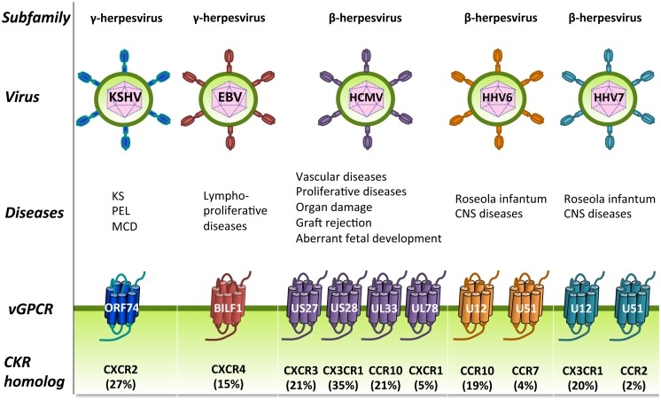
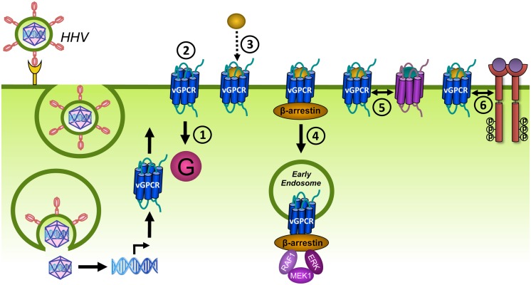
Human herpesviruses (HHVs) are widespread infectious pathogens that have been associated with proliferative and inflammatory diseases. During viral evolution, HHVs have pirated genes encoding viral G protein-coupled receptors (vGPCRs), which are expressed on infected host cells. These vGPCRs show highest homology to human chemokine receptors, which play a key role in the immune system. Importantly, vGPCRs have acquired unique properties such as constitutive activity and the ability to bind a broad range of human chemokines. This allows vGPCRs to hijack human proteins and modulate cellular signaling for the benefit of the virus, ultimately resulting in immune evasion and viral dissemination to establish a widespread and lifelong infection. Knowledge on the mechanisms by which herpesviruses reprogram cellular signaling might provide insight in the contribution of vGPCRs to viral survival and herpesvirus-associated pathologies.

Keywords: EBV; HCMV; KSHV; chemokine; chemokine receptor; human herpesvirus; review; viral GPCR.


Figures

FIGURE 1
FIGURE 1
The general architecture of class A G protein-coupled receptors (GPCRs). Shown are the three extracellular loops (ECL1-3) and the N-terminus in the EC region and the three intracellular loops (ICL1-3) and the C-terminus in the IC region. The seven transmembrane (TM) helices are arranged in a counter-clockwise manner and contain a number of proline-dependent kinks that divide the GPCR into the ligand binding module and the module that binds downstream effectors such as G proteins. The C-terminus of many GPCRs is folded into an eighth helix that runs parallel to the plasma membrane and is often anchored to the membrane via a palmitoylation site. Image is based on (Katritch et al., 2012).
FIGURE 2
FIGURE 2
G protein-dependent signaling. Gα proteins are divided into Gαs, Gαi, Gαq, and Gα12/13 protein families that regulate different effector proteins such as AC and PLC. Effector proteins produce second messengers (e.g., cAMP) that subsequently activate transcription factors such as CRE, NFAT and SRF. AC, adenylyl cyclase; ATP, adenosine triphosphate; cAMP, cyclic adenosine monophosphate; CRE, cAMP-responsive element; DAG, diacylglycerol; GEF, guanine nucleotide exchange factor; IP3, inositol 1,4,5-triphosphate; NFAT, nuclear factor of activated T-cells; PIP2, phosphatidylinositol-4,5-bisphosphate; PKA, protein kinase A; PKC, protein kinase C; PLCβ, phospholipase Cβ; RhoA, Ras homolog gene family A; ROCK, RhoA kinase; SRF, serum response factor.
FIGURE 3
FIGURE 3
Chemokine subclasses. Chemokines are divided into four families according to the number and spatial organization of conserved cysteine residues in their N-terminus. Disulfide bridges are shown as black lines. The transmembrane domain of CX3CL1 is depicted by lipids (in gray).
FIGURE 4
FIGURE 4
Chemokines and their human and viral receptors. The chemokines (vertical) are divided into four families (colors match with Figure 3) and the virus-encoded chemokines are also included at the bottom in black. Human chemokine receptors (horizontal) are classified according to the chemokines they bind and the a-typical chemokine receptors-5 (ACKR1-5) are also included. Viral receptors are depicted on the right. A colored dot represents the pairing of a chemokine to a specific receptor. One receptor can bind multiple chemokines and vice versa. No receptor has hitherto been identified for CXCL14 and the vGPCRs BILF1, US27, UL33, and UL78 are classified as orphan receptors as no chemokines have been identified to bind these receptors. The distribution of the colored dots shows that human chemokine receptors only bind chemokines within their own class. However, ACKR1 and some vGPCRs cross this boundary as they bind CC, CXC, and CX3CL1 chemokines. Moreover, KSHV-encoded vCCL2 binds promiscuously to XC, CC, CXC, and CX3C chemokine receptors. The diagram is based on (Bachelerie et al., 2014; Steen et al., 2014).
FIGURE 5
FIGURE 5
Human herpesviruses (HHVs)-encoded GPCRs. HHVs are divided into three subfamilies: the γ-herpesviruses (left), the β-herpesviruses (right), and the α-herpesviruses (not shown) and have been associated with several human diseases, including proliferative diseases. HHVs from the β and γ subfamilies encode one or more vGPCRs that show closest sequence identity to cellular chemokine receptors (percentage amino acid identity is shown between brackets). These vGPCRs have most likely been pirated from the human genome during viral evolution and function to modify cellular signaling. CKR, chemokine receptor; CNS, central nervous system; KS, Kaposi’s sarcoma; MCD, multicentric Castleman’s disease; PEL, Primary effusion lymphoma.
FIGURE 6
FIGURE 6
Desensitization and trafficking of GPCRs. Upon ligand binding, GPCRs traditionally signal via G proteins (1). In addition, GPCRs are phosphorylated on S and T residues in their C-terminus or ICLs by GRKs (2). β-arrestins bind to phosphorylated GPCRs and prevent further coupling of G proteins, a process known as desensitization (3). β-arrestins target phosphorylated GPCRs for endocytosis via clathrin-coated pits (CCPs) by scaffolding proteins of the internalization-machinery (4). Internalized receptors may activate β-arrestin-dependent signaling (5). Internalized GPCRs are subsequently sorted to recycling endosomes (6) or to lysosomes for degradation (7).
FIGURE 7
FIGURE 7
Sequences of the C-terminus of the different HHV-encoded vGPCRs. Sequences start at the conserved NPxxY motif. The start of the C-terminus of UL78 and BILF1, which lack the NPxxY motif, have been determined by sequence alignment with the other vGPCRs. S/T residues are underlined, Y residues are bold, and di-leucine motifs are italic. With the exception of UL78, all HCMV-encoded vGPCRs contain a di-leucine motif in their C-terminus. Although the C-terminus of the different vGPCRs differ in length, all receptors contain serine (S)/threonine (T) residues in their C-terminus. Only UL78 contains multiple S/T clusters (three or more S/T residues in a row). Most vGPCRs contain at least one Y residue. For some receptors, this Y residue is part of the NPxxY motif and unlikely to directly interact with proteins such as AP-2 as the NPxxY motif is located in TM7.
FIGURE 8
FIGURE 8
G protein-coupled receptors can modulate each other’s function via different mechanisms. (A) GPCRs can positively (+) or negatively (–) modulate (dashed black arrows) ligand binding, signaling (solid black arrow), or trafficking via allosteric interactions (dashed white arrow) within a heterodimer. (B) Crosstalk can be the result of signals (solid black arrows) that integrate downstream of two GPCRs. (C) Scavenging of a limited pool of shared signaling or scaffolding proteins (curved black arrow) might modulate signaling or ligand binding (dashed black arrow) of co-expressed GPCRs. (D) GPCRs can regulate the expression levels of other GPCRs, their ligands or signaling proteins.
FIGURE 9
FIGURE 9
Different mechanisms of receptor tyrosine kinase (RTK) transactivation. (A) GPCR-induced activation of a membrane-anchored metalloproteinase results in the release of a growth factor which activates its cognate RTK in a autocrine and/or paracrine manner. (B) GPCRs regulate the expression and secretion of growth factors that transactivate RTKs in a autocrine and/or paracrine manner. (C) Ligand-independent transactivation of RTKs via the GPCR-induced activation (black arrow) or inhibition of tyrosine kinases or phosphatases, respectively. (D) GPCRs transactivate RTKs within a protein complex, possibly via allosteric interactions (dashed white arrow).
FIGURE 10
FIGURE 10
Different mechanisms of GPCR transactivation. (A) RTK-induced de novo synthesis of GPCR ligands that activate their cognate receptor in an autocrine and/or paracrine manner. (B) RTK-induced enzymatic activation and secretion of GPCR ligands that activate their cognate receptor in an autocrine and/or paracrine manner. (C) RTKs might transactivate interacting GPCRs from internalized vesicles, leading to extracellular-signal-regulated kinases (ERK) activation. The exact mechanism and requirement of the RTK/GPCR interaction for transactivation is not clear.
FIGURE 11
FIGURE 11
Viral G protein-coupled receptors can modulate cellular signaling by means of different mechanisms. vGPCR are expressed at the cell surface of HHV-infected cells. For most vGPCRs, canonical mechanisms of host cell modulation has been studied in detail and shows that vGPCRs can signal through G proteins (1) in a constitutively active manner (2). Chemokine binding might modulate constitutive signaling (3). Less knowledge is available on non-canonical mechanisms that involve the interaction of vGPCRs with proteins from the endocytic machinery (e.g., β-arrestin) (4). In addition, vGPCR may modulate the function of human GPCRs (5) or RTKs (6).

Similar articles

Cited by

References

    1. Akekawatchai C., Holland J. D., Kochetkova M., Wallace J. C., Mccoll S. R. (2005). Transactivation of CXCR4 by the insulin-like growth factor-1 receptor (IGF-1R) in human MDA-MB-231 breast cancer epithelial cells. J. Biol. Chem. 280 39701–39708 10.1074/jbc.M509829200 - DOI - PubMed
    1. Akula S. M., Pramod N. P., Wang F. Z., Chandran B. (2002). Integrin alpha3beta1 (CD 49c/29) is a cellular receptor for Kaposi’s sarcoma-associated herpesvirus (KSHV/HHV-8) entry into the target cells. Cell 108 407–419 10.1016/S0092-8674(02)00628-1 - DOI - PubMed
    1. Alderton F., Rakhit S., Kong K. C., Palmer T., Sambi B., Pyne S., et al. (2001). Tethering of the platelet-derived growth factor beta receptor to G-protein-coupled receptors. A novel platform for integrative signaling by these receptor classes in mammalian cells. J. Biol. Chem. 276 28578–28585 10.1074/jbc.M102771200 - DOI - PubMed
    1. Allen J. A., Halverson-Tamboli R. A., Rasenick M. M. (2007). Lipid raft microdomains and neurotransmitter signalling. Nat. Rev. Neurosci. 8 128–140 10.1038/nrn2059 - DOI - PubMed
    1. Arasteh K., Hannah A. (2000). The role of vascular endothelial growth factor (VEGF) in AIDS-related Kaposi’s sarcoma. Oncologist 5(Suppl. 1) 28–31 10.1634/theoncologist.5-suppl_1-28 - DOI - PubMed

LinkOut - more resources