. 2015 May 28;125(22):3491-500.
doi: 10.1182/blood-2014-11-612762. Epub 2015 Mar 20.

Third-party CD4+ invariant natural killer T cells protect from murine GVHD lethality

Affiliations

Third-party CD4+ invariant natural killer T cells protect from murine GVHD lethality

Dominik Schneidawind et al. Blood. .

Abstract

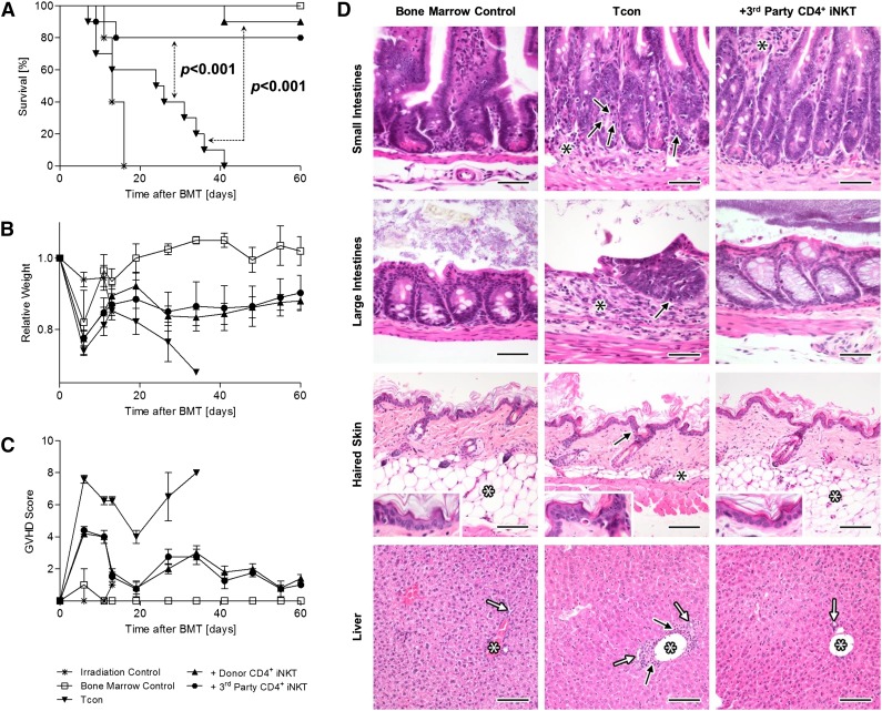
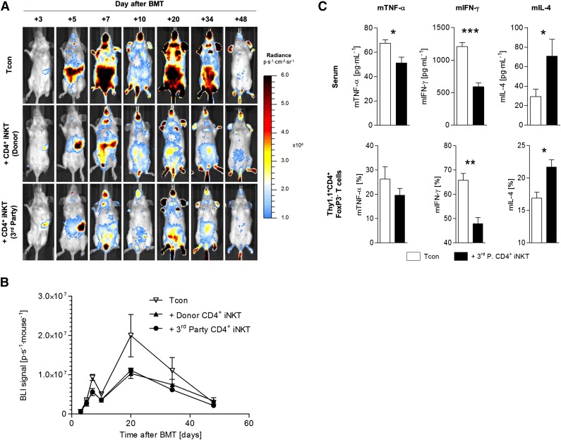
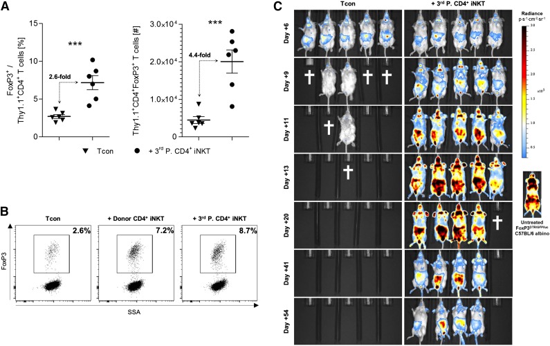
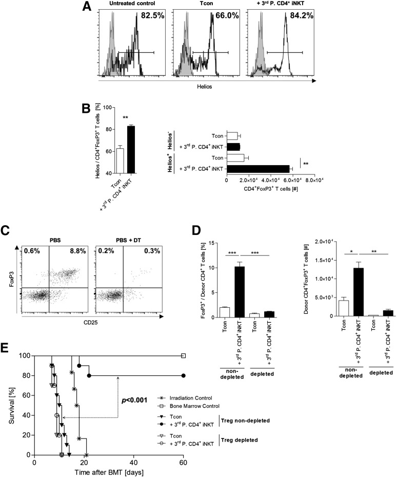
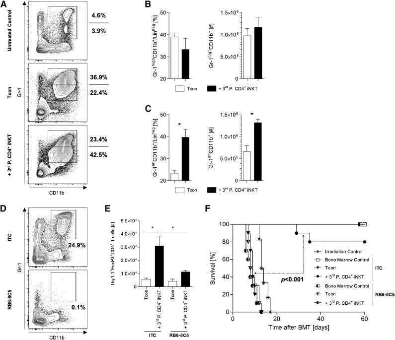
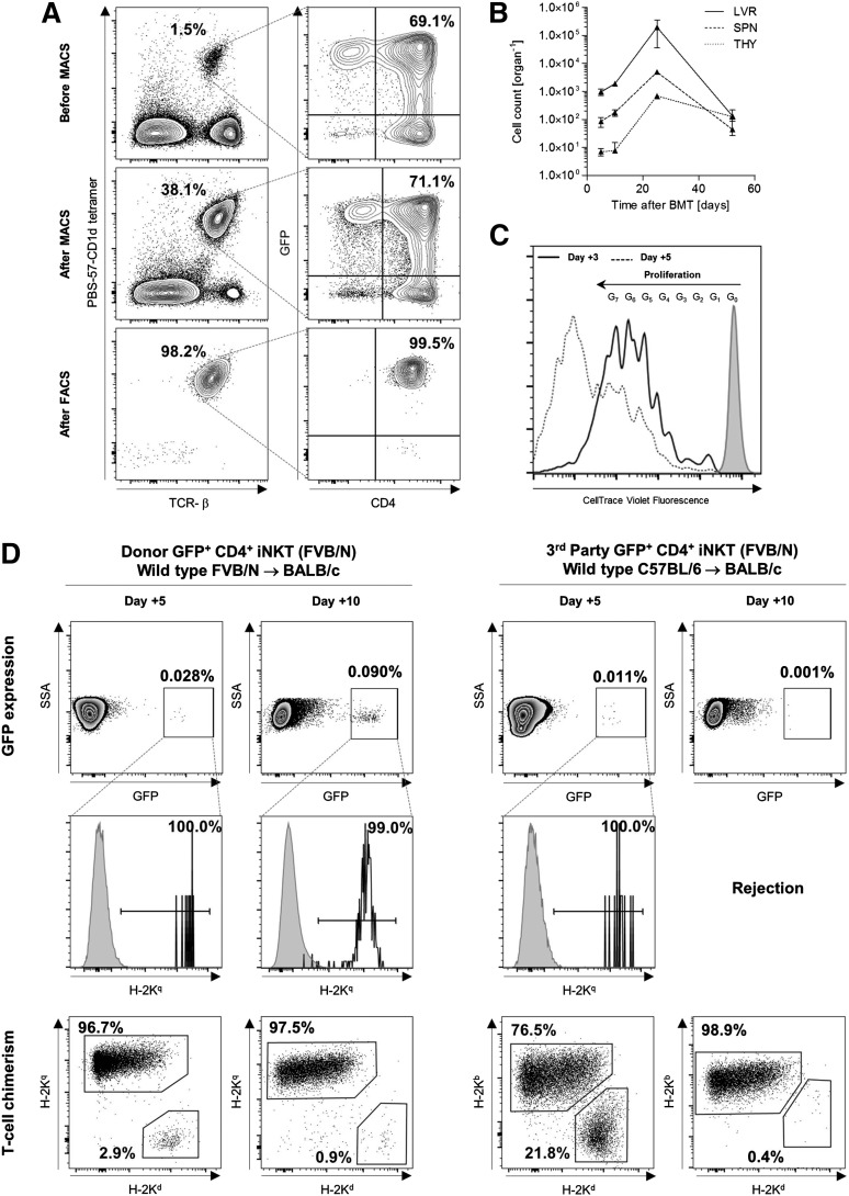
Graft-versus-host disease (GVHD) is driven by extensive activation and proliferation of alloreactive donor T cells causing significant morbidity and mortality following allogeneic hematopoietic cell transplantation (HCT). Invariant natural killer T (iNKT) cells are a potent immunoregulatory T-cell subset in both humans and mice. Here, we explored the role of adoptively transferred third-party CD4(+) iNKT cells for protection from lethal GVHD in a murine model of allogeneic HCT across major histocompatibility barriers. We found that low numbers of CD4(+) iNKT cells from third-party mice resulted in a significant survival benefit with retained graft-versus-tumor effects. In vivo expansion of alloreactive T cells was diminished while displaying a T helper cell 2-biased phenotype. Notably, CD4(+) iNKT cells from third-party mice were as protective as CD4(+) iNKT cells from donor mice although third-party CD4(+) iNKT cells were rejected early after allogeneic HCT. Adoptive transfer of third-party CD4(+) iNKT cells resulted in a robust expansion of donor CD4(+)CD25(+)FoxP3(+) regulatory T cells (Tregs) that were required for protection from lethal GVHD. However, in vivo depletion of myeloid-derived suppressor cells abrogated both Treg expansion and protection from lethal GVHD. Despite the fact that iNKT cells are a rare cell population, the almost unlimited third-party availability and feasibility of in vitro expansion provide the basis for clinical translation.


Figures

Figure 1
Figure 1
Third-party CD4+ iNKT cells protect from lethal GVHD. BALB/c recipient mice were irradiated with 2 × 4 Gy, followed by transplantation of 5.0 × 10⁶ TCD-BM cells and 1.0 × 10⁶ Tcons from C57BL/6 donor mice. In addition, 5.0 × 10⁴ CD4+ iNKT cells from C57BL/6 donor or FVB/N third-party mice were transferred together with the graft. (A) Overall survival pooled from 2 independent experiments with 10 mice per group except irradiation control (n = 5). (B) Weight and (C) GVHD score from 1 of 2 independent experiments with 5 animals per group except irradiation control (n = 3). Error bars indicate standard error of the mean (SEM). (D) Photomicrographs of small intestines, large intestines, haired skin, and liver at day +25. Small intestinal histology is normal in BM mice with presence of regularly-shaped and -sized villi, regularly-spaced and -sized intestinal crypts, presence of a regular complement of enterocytes (including goblet cells), and absence of any significant inflammation in the lamina propria and submucosa. In Tcon mice, there are many apoptotic cells in the crypt epithelium (black arrows), which appear as shrunken, hypereosinophilic, round bodies with pyknotic nuclei. Mild-to-moderate lymphoplasmacytic inflammation (black asterisks) is also present in the lamina propria and/or submucosa of the same mice, causing mild separation of the intestinal crypts. Third-party CD4+ iNKT-cell–treated mice have essentially normal small intestines when compared with the BM control mice, except for the presence of mild inflammatory infiltrates in the lamina propria (black asterisks). H&E stain; original magnification ×400; bar = 50 µm. Large intestinal histology is normal in BM control and third-party CD4+ iNKT-cell–treated mice with presence of regularly-spaced and -sized intestinal glands, presence of a regular complement of enterocytes (including goblet cells), and absence of any significant inflammation in the lamina propria and submucosa. In Tcon mice, there is marked lymphoplasmacytic inflammation (black asterisks) in the lamina propria and/or submucosa associated with the loss of intestinal glands. The remaining intestinal glands are abnormal with loss of goblet cells and presence of many apoptotic cells (black arrow), which appear as shrunken, hypereosinophilic, round bodies with pyknotic nuclei. H&E stain; original magnification ×400; bar = 50 µm. The BM control and third-party CD4+ iNKT-cell–treated mice have normal histology of the skin with an intact, thin epidermis and a thick layer of subcutaneous adipose tissue (white asterisks). In the Tcon mice however, there is mild interface damage of the epidermis (as evidenced by apoptotic keratinocytes; black arrows) and mild-to-complete atrophy of the subcutaneous adipose tissue (black asterisks). A mild dermal inflammatory infiltrate is also noted within the Tcon mice. H&E stain; original magnification ×200 (inset magnification ×400); bar = 100 µm. The BM control and third-party CD4+ iNKT-cell–treated mice have normal liver histology, with portal triads containing a single portal vein (white asterisks), and one or two bile ductules (white arrows). The livers of Tcon mice also appear essentially normal, except for some minimal lymphoplasmacytic inflammation in periportal interstitial tissues (black arrows) and mild proliferation/hyperplasia of bile ductules. H&E stain; original magnification ×200; bar = 100 µm. BMT, bone marrow transplantation.
Figure 2
Figure 2
Third-party CD4+ iNKT cells inhibit Tcon proliferation and induce a Th2 immune phenotype. Photon emission of Tcons from luc+ C57BL/6 donor mice was measured by BLI to assess the in vivo expansion capacity of alloreactive T cells in donor and third-party CD4+ iNKT-cell–treated BALB/c recipient mice. (A) Representative serial bioluminescence images. (B) Bioluminescence signal intensity time course. Error bars indicate SEM. Shown is 1 of 2 independent experiments with 5 mice per group. (C) To determine alterations in immune polarization in third-party CD4+ iNKT-cell–treated BALB/c recipient mice, murine sera, and Thy1.1+CD4+FoxP3 T cells from spleens were analyzed at day +10 by multiplex assay and ICS, respectively. Error bars indicate SEM. Pooled data from 2 independent experiments with 6 mice per group are shown. *P ≤ .05; **P ≤ .01; ***P ≤ .001.
Figure 3
Figure 3
Third-party CD4+ iNKT cells promote an expansion of donor Tregs. (A) Relative and absolute numbers of donor Tregs from spleens of BALB/c recipient mice at day +10. Error bars indicate SEM. Pooled data from 2 independent experiments with 6 mice per group are shown. (B) Representative dot plots gated on live Thy1.1+CD4+ donor T cells re-isolated at day +10 from BALB/c recipient spleens. Shown is 1 of 2 independent experiments. (C) BALB/c recipient mice received Tcons from FoxP3DTR/GFP/luc C57BL/6 albino mice with and without CD4+ iNKT cells from FVB/N third-party mice. Bioluminescence signals derive from luc+FoxP3+ donor T cells. Shown is 1 of 2 independent experiments with 5 mice per group, as well as a representative bioluminescence image from one untreated FoxP3DTR/GFP/luc C57BL/6 albino mouse. The † indicates that all animals from the respective group died or needed to be euthanized. ***P ≤ .001. SSA, side scatter area.
Figure 4
Figure 4
Donor Tregs expand from the graft and are required for protection from GVHD lethality. (A) Representative histograms and (B) relative and absolute numbers of Helios-expressing donor Tregs re-isolated from recipient livers at day +10. Error bars indicate SEM. Shown are 3 mice per group from 1 of 3 independent experiments. (C) FoxP3DTR/GFP/luc C57BL/6 albino donor mice were injected IP with DT to deplete these mice of Tregs. Dot plots are gated on live TCR-β+CD4+ T cells and show representative examples of Tcons prepared from mice treated with PBS only or DT dissolved in PBS. (D) Relative and absolute numbers of donor Tregs re-isolated from spleens of BALB/c mice at day +10. Mice received either a Treg-nondepleted or Treg-depleted BM graft with or without adoptive transfer of 5.0 × 10⁴ FVB/N third-party CD4+ iNKT cells. TCD-BM and third-party CD4+ iNKT cells derive from untreated WT C57BL/6 mice. Error bars indicate SEM. Three animals per group from 1 of 3 independent experiments are shown. (E) Overall survival of BALB/c recipient mice receiving either a Treg-nondepleted or Treg-depleted graft from C57BL/6 donor mice with or without adoptive transfer of 5.0 × 10⁴ FVB/N third-party CD4+ iNKT cells. TCD-BM was derived from untreated WT C57BL/6 mice. Ten mice per group except irradiation control (n = 6). Pooled data from 2 independent experiments are shown. *P ≤ .05; **P ≤ .01; ***P ≤ .001.
Figure 5
Figure 5
MDSCs are required for Treg expansion and protection from lethal GVHD. (A) Representative dot plots gated on live donor (CD45.1+) and lineage negative (CD3CD19CD49bTER-119) cells from spleens of BALB/c recipient mice at day +10 after allogeneic HCT. Absolute and relative cell numbers of donor (B) CD11b+Gr-1high and (C) CD11b+Gr-1int MDSCs. Error bars indicate SEM. Three animals per group from 1 of 3 independent experiments are shown. (D) Representative dot plots gated on live and lineage negative (CD3-CD19-CD49b-TER-119-) cells from spleens of BALB/c mice injected with ITC or RB6-8C5. (E) Absolute number of donor Tregs re-isolated from spleens of BALB/c mice at day +10. Error bars indicate SEM. Three animals per group from 1 of 2 independent experiments are shown. (F) Overall survival of BALB/c recipient mice either depleted (RB6-8C5) or nondepleted (ITC) of MDSCs. Ten mice per group except irradiation control (n = 6). Pooled data from 2 independent experiments are shown. *P ≤ .05. ITC, isotype control antibody.
Figure 6
Figure 6
Third-party CD4+ iNKT cells are rejected early after allogeneic HCT. (A) GFP+CD4+ iNKT cells were enriched (magnetic-activated cell sorting) and sorted (fluorescence-activated cell sorting) from whole splenocytes of GFP-expressing FVB/N mice enabling distinct re-isolation from BALB/c recipient mice after adoptive transfer. Representative dot plots gated on live cells. (B) Absolute cell numbers of adoptively transferred FVB/N donor GFP+CD4+ iNKT cells re-isolated at different time points from various organs of BALB/c mice that received TCD-BM and Tcons from WT FVB/N donor mice. No GFP+CD4+ iNKT cells could be re-isolated from lymph nodes, gut, and skin (not shown). Error bars indicate SEM. Shown are cell numbers from 3 mice per time point from 1 of 2 independent experiments. (C) Fluorescence intensity deriving from 1.5 × 10⁶ adoptively transferred FVB/N donor GFP+CD4+ iNKT cells labeled with CellTrace Violet and re-isolated at days +3 and +5. Histogram gated on GFP+ cells pooled from the spleen and liver of one BALB/c mouse that received TCD-BM and Tcons from one WT FVB/N donor mouse. Shown is 1 of 3 independent experiments. (D) Donor and third-party model of allogeneic HCT with BALB/c (H-2Kd) mice receiving TCD-BM and Tcons from WT FVB/N (H-2Kq) and C57BL/6 (H-2Kb) mice, respectively. Adoptively transferred CD4+ iNKT cells derived from GFP-expressing FVB/N (H-2Kq) mice and were re-isolated from recipient livers at days +5 and +10. Shown are representative dot plots gated on live cells from 1 of 2 independent experiments. The third row shows T-cell chimerism from the same sample. Comparable results were obtained for re-isolation of GFP+ cells from recipient spleens (not shown). LVR, liver; SPN, spleen; THY, thymus.

Comment in

Similar articles

Cited by

References

    1. Ferrara JL, Deeg HJ. Graft-versus-host disease. N Engl J Med. 1991;324(10):667–674. - PubMed
    1. Shlomchik WD, Couzens MS, Tang CB, et al. Prevention of graft versus host disease by inactivation of host antigen-presenting cells. Science. 1999;285(5426):412–415. - PubMed
    1. Blazar BR, Murphy WJ, Abedi M. Advances in graft-versus-host disease biology and therapy. Nat Rev Immunol. 2012;12(6):443–458. - PMC - PubMed
    1. Magenau J, Reddy P. Next generation treatment of acute graft-versus-host disease. Leukemia. 2014;28(12):2283–2291. - PubMed
    1. Lantz O, Bendelac A. An invariant T cell receptor alpha chain is used by a unique subset of major histocompatibility complex class I-specific CD4+ and CD4-8- T cells in mice and humans. J Exp Med. 1994;180(3):1097–1106. - PMC - PubMed

Publication types

MeSH terms