

Translation. An RNA biosensor for imaging the first round of translation from single cells to living animals
- PMID: 25792328
- PMCID: PMC4451088
- DOI: 10.1126/science.aaa3380
Translation. An RNA biosensor for imaging the first round of translation from single cells to living animals
Abstract
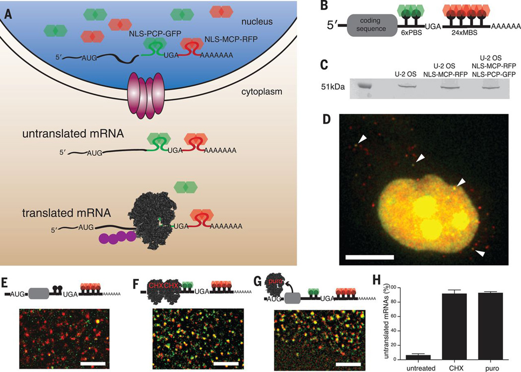
Analysis of single molecules in living cells has provided quantitative insights into the kinetics of fundamental biological processes; however, the dynamics of messenger RNA (mRNA) translation have yet to be addressed. We have developed a fluorescence microscopy technique that reports on the first translation events of individual mRNA molecules. This allowed us to examine the spatiotemporal regulation of translation during normal growth and stress and during Drosophila oocyte development. We have shown that mRNAs are not translated in the nucleus but translate within minutes after export, that sequestration within P-bodies regulates translation, and that oskar mRNA is not translated until it reaches the posterior pole of the oocyte. This methodology provides a framework for studying initiation of protein synthesis on single mRNAs in living cells.
Copyright © 2015, American Association for the Advancement of Science.
Figures


Comment in
-
RNA. A TRICK'n way to see the pioneer round of translation.Science. 2015 Mar 20;347(6228):1316-7. doi: 10.1126/science.aaa9484. Science. 2015. PMID: 25792317 No abstract available.
Similar articles
-
TRICK: A Single-Molecule Method for Imaging the First Round of Translation in Living Cells and Animals.Methods Enzymol. 2016;572:123-57. doi: 10.1016/bs.mie.2016.02.027. Epub 2016 Apr 29. Methods Enzymol. 2016. PMID: 27241753
-
Localization-dependent oskar protein accumulation; control after the initiation of translation.Dev Cell. 2004 Jul;7(1):125-31. doi: 10.1016/j.devcel.2004.06.009. Dev Cell. 2004. PMID: 15239960
-
RNA. A TRICK'n way to see the pioneer round of translation.Science. 2015 Mar 20;347(6228):1316-7. doi: 10.1126/science.aaa9484. Science. 2015. PMID: 25792317 No abstract available.
-
Microtubule-based motor-mediated mRNA localization in Drosophila oocytes and embryos.Biochem Soc Trans. 2011 Oct;39(5):1197-201. doi: 10.1042/BST0391197. Biochem Soc Trans. 2011. PMID: 21936788 Review.
-
Localization, anchoring and translational control of oskar, gurken, bicoid and nanos mRNA during Drosophila oogenesis.Fly (Austin). 2009 Jan-Mar;3(1):15-28. doi: 10.4161/fly.3.1.7751. Epub 2009 Jan 2. Fly (Austin). 2009. PMID: 19182536 Review.
Cited by
-
Making many from few: IL-12p40 as a model for the combinatorial assembly of heterodimeric cytokines.Cytokine. 2015 Nov;76(1):53-7. doi: 10.1016/j.cyto.2015.07.026. Epub 2015 Aug 1. Cytokine. 2015. PMID: 26242928 Free PMC article. Review.
-
Single-Molecule RNA Imaging Using Mango II Arrays.Methods Mol Biol. 2022;2404:267-280. doi: 10.1007/978-1-0716-1851-6_14. Methods Mol Biol. 2022. PMID: 34694614
-
Emerging Roles for 3' UTRs in Neurons.Int J Mol Sci. 2020 May 12;21(10):3413. doi: 10.3390/ijms21103413. Int J Mol Sci. 2020. PMID: 32408514 Free PMC article. Review.
-
Subcellular Transcriptomics and Proteomics: A Comparative Methods Review.Mol Cell Proteomics. 2022 Feb;21(2):100186. doi: 10.1016/j.mcpro.2021.100186. Epub 2021 Dec 16. Mol Cell Proteomics. 2022. PMID: 34922010 Free PMC article. Review.
-
Following the messenger: Recent innovations in live cell single molecule fluorescence imaging.Wiley Interdiscip Rev RNA. 2020 Jul;11(4):e1587. doi: 10.1002/wrna.1587. Epub 2020 Jan 28. Wiley Interdiscip Rev RNA. 2020. PMID: 31990126 Free PMC article. Review.
References
-
- Schwanhäusser B, Busse D, Li N, Dittmar G, Schuchhardt J, Wolf J, Chen W, Selbach M. Global quantification of mammalian gene expression control. Nature. 2011;473:337–342. - PubMed
Publication types
MeSH terms
Substances
Grants and funding
LinkOut - more resources
Full Text Sources
Other Literature Sources
Molecular Biology Databases
Research Materials
