Strengthening of the intestinal epithelial tight junction by Bifidobacterium bifidum
- PMID: 25780093
- PMCID: PMC4393161
- DOI: 10.14814/phy2.12327
Strengthening of the intestinal epithelial tight junction by Bifidobacterium bifidum
Abstract
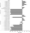
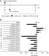
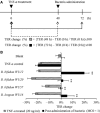
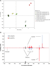
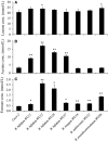
Epithelial barrier dysfunction has been implicated as one of the major contributors to the pathogenesis of inflammatory bowel disease. The increase in intestinal permeability allows the translocation of luminal antigens across the intestinal epithelium, leading to the exacerbation of colitis. Thus, therapies targeted at specifically restoring tight junction barrier function are thought to have great potential as an alternative or supplement to immunology-based therapies. In this study, we screened Bifidobacterium, Enterococcus, and Lactobacillus species for beneficial microbes to strengthen the intestinal epithelial barrier, using the human intestinal epithelial cell line (Caco-2) in an in vitro assay. Some Bifidobacterium and Lactobacillus species prevented epithelial barrier disruption induced by TNF-α, as assessed by measuring the transepithelial electrical resistance (TER). Furthermore, live Bifidobacterium species promoted wound repair in Caco-2 cell monolayers treated with TNF-α for 48 h. Time course (1)H-NMR-based metabonomics of the culture supernatant revealed markedly enhanced production of acetate after 12 hours of coincubation of B. bifidum and Caco-2. An increase in TER was observed by the administration of acetate to TNF-α-treated Caco-2 monolayers. Interestingly, acetate-induced TER-enhancing effect in the coculture of B. bifidum and Caco-2 cells depends on the differentiation stage of the intestinal epithelial cells. These results suggest that Bifidobacterium species enhance intestinal epithelial barrier function via metabolites such as acetate.
Keywords: 1H‐NMR; intestinal epithelial permeability; metabonomics; probiotics; tight junctions.
© 2015 The Authors. Physiological Reports published by Wiley Periodicals, Inc. on behalf of the American Physiological Society and The Physiological Society.
Figures





Similar articles
-
Secretions of Bifidobacterium infantis and Lactobacillus acidophilus Protect Intestinal Epithelial Barrier Function.J Pediatr Gastroenterol Nutr. 2017 Mar;64(3):404-412. doi: 10.1097/MPG.0000000000001310. J Pediatr Gastroenterol Nutr. 2017. PMID: 28230606
-
Bifidobacterium bifidum Enhances the Intestinal Epithelial Tight Junction Barrier and Protects against Intestinal Inflammation by Targeting the Toll-like Receptor-2 Pathway in an NF-κB-Independent Manner.Int J Mol Sci. 2021 Jul 28;22(15):8070. doi: 10.3390/ijms22158070. Int J Mol Sci. 2021. PMID: 34360835 Free PMC article.
-
Protective Effects of Bifidobacterium on Intestinal Barrier Function in LPS-Induced Enterocyte Barrier Injury of Caco-2 Monolayers and in a Rat NEC Model.PLoS One. 2016 Aug 23;11(8):e0161635. doi: 10.1371/journal.pone.0161635. eCollection 2016. PLoS One. 2016. PMID: 27551722 Free PMC article.
-
Bioactive factors secreted by Bifidobacterium breve B-3 enhance barrier function in human intestinal Caco-2 cells.Benef Microbes. 2019 Feb 8;10(1):89-100. doi: 10.3920/BM2018.0062. Epub 2018 Oct 24. Benef Microbes. 2019. PMID: 30353739
-
Galacto-oligosaccharides Protect the Intestinal Barrier by Maintaining the Tight Junction Network and Modulating the Inflammatory Responses after a Challenge with the Mycotoxin Deoxynivalenol in Human Caco-2 Cell Monolayers and B6C3F1 Mice.J Nutr. 2015 Jul;145(7):1604-13. doi: 10.3945/jn.114.209486. Epub 2015 May 27. J Nutr. 2015. PMID: 26019243
Cited by
-
Gut microbiome changes due to sleep disruption in older and younger individuals: a case for sarcopenia?Sleep. 2022 Dec 12;45(12):zsac239. doi: 10.1093/sleep/zsac239. Sleep. 2022. PMID: 36183306 Free PMC article.
-
A Potential Probiotic Lactobacillus plantarum JBC5 Improves Longevity and Healthy Aging by Modulating Antioxidative, Innate Immunity and Serotonin-Signaling Pathways in Caenorhabditis elegans.Antioxidants (Basel). 2022 Jan 28;11(2):268. doi: 10.3390/antiox11020268. Antioxidants (Basel). 2022. PMID: 35204151 Free PMC article.
-
Characterization of the fecal microbiome in cats with inflammatory bowel disease or alimentary small cell lymphoma.Sci Rep. 2019 Dec 16;9(1):19208. doi: 10.1038/s41598-019-55691-w. Sci Rep. 2019. PMID: 31844119 Free PMC article.
-
Safety Assessment of Potential Probiotic Lactobacillus fermentum MTCC-5898 in Murine Model after Repetitive Dose for 28 Days (Sub-Acute Exposure).Probiotics Antimicrob Proteins. 2020 Mar;12(1):259-270. doi: 10.1007/s12602-019-09529-6. Probiotics Antimicrob Proteins. 2020. PMID: 30847835
-
Exogenous Fecal Microbiota Transplantation from Local Adult Pigs to Crossbred Newborn Piglets.Front Microbiol. 2018 Jan 9;8:2663. doi: 10.3389/fmicb.2017.02663. eCollection 2017. Front Microbiol. 2018. PMID: 29375527 Free PMC article.
References
-
- Balda MS. Matter K. Tight junctions at a glance. J. Cell Sci. 2008;121:3677–3682. - PubMed
-
- Balda MS, Whitney JA, Flores C, Gonzalez S, Cereijido M. Matter K. Functional dissociation of paracellular permeability and transepithelial electrical resistance and disruption of the apical-basolateral intramembrane diffusion barrier by expression of a mutant tight junction membrane protein. J. Cell Biol. 1996;134:1031–1049. - PMC - PubMed
-
- Brown AJ, Goldsworthy SM, Barnes AA, Eilert MM, Tcheang L, Daniels D, et al. The orphan G protein-coupled receptors GPR41 and GPR43 are activated by propionate and other short chain carboxylic acids. J. Biol. Chem. 2003;278:11312–11319. - PubMed
LinkOut - more resources
Full Text Sources
Other Literature Sources
Research Materials

