. 2015 May;89(10):5615-32.
doi: 10.1128/JVI.00305-15. Epub 2015 Mar 11.

Dynamics of Human Cytomegalovirus Infection in CD34+ Hematopoietic Cells and Derived Langerhans-Type Dendritic Cells

Affiliations

Dynamics of Human Cytomegalovirus Infection in CD34+ Hematopoietic Cells and Derived Langerhans-Type Dendritic Cells

Roxanne Coronel et al. J Virol. 2015 May.

Abstract

Acquisition of human cytomegalovirus (CMV) usually occurs by contact between contaminated bodily fluids, such as urine and saliva, and host mucosal cells. Langerhans-type dendritic cells (LC) are the only type of immune cells found in the outermost layers of the oral mucosae, where they not only provide a first line of defense against CMV but can easily be targeted by orally administered vaccines, while their bone marrow resident progenitors are important sites of virus latency. In this work, we tracked the progress of infection in CD34(+) progenitor cells, immature LC (iLC), and mature LC (mLC) exposed to the clinical-like strain TB40-BAC4 or to the vaccine strain AD169varATCC, prior to their long-term maintenance under either immature or mature conditions. We show that the genomes of both strains are efficiently maintained in CD34(+) cells during their differentiation into iLC, although this requires the presence of larger amounts of input AD169varATCC DNA. Lipopolysaccharide- and CD40 ligand-induced maturation of iLC derived from latently infected progenitors was not associated with robust viral genome replication and progeny production, while maturation of directly infected iLC increased and prolonged expression of the viral immediate early proteins. While effective replication of viral genomes from both strains occurred only in mLC, both iLC and mLC produced viral progeny, suggesting that both types of LC may contribute to CMV horizontal transmission in vivo.

Importance: Human CMV is usually acquired via the oral and nasal mucosae. Langerhans-type dendritic cells (LC) are the only type of immune cells found in the outermost layers of these tissues. Understanding how CMV interacts with LC and their hematopoietic progenitors is thus essential to develop innovative means of defense against this virus. Here we show that the genomes of a virulent and an attenuated strain of CMV are maintained in hematopoietic progenitor cells during their differentiation into immature LC and that maturation of these cells by exposure to lipopolysaccharide and CD40 ligand is not sufficient to trigger virus reactivation. While the extents of viral protein expression and genome replication were broadest in directly infected mature LC populations, similar amounts of viral progeny were detected in the supernatants of immature and mature LC, suggesting that these immune cells of the oral mucosa are likely to be important for CMV transmission within the human population.


Figures

FIG 1
FIG 1
Schematic representation of the methods used to generate the five different types of dendritic cells analyzed in this work. Umbilical cord blood-derived CD34+ hematopoietic progenitor cells from single donors were partitioned into two groups immediately after thawing. One set was left uninfected (B, white oval), while the other was exposed to TB40-BAC4 or AD169varATCC virions for 4 h (A, light gray oval). Infected and uninfected CD34+ cells were then cultured in iLC differentiation medium for 8 days, with supernatants and cells collected every 2 days. (A) Immature LC derived from infected progenitors were harvested on day eight and replated in either fresh iLC differentiation medium (34i) or maturation medium (34m) for 14 days, with collection of cells and supernatants every 2 days. (B) Immature LC derived from uninfected progenitors were harvested on day eight and exposed to TB40-BAC4 or AD169varATCC virions for 4 h prior to culture in iLC differentiation medium (iiLC) or in maturation medium (imLC) for 14 days, with collection of cells and supernatants every 2 days. (C) A portion of the iLC differentiated from uninfected CD34+ cells was cultured in maturation medium for 2 days to generate mLC, which were then exposed to TB40-BAC4 or AD169varATCC virions for 4 h prior to culture in maturation medium (mmLC) for 14 days, with supernatants and cells collected every 2 days. White ovals and star-shaped outlines represent uninfected progenitor and dendritic cells, respectively, while light gray, dark gray, and black outlines represent infected cells. Black circles enclosing a black hexagon depict CMV virions. GM-CSF, granulocyte-macrophage-colony-stimulating factor; SCF, stem cell factor; Flt3L, Flt3 ligand; TNF-α, tumor necrosis factor alpha; TGF-β, transforming growth factor β1; CD40L, CD40 ligand; LPS, lipopolysaccharide.
FIG 2
FIG 2
TB40-BAC4 growth kinetics in confluent HFF infected at a low MOI. HFF were exposed to TB40-BAC4 virions at an MOI of 0.05 for 4 h prior to washing and further culture in the presence or absence of 300 μg/ml of the viral polymerase inhibitor foscarnet for 14 days. Cells and supernatants collected at days 2, 4, 6, 8, 10, and 14 were used to determine the proportion of cells expressing the IE1/IE2 proteins relative to that at day two (fold day 2) (A and E), the proportions of cells per well (B and F) and of viral genomes per well (C and G) present at each time point relative to the beginning of infection at 4 hpi (fold 4 hpi), and the amount of virus released into the supernatant (PFU/ml) (D and H). The dotted horizontal line marks the 1-fold point on the y axis.
FIG 3
FIG 3
Kinetics of TB40-BAC4 and AD169varATCC infection in proliferating/differentiating CD34+ cells and in derived LC maintained under immature (34i) or mature (34m) conditions. Immediately after thawing, CD34+ progenitor cells were exposed to TB40-BAC4 (A and C) or AD169varATCC (E and G) virions for 4 h prior to washing and culture in iLC differentiation medium containing (C) or not (A, E and G) 300 μg/ml of foscarnet. At the end of the iLC differentiation period on day eight, cells were harvested, counted, and replated in either iLC differentiation medium (34i) or maturation medium (34m) for 14 days, with (D) or without (B, F, and H) foscarnet. Real-time quantitative genomic PCR was used to determine the proportion of cells per well (light gray diamonds for CD34+ cells and 34i, black diamonds for 34m) and of viral genomes per well (light gray circles for CD34+ cells and 34i, black circles for 34m) present at each time point relative to the beginning of infection (fold 4 hpi). Panels A to F show results obtained from cultures containing 105 to 106 copies of viral DNA per well at 4 hpi, while panels G and H show results obtained from AD169varATCC-infected cells containing 106 to 107 copies of viral DNA per well at 4 hpi. Graphs show median and median absolute deviation values of data collected from six different CD34+ cell donors in six independent experiments (A to D), four different donors in three independent experiments (E and F), and three different donors in two independent experiments (G and H). The dotted line marks the 1-fold point, while the solid line marks the limit of detection point on the y axis. D8P refers to the proportion of cells per well or genomes per well replated on day eight relative to 4 hpi.
FIG 4
FIG 4
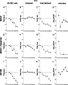
Kinetics of TB40-BAC4 and AD169varATCC infection in iiLC. Immature LC differentiated from uninfected CD34+ progenitors were exposed to TB40-BAC4 (A to H) or AD169varATCC (I to L) virions for 4 h prior to washing and culture under immature conditions in the presence (E to H) or not of 300 μg/ml of foscarnet. Cells and supernatants were collected every 2 days. The proportion of cells expressing the viral IE1/IE2 proteins at each time postinfection relative to that at day two (A, E, and I) (fold day two) was determined by immunofluorescence staining analyses of cytospin preparations, while real-time, quantitative genomic PCR was used to determine the proportions of cells per well (B, F, and J) and of viral genomes per well (C, G, and K) present at each time point relative to the beginning of infection at 4 hpi (fold 4 hpi). The amount of virus present in the supernatants was quantified by immunofluorescence staining analyses of HFF collected at 24 h postexposure to each medium (D, H, and L). The “IE1/IE2+ cells” and “viral titers” graphs show mean and standard deviation values of data collected from five donors in four (A) and three (E) independent experiments and from three donors in three (I), four (D and L), and two (H) independent experiments. The “cells per well” and “viral DNA/well” graphs show median and median absolute deviation values of data collected from five donors in five independent experiments (B and C), five donors in three independent experiments (F and G), and three donors in four independent experiments (J and K). The dotted line marks the 1-fold point on the y axis.
FIG 5
FIG 5
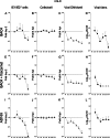
Kinetics of TB40-BAC4 and AD169varATCC infection in imLC. Immature LC differentiated from uninfected CD34+ progenitors were exposed to TB40-BAC4 (A to H) or AD169varATCC (I to L) virions for 4 h, prior to washing and culture in maturation medium in the presence (E to H) or not of 300 μg/ml of foscarnet. Cells and supernatants were collected every 2 days. Data collection and values shown in the graphs are as described in the legend to Fig. 4. The dotted line marks the 1-fold point on the y axis.
FIG 6
FIG 6
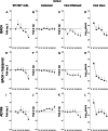
Kinetics of TB40-BAC4 and AD169varATCC infection in mmLC. Mature LC obtained after 2 days of LPS and CD40L stimulation of iLC differentiated from uninfected CD34+ progenitors were exposed to TB40-BAC4 (A to H) or AD169varATCC (I to L) virions for 4 h prior to washing and further culture in maturation medium in the presence (E to H) or not of 300 μg/ml of foscarnet. Cells and supernatants were collected every 2 days. Data were collected as described in the legend to Fig. 4. The “IE1/IE2+ cells” and “viral titers” graphs show mean and standard deviation values of data collected from five donors in five (A), four (E), and six (D) independent experiments, from four donors in four independent experiments (L), and from three donors in three (I) and two (H) independent experiments. The “cells per well” and “viral DNA/well” graphs show median and median absolute deviation values of data collected from six donors in six independent experiments (B and C), five donors in four independent experiments (F and G), and three donors in four independent experiments (J and K). The dotted line marks the 1-fold point on the y axis.
FIG 7
FIG 7
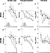
Comparison of TB40-BAC4 and AD169varATCC infection kinetics in iiLC, imLC, and mmLC. The data shown in this figure are the same as in Fig. 4, 5, and 6 but rearranged for direct comparison of the proportions of IE1/IE2+ cells (A and D) and of viral genomes per well (B and E) present at each time point relative to those at day two and at 4 hpi, respectively, and of the amounts of viral particles released in the culture supernatants (C and F) after TB40-BAC4 or AD169varATCC infection of iiLC (white symbols), imLC (gray symbols), and mmLC (black symbols). The dotted line marks the 1-fold point on the y axis.

Similar articles

Cited by

References

    1. Mendelson M, Monard S, Sissons P, Sinclair J. 1996. Detection of endogenous human cytomegalovirus in CD34+ bone marrow progenitors. J Gen Virol 77:3099–3102. doi:10.1099/0022-1317-77-12-3099. - DOI - PubMed
    1. Khaiboullina SF, Maciejewski JP, Crapnell K, Spallone PA, Dean Stock A, Pari GS, Zanjani ED, Jeor SS. 2004. Human cytomegalovirus persists in myeloid progenitors and is passed to the myeloid progeny in a latent form. Br J Haematol 126:410–417. doi:10.1111/j.1365-2141.2004.05056.x. - DOI - PubMed
    1. Minton EJ, Tysoe C, Sinclair JH, Sissons JG. 1994. Human cytomegalovirus infection of the monocyte/macrophage lineage in bone marrow. J Virol 68:4017–4021. - PMC - PubMed
    1. Zhuravskaya T, Maciejewski JP, Netski DM, Bruening E, Mackintosh FR, St Jeor S. 1997. Spread of human cytomegalovirus (HCMV) after infection of human hematopoietic progenitor cells: model of HCMV latency. Blood 90:2482–2491. - PubMed
    1. Slobedman B, Mocarski ES. 1999. Quantitative analysis of latent human cytomegalovirus. J Virol 73:4806–4812. - PMC - PubMed

Publication types

MeSH terms