. 2015 Mar 11;10(3):e0117923.
doi: 10.1371/journal.pone.0117923. eCollection 2015.

A novel therapy for melanoma developed in mice: transformation of melanoma into dendritic cells with Listeria monocytogenes

Affiliations

A novel therapy for melanoma developed in mice: transformation of melanoma into dendritic cells with Listeria monocytogenes

Lucia Bronchalo-Vicente et al. PLoS One. .

Abstract

Listeria monocytogenes is a gram-positive bacteria and human pathogen widely used in cancer immunotherapy because of its capacity to induce a specific cytotoxic T cell response in tumours. This bacterial pathogen strongly induces innate and specific immunity with the potential to overcome tumour induced tolerance and weak immunogenicity. Here, we propose a Listeria based vaccination for melanoma based in its tropism for these tumour cells and its ability to transform in vitro and in vivo melanoma cells into matured and activated dendritic cells with competent microbicidal and antigen processing abilities. This Listeria based vaccination using low doses of the pathogen caused melanoma regression by apoptosis as well as bacterial clearance. Vaccination efficacy is LLO dependent and implies the reduction of LLO-specific CD4+ T cell responses, strong stimulation of innate pro-inflammatory immune cells and a prevalence of LLO-specific CD8+ T cells involved in tumour regression and Listeria elimination. These results support the use of low doses of pathogenic Listeria as safe melanoma therapeutic vaccines that do not require antibiotics for bacterial removal.


Conflict of interest statement

Competing Interests: The authors have declared that no competing interests exist.

Figures

Fig 1
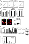
Fig 1. Listeria induced transformation of melanoma into dendritic cells.
A, Kinetic analysis of BMDC, murine (B16F10) and human (A-375 and Mel-H0) melanoma cells infected with different LM strains (LMWT, LMΔLLO). Results are expressed as CFU (mean ± SD) obtained with triplicate samples from three independent experiments (P<0.05). B, Different phagocytic parameters analysed in melanoma and BMDC: phagocytic rates after incubation with [35S]-labelled LM strains for 45 min (left plot). Radioactivity associated with cell lysates (CPM) was quantified in a β2 counter as the bacterial phagocytic rates. Results are expressed as cpm of internalized bacteria (mean ± SD) (p < 0.05). Replication indexes (RI) analysis is shown in middle plot. RIs were calculated as the ratio of the number of CFU at 16 h divided by the amount of CFU at 0 h. This parameter was considered as an indicator of bacterial growth. Results are expressed as CFU (mean ± SD) (p < 0.05). The percentages of cytosolic fractions are shown in right plot after purification of phagosomal and cytosolic fractions as in Material and Methods. Results are expressed as percentages of total internalized CFU in PNS (mean ± SD) (p < 0.05). C, Images correspond to confocal microscopy examination of melanoma and BMDC infected with GFP-LMWT. GFP-LMWT (green channel) co-localize with MHC-II molecules (red channel). Western blots correspond to the analysis in purified phagosomes for different MIIC markers: a/b stable MHC-II chains; Rab5a and LLO forms bound to MHC-class II molecules. CFU values of purified phagosomes are shown below western blots. D, BMDC and B16F10 infected with LM strains or non-infected (NI) were surface stained for the following markers: CD11c-PE, CD11b-FITC, F4/80-PE, CD40-PE, Gr-1-FITC and anti-MHC-II-APC. Samples were acquired using FACSCanto flow cytometer and percentages of positive cells for each antibody are shown. Results are expressed as the mean ± SD of triplicates (p<0.05). E, Same melanoma cells infected with different LM strains or non-infected (NI) as in D for 24 hours. Supernatants were recovered, filtered through 3 μm syringe to discard bacteria and the levels of pro-inflammatory cytokines MCP-1, TNF-alfa, IL-6, IL-10 or IL-12 were analysed using the CBA kit (Becton Dickinson) by flow cytometry. Results were expressed as cytokine concentration (pg/ml of mean ± SD, P<0,05).
Fig 2
Fig 2. Listeria vaccination of melanoma shows a dual action, tumour regression by apoptosis and bacterial clearance.
A, C57BL/6 female were inoculated i.p. with 5 x 10⁵ B16F10/mice (n = 5) for 7 (7-D) or 15 days (15-D). Mice were bled, sacrificed and treated for histological analysis as described in Material and Methods. Images correspond to sections of peritoneum infiltrates or liver metastases. B, C57BL/6 female were inoculated i.p. with 5 x 10⁵ B16F10/mice (n = 5) as in A for none (NT), 7 (7-D) or 15 days (15-D). Spleens from sacrificed mice (n = 5) were stained for histological analysis using different antibodies as described in Material and Methods and images correspond to sections. Results are expressed as percentages of positive cells (mean ± SD) (P < 0.05). C, C57BL/6 female were inoculated i.p. with 5 x 10⁵ B16F10/mice (n = 5) for 7 days and next injected i.p. or not (NT) with 5 x 10³ bc/mice of different LM strains (LMWT or LMΔLLO) for 5 additional days. Mice were sacrificed, bled to collect sera and photographed before collecting melanoma and lungs. Images correspond to the peritoneum of mice and the recovered melanoma. Plots correspond to measurements of diameters of collected melanoma. Results are expressed as the mean ± SD (P < 0,05). D, Melanoma recovered from LMWT or LMΔLLO vaccinated mice or from non-vaccinated mice (NV) as in C were analysed for early and late apoptosis by FACS according to Materials and Methods after double staining with 7-AAD (IP labelled) and annexin V (anexina labelled). Results are expressed as the percentages of late apoptotic cells, necrotic death, (Q2 area corresponding to double positive for 7-AAD and annexin V cells) and the percentages of early apoptotic cells, programmed cell death (Q4 area corresponding to annexin V positive while 7-AAD negative cells) (mean ± SD) (p < 0.05).
Fig 3
Fig 3. Efficiency of Listeria vaccination of melanoma is mediated by activation of LLOrec specific CD8+ T cells and inhibition of LLOrec specific CD4+ T cells.
C57BL/6 female were inoculated i.p. with 5 x 10⁵ B16F10/mice (n = 5) for 7 days and next injected i.p. or not (NT) with 5 x 10³ bc/mice of GFP-LMWT strain for 3 additional days. Mice were bled and sacrificed. A, Immune cells plot (left) corresponds with spleens were homogenized and cell populations were analysed by FACS. Results were expressed as the mean of the percentages of positive cells ± SD. LM growth plot (right) corresponds with spleen homogenates examined for CFU in blood-agar plates. Results are expressed as CFU (mean ± SD) obtained with triplicate samples from three independent experiments (P< 0,05). B, Levels of pro-inflammatory cytokines (MCP-1, TNF-alfa, IFN-gamma, IL-6, IL-10, IL-12) were analysed in sera of mice using the CBA kit (Becton Dickinson) by flow cytometry. Results were expressed as cytokine concentration (pg/ml of mean ± SD, P<0,05). C, Spleen cells obtained from homogenates after inoculation with melanoma B16F10 (5 x 10⁵ cells/mice) for 7 days and vaccination with LMWT for 5 days (LMWT-MEL). Cells were stimulated 5 h with recombinant LLO (0.1 μg/ml) in the presence of brefeldin A for intracellular cytokine staining. LLO-stimulated spleen cell surface was stained for CD4 or CD8 and fixed and permeabilized using cytofix/cytoperm kit. Stimulated cells were surface stained for CD4 or CD8 using anti-CD4+FITC-labeled or anti-CD8+APC-labelled and data gated to include histograms show the percentages of LLO-CD4+ and IFN-gamma producers (lower left) and LLO-CD8+ and IFN-gamma producers (lower right) (R2 and R3 gates). Experiments were performed in triplicate and results are expressed as the mean ± SD (p < 0.05). D, Spleen cells obtained from homogenates after inoculation with melanoma B16F10 pre-infected with LMWT (5 x 10⁵ cells/mice) for 7 days. Cells were stimulated 5 h with recombinant LLO (0.1 μg/ml) in the presence of brefeldin A for intracellular cytokine staining. Procedures were performed as in C and results expressed as the mean ± SD (p < 0.05).
Fig 4
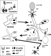
Fig 4. Model of action of our LMWT vaccination of melanoma.
After LMWT vaccination of melanoma induce mice, bacteria would infect the melanoma and transformed them into matured dendritic-like cells (MEL-DCm) (step 4) with APC competence to process LLO and generate LLO189–201/CD4+-restricted and LLO91–99/CD8+ restricted peptides. Melanoma transformation into DCm generates exaggerated levels of the pro-inflammatory cytokines/chemokines, MCP-1, TNF-alfa and IL-12 (step 5), amplifies the activation of LLO91–99/CD8+-restricted T cells (step 5) and blocks the stimulation of LLO189–201/CD4+-restricted T cells (step 5). The exaggerated levels of pro-inflammatory cytokines and the amplification of LLO91–99/CD8+-restricted T cells producing high levels of IFN-gamma, strongly activated innate cells with tumoricidal potential. The amplification of LLO91–99/CD8+-restricted T cells would cause a LLO-driven tumour destruction by programmed cell death (step 6). Also these LLO91–99/CD8+-restricted T cells producing high levels of IFN-gamma would activate the LMWT infected melanoma (MEL-Dm) to a fully activated state (MEL-DCma) that might eliminate the pathogen (step 6) with the advantage of not requiring antibiotic treatment to destroy this low dose of the pathogen used as therapeutic vaccine.

Similar articles

Cited by

References

    1. Coley WB. The Treatment of Inoperable Sarcoma by Bacterial Toxins (the Mixed Toxins of the Streptococcus erysipelas and the Bacillus prodigiosus). Proc R Soc Med. 1910; 3: 1–48. - PMC - PubMed
    1. Shahabi V, Maciag PC, Rivera S, Wallecha A. Live, attenuated strains of Listeria and Salmonella as vaccine vectors in cancer treatment. Bioeng Bugs. 2010; 1: 235–243. 10.4161/bbug.1.4.11243 - DOI - PMC - PubMed
    1. Patyar S, Joshi R, Prasad Byrav DS, Prakash A, Medhi B, Das BK. Bacteria in cancer therapy: a novel experimental strategy. J Biomed Sci. 2010. 17: 1–9. 10.1186/1423-0127-17-1 - DOI - PMC - PubMed
    1. Maciag PC, Radulovic S, Rothman J. The first clinical used of live-attenuated Listeria monocytogenes vaccine: a Phase I safety study of Lm-LLO-E7 in patients with advanced carcinoma of cervix. Vaccine 2009; 27: 3975–3983. 10.1016/j.vaccine.2009.04.041 - DOI - PubMed
    1. Kim SH, Castro F, Paterson Y, Gravekamp C. High efficacy of a Listeria-based vaccine against metastatic breast cancer reveals a dual mode of action. Cancer Res. 2009; 69: 5860–5866. 10.1158/0008-5472.CAN-08-4855 - DOI - PMC - PubMed

Publication types

MeSH terms

Substances

Grants and funding

This work was supported by grants SAF2009-08695 and SAF2012-34203. The funders had no role in the study design, data collection and analysis, decision to publish, or preparation of the manuscript.