. 2015 Apr 6;212(4):569-80.
doi: 10.1084/jem.20141510. Epub 2015 Mar 9.

Lin28b promotes fetal B lymphopoiesis through the transcription factor Arid3a

Affiliations

Lin28b promotes fetal B lymphopoiesis through the transcription factor Arid3a

Yan Zhou et al. J Exp Med. .

Abstract

Mouse B cell precursors from fetal liver and adult bone marrow (BM) generate distinctive B cell progeny when transplanted into immunodeficient recipients, supporting a two-pathway model for B lymphopoiesis, fetal "B-1" and adult "B-2." Recently, Lin28b was shown to be important for the switch between fetal and adult pathways; however, neither the mechanism of Lin28b action nor the importance of B cell antigen receptor (BCR) signaling in this process was addressed. Here, we report key advances in our understanding of the regulation of B-1/B-2 development. First, modulation of Let-7 in fetal pro-B cells is sufficient to alter fetal B-1 development to produce B cells resembling the progeny of adult B-2 development. Second, intact BCR signaling is required for the generation of B1a B cells from Lin28b-transduced BM progenitors, supporting a requirement for ligand-dependent selection, as is the case for normal B1a B cells. Third, the VH repertoire of Lin28b-induced BM B1a B cells differs from that of normal B1a, suggesting persisting differences from fetal progenitors. Finally, we identify the Arid3a transcription factor as a key target of Let-7, whose ectopic expression is sufficient to induce B-1 development in adult pro-B cells and whose silencing by knockdown blocks B-1 development in fetal pro-B cells.


Figures

Figure 1.
Figure 1.
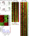
Fetal and adult pro-B cells generate B cells with distinct phenotype and show differential expression of mRNAs and miRNAs. (A) Pro-B cells were sorted from FL and BM and then transferred i.v. into immunodeficient SCID mice. Peritoneal cavity washout cells (PerC) were analyzed 3 wk later for the indicated surface proteins by flow cytometry. Data are representative of five independent transfer experiments with two mice per group. (B) Heat map displays of mRNA fold change comparing fetal and adult pre-pro-B (Fr. A) and pro-B (Fr. BC) stage cell samples by microarray of genes higher in FL or in BM using both the Affymetrix (C57BL/6J) and Agilent Technologies (BALB/c ICR) platforms. This analysis identifies 219 differentially expressed genes. (C) Heat map display of microarray data (top) and Q-PCR analysis (bottom) of miRNA species differentially expressed between adult BM and FL pro-B cells (Fr. BC). miRNAs in the heat map that are also analyzed by Q-PCR are indicated by a red asterisk. Q-PCR data were normalized to SNO202 expression. All expression analyses were performed in duplicate or triplicate, with each sample representing RNA or miRNA from at least four mice. Error bars represent ±SEM.
Figure 2.
Figure 2.
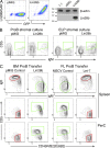
Lin28b and Let-7 miRNA reciprocally redirect adult and fetal B cell development. (A) The Ret02 pro-B stage cell line, which lacks expression of Lin28b, was transduced with a Lin28b-containing retrovirus or with empty vector (pMIG). 1 d after transduction, cells were examined for Lin28b expression by cytoplasmic staining using flow cytometry (left) and Western blotting (right). Molecular mass is indicated in kilodaltons. (B) BM pro-B and ELP stage cells, 1 d after initiation of stromal cell culture, were transduced with empty vector or a Lin28b-containing retrovirus. After 11 (pro-B) or 15 d (ELP), lymphoid cells were recovered from cultures and analyzed by flow cytometry. CD19+GFP+ cells (pro-B) or CD19+Cherry+ cells (for ELP) are shown in the plots. 20–40% of IgM+ cells were CD5+ in cultures of Lin28b-transduced BM, whereas <5% were positive with pMIG. Representative data from three independent experiments are shown in flow cytometry plots. (C) Analysis of B cells generated in SCID mice, 3 wk after transfer of Lin28b (or pMIG empty vector)-transduced BM pro-B or Let-7 (or MSCV empty vector)–transduced FL pro-B cells. Lin28b-induced fetal-type B cells from BM (green regions of interest; IgM2+IgD+/−CD45R+ CD5+) and empty vector B cells (follicular/B2 phenotype; red regions of interest; IgM+IgD2+CD45R2+CD5) are shown. Plots are gated for CD19+GFP+ cells. Data are representative of five independent experiments, with two mice per group. PerC, peritoneal cavity washout cells.
Figure 3.
Figure 3.
Phenotype of Lin28b-transduced BM and Let-7–transduced FL generates B cells in recipient spleen that resemble B1 and FO/B2. CD19+GFP+ mature (CD93) B cells in spleen were generated from transfer of the indicated pro-B–transduced cells, 3 wk after transfer in SCID mice, and analyzed by flow cytometry using the indicated markers. The red boxes indicate an adult-type and the green boxes indicate a fetal-type phenotype. The blue gates represent IgMhiCD21hi cells, a marginal zone B cell type. Data are representative of five independent transfer experiments with two mice per group.
Figure 4.
Figure 4.
Phenotype of Lin28b-transduced BM and Let-7–transduced FL generates B cells in recipient peritoneal cavity that resemble B1 and B2. CD19+GFP+ cells in the peritoneal cavity were generated from transfer of the indicated pro-B–transduced cells, 3 wk after transfer in SCID mice, and analyzed by flow cytometry using the indicated markers. The red boxes indicate an adult-type and the green boxes indicate a fetal-type phenotype. Data are representative of five independent transfer experiments with two mice per group.
Figure 5.
Figure 5.
Lin28b BM–generated CD5+ B cells require intact BCR signaling but do not express the usual B1a repertoire, regardless of TdT expression. (A) Btk-deficient BM pro-B cells, transduced with Lin28b or empty (pMIG) retrovirus, were transferred to SCID mice, and spleen cells were analyzed 3 wk after transfer by flow cytometry using the indicated markers. Plots are gated for CD19+GFP+ cells. Data are representative of two independent transfer experiments with two mice per group. (B) The level of N-addition and distribution of VH gene usage was analyzed in individual sorted follicular/B2 (Fo) and CD5+ B1 (B1a) B cells from intact mice and in B cells from SCID mice repopulated with BM pro-B cells (transduced with empty vector [pMIG] or Lin28b-containing retrovirus). Data were combined from three separate experiments for each cell type, each experiment using two mice for each sample. (C) The VH gene distribution in B cells from SCID mice repopulated with BM pro-B cells from TdT mice (transduced as in B) is illustrated in pie charts. Data were combined from three separate experiments for each cell type, each experiment using two mice for each sample.
Figure 6.
Figure 6.
Analysis of mRNAs regulated by the Lin28b–Let-7 axis in pro-B cells identified Arid3a as a key regulator of B cell fate. (A) Venn diagram illustrating the identification of a set of 17 genes whose expression is perturbed reciprocally by alteration of Lin28b–Let-7 in pro-B stage cells. BM pro-B cells sorted from BALB/c mice were transduced after 1 d in culture with either Lin28b or empty vector retrovirus and then cultured an additional 4 d and finally sorted as CD19+GFP+ cells. FL pro-B cells from the same strain were treated similarly, except that transduction was with Let-7b or empty retrovirus and cells were sorted as CD19+GFP+ (results are incorporated from B). (B) Heat map of mRNA expression fold change perturbations (relative to control-transduced samples) induced by retroviral transduction of Lin28b–Let-7b (as diagrammed in A). Two samples representative of expression changes in each cell type are shown. For BM cells, genes normally high (red) switch to green, whereas those normally low (green) switch to red (left). A reciprocal change is seen with FL cells (right). Data from unperturbed pre-pro-B (Fr. A) and pro-B (Fr. BC) microarray analysis of FL and BM, using both the Agilent (Ag) and Affymetrix (Af) platforms, are included for comparison. Results are representative of two independent experiments. (C) ARID3a, one of the genes whose expression was increased by Lin28b and decreased by Let-7 miRNA, has Let-7 and miR-125b target seed sites in its 3′-UTR. The Arid3a diagram was produced in MacVector, and species alignments were obtained from the TargetScan website, Whitehead Institute for Biomedical Research.
Figure 7.
Figure 7.
Arid3a induces fetal-type B cell generation from BM pro-B cells. (A) BM pro-B cells were transduced with an Arid3a retrovirus or pMIG control and then transferred to SCID recipients. 3 wk later, spleen or PerC B cells, gated as CD19+GFP+, were analyzed by flow cytometry. The green regions indicate a fetal phenotype. Data are representative of three independent experiments using two mice per group. (B) The expression of other markers on PerC B cells generated from Arid3a-reprogrammed BM pro-B cells was analyzed by flow cytometry. Cells were gated as CD19+GFP+. The red regions indicate an adult-type and the green regions indicate a fetal-type phenotype. Data are representative of three independent experiments using two mice per group.
Figure 8.
Figure 8.
shRNA knockdown of Arid3a inhibits FL pro-B generation of B1a B cells. (A) FL pro-B cells or the N38 Abelson mouse line was transduced with empty vector (LMP) or vector containing each of five different shRNAs. 3 d after transduction, GFP+ cells were sorted for RNA. ARID expression was quantified by Q-PCR. Arid3a expression was first normalized to β-actin, and then the level in control vector (LMP) was set to 100. Error bars represent ±SEM. (B) Phenotype of CD19+GFP+ B cells generated in SCID mice 3 wk after transfer of fetal pro-B cells, transduced with empty vector or a pool of three shRNA constructs. The red regions indicate an adult-type and the green boxes indicate a fetal-type phenotype. Data are representative of three independent experiments using two mice per group.

Similar articles

Cited by

References

    1. Carmack C.E., Shinton S.A., Hayakawa K., and Hardy R.R.. 1990. Rearrangement and selection of VH11 in the Ly-1 B cell lineage. J. Exp. Med. 172:371–374 10.1084/jem.172.1.371 - DOI - PMC - PubMed
    1. Dickins R.A., Hemann M.T., Zilfou J.T., Simpson D.R., Ibarra I., Hannon G.J., and Lowe S.W.. 2005. Probing tumor phenotypes using stable and regulated synthetic microRNA precursors. Nat. Genet. 37:1289–1295 10.1038/ng1651 - DOI - PubMed
    1. Dickins R.A., McJunkin K., Hernando E., Premsrirut P.K., Krizhanovsky V., Burgess D.J., Kim S.Y., Cordon-Cardo C., Zender L., Hannon G.J., and Lowe S.W.. 2007. Tissue-specific and reversible RNA interference in transgenic mice. Nat. Genet. 39:914–921 10.1038/ng2045 - DOI - PMC - PubMed
    1. Förster I., Gu H., and Rajewsky K.. 1988. Germline antibody V regions as determinants of clonal persistence and malignant growth in the B cell compartment. EMBO J. 7:3693–3703. - PMC - PubMed
    1. Goebel P., Montalbano A., Ayers N., Kompfner E., Dickinson L., Webb C.F., and Feeney A.J.. 2002. High frequency of matrix attachment regions and cut-like protein x/CCAAT-displacement protein and B cell regulator of IgH transcription binding sites flanking Ig V region genes. J. Immunol. 169:2477–2487 10.4049/jimmunol.169.5.2477 - DOI - PubMed

Publication types

MeSH terms