. 2015 Mar 6;11(3):e1004702.
doi: 10.1371/journal.ppat.1004702. eCollection 2015 Mar.

γδ T cells confer protection against murine cytomegalovirus (MCMV)

Affiliations

γδ T cells confer protection against murine cytomegalovirus (MCMV)

Camille Khairallah et al. PLoS Pathog. .

Abstract

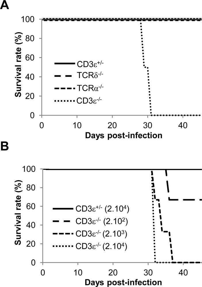
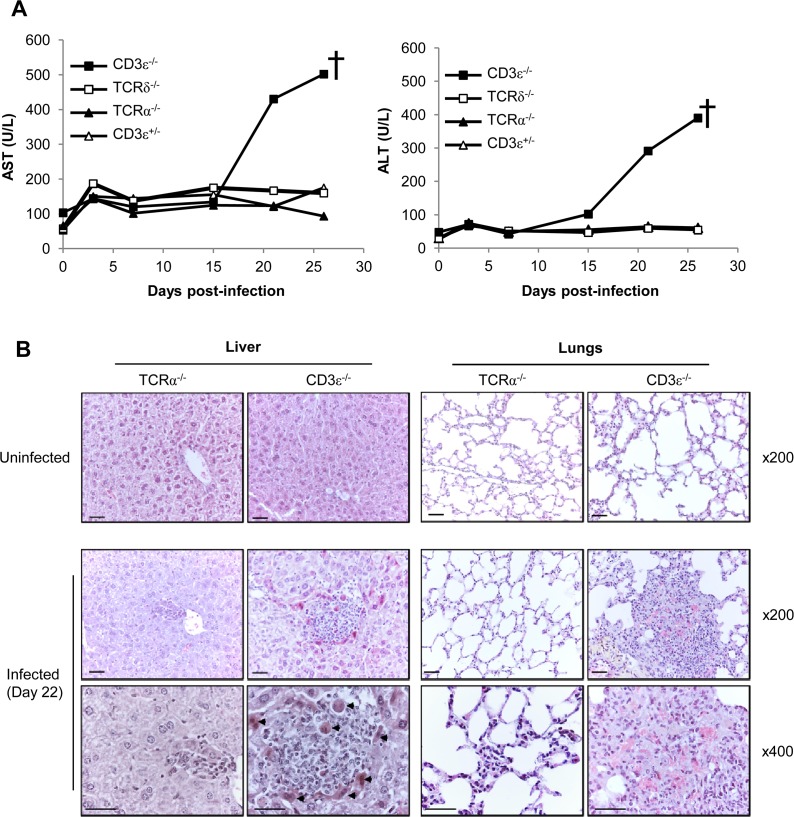
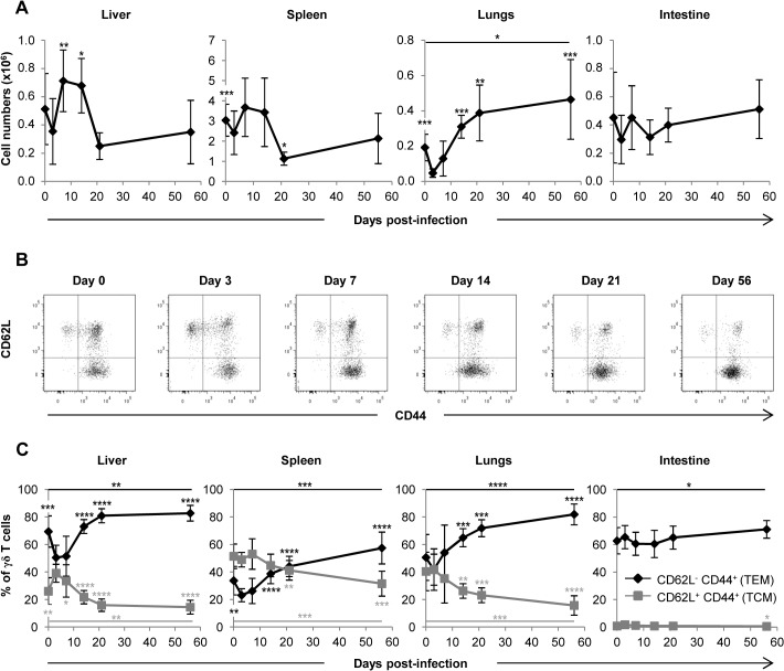
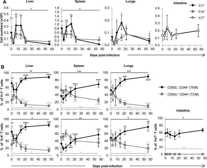
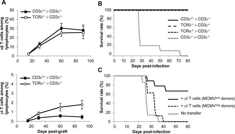
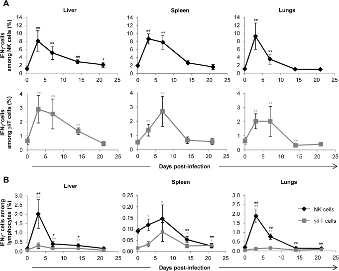
Cytomegalovirus (CMV) is a leading infectious cause of morbidity in immune-compromised patients. γδ T cells have been involved in the response to CMV but their role in protection has not been firmly established and their dependency on other lymphocytes has not been addressed. Using C57BL/6 αβ and/or γδ T cell-deficient mice, we here show that γδ T cells are as competent as αβ T cells to protect mice from CMV-induced death. γδ T cell-mediated protection involved control of viral load and prevented organ damage. γδ T cell recovery by bone marrow transplant or adoptive transfer experiments rescued CD3ε-/- mice from CMV-induced death confirming the protective antiviral role of γδ T cells. As observed in humans, different γδ T cell subsets were induced upon CMV challenge, which differentiated into effector memory cells. This response was observed in the liver and lungs and implicated both CD27+ and CD27- γδ T cells. NK cells were the largely preponderant producers of IFNγ and cytotoxic granules throughout the infection, suggesting that the protective role of γδ T cells did not principally rely on either of these two functions. Finally, γδ T cells were strikingly sufficient to fully protect Rag-/-γc-/- mice from death, demonstrating that they can act in the absence of B and NK cells. Altogether our results uncover an autonomous protective antiviral function of γδ T cells, and open new perspectives for the characterization of a non classical mode of action which should foster the design of new γδ T cell based therapies, especially useful in αβ T cell compromised patients.


Conflict of interest statement

The authors have declared that no competing interests exist.

Figures

Fig 1
Fig 1. γδ T cells prevent mice from MCMV-induced mortality.
A. TCRδ−/−, TCRα−/− CD3ε+/− and CD3ε−/− mice (10 of each) were infected i.p. with 1.105 PFU of MCMV at day 0 and monitored every other day for mortality. Data are from one representative of 3 independent experiments. B. CD3ε+/− and CD3ε−/− mice (4 of each) were infected i.p. with indicated doses of MCMV at day 0 and monitored every day for mortality. Data are from one experiment.
Fig 2
Fig 2. MCMV dissemination in lungs, spleen, liver, intestine and salivary glands from T cell competent and T cell deficient mice.
TCRδ−/−, TCRα−/−, CD3ε+/−, and CD3ε−/− mice were infected i.p. with 2.103 PFU of MCMV. At indicated days post-infection, 4 mice of each mouse line were dissected and MCMV gB was quantified in organs by real time PCR. The experiment was repeated 3 times under similar conditions. Histograms represent means of MCMV DNA copy number (per 100 ng genomic DNA) ± SD of all mice from the three experiments (n = 4x3 mice). Statistical differences between viral loads in TCRα−/− versus CD3ε+/− mice, and in TCRα−/− versus CD3ε−/− mice are shown.
Fig 3
Fig 3. γδ T cell control of MCMV infection is associated with reduced organ damage.
A. TCRδ−/−, TCRα−/−, CD3ε+/− and CD3ε−/− mice were infected i.p. as indicated in Fig. 1A. 3 mice/group were bled at days 0, 3, 7, 15, 21 and just before death for biochemical analyses of AST and ALT in serums. The experiment was repeated twice and data obtained for one representative mouse/group are shown. † death of CD3ε−/−. B. TCRα−/− and CD3ε−/− mice were uninfected, or i.p. infected with 2.103 pfu of MCMV. Uninfected and Day 22-infected mice were sacrificed and the liver and lungs were embedded in paraffin for HES staining. Apoptotic hepatocytes are shown (arrowheads). Scale bar = 200 mm. Magnifications are indicated in the right-hand side of the figure. The data are from one representative of 3 mice for each condition.
Fig 4
Fig 4. Mobilization of γδ T cells in MCMV-infected organs from TCRα−/− mice.
TCRα−/− mice were infected i.p. with 2.103 PFU of MCMV. At indicated post-infection days, 5–9 mice were sacrificed, immune cells prepared from each organ and γδ T cells stained as shown in S2 Fig. A. Kinetics of absolute γδ T cell numbers determined as described in methods. Presented data are mean ± SEM of 8–9 mice from one representative of 2 experiments. B. CD62L and CD44 expression by lymphocytes was evaluated by flow cytometry, with the presented gating strategy (lungs shown as example). C. Longitudinal analysis of γδ T cell phenotype in all organs. Results are pooled from 2 independent experiments representing a total of 13–14 mice (means ± SEM). Statistical differences of cell numbers and percentages between day 3 and other time points are shown, as well as statistical differences between days 0 and 56 (solid line).
Fig 5
Fig 5. Both Vγ1 and Vγ4 subset are involved in γδ T cell response to MCMV.
TCRα−/− mice were infected i.p. with 2.103 PFU of MCMV. At indicated days post-infection, 5–9 mice were sacrificed and immune cells were prepared from each organ. Expression of Vγ1, Vγ4 and Vγ7 chains by lymphocytes was evaluated by flow cytometry (S2 Fig.). A. Kinetics of absolute cell numbers for each subset. Presented data are mean ± SEM of 8–9 mice from one representative of 2 experiments. B. CD62L and CD44 expression by γδ T cell subsets was evaluated by flow cytometry in all organs. Results are pooled from 2 independent experiments representing a total of 13–14 mice (means ± SEM). Statistical differences of cell numbers and percentages between day 3 and other time points are shown, as well as statistical differences between days 0 and 56 (solid line).
Fig 6
Fig 6. γδ T cell recovery rescues CD3ε−/− mice from MCMV-induced death.
A. Bone marrows (BM) from TCRδ−/−, TCRα−/−, CD3ε+/− and CD3ε−/− mice (10 of each) were transferred into CD3ε−/− recipient mice at day 0 (1 donor BM/recipient). At days 15, 30, 60 and 90, blood samples were collected (5 for each grafted mouse line) in order to follow αβ/γδ T cell reconstitution in peripheral blood. The evolution of the proportions of αβ T cells in CD3ε+/− > CD3ε−/− and TCRδ−/− > CD3ε−/− mice are shown (top) as well as the evolution of γδ T cells in CD3ε+/− > CD3ε−/− and TCRα−/− > CD3ε−/− mice (bottom). Results are expressed as percentages among peripheral blood lymphocytes ± SD. B. Three months post-graft, CD3ε+/− > CD3ε−/−, TCRα−/− > CD3ε−/−, TCRδ−/− > CD3ε−/− and CD3ε−/− > CD3ε−/− mice (10 of each) were infected i.p. with 2.103 PFU of MCMV and monitored daily for mortality. This experiment was repeated twice with concordant results. C. γδ T cells from uninfected or 14-days infected TCRα−/− mice were purified and i.v. transferred (8–9.105 cells, 92–93% purity) into CD3ε−/− mice (8–9 recipients). 24h after transfer, reconstituted CD3ε−/− mice were challenged with 2.103 PFU of MCMV and monitored daily for mortality. 7 untransferred CD3ε−/− were used as controls. This experiment was repeated twice.
Fig 7
Fig 7. γδ T cells are not the main producers of IFNγ during early acute MCMV infection.
TCRα−/− mice were infected i.p. with 2.103 PFU of MCMV. At indicated days post-infection, 6–8 mice were sacrificed and immune cells were isolated from each organ for ex-vivo analysis of IFNγ production by live (7AAD) CD3εNKp46+ and CD3ε+γδ+ cells. A. Proportions of IFNγ producing cells for each NK or γδ T cell subtype are shown. B. Percentages of IFNγ producing NK and γδ T cells among lymphocytes. Data are from 1 representative of 2 independent experiments and are expressed as mean percentages ± SEM of 6–8 mice. Statistical differences between day 0 and other time points are shown.
Fig 8
Fig 8. NK-independent antiviral protective effect of γδ T cells.
A. TCRα−/− and CD3ε−/− mice were uninfected (Day 0) or infected i.p. with 2.103 PFU of MCMV. At indicated days post-infection, 5–6 mice were sacrificed and immune cells were isolated from each organ for flow cytometry analysis. Absolute numbers of NK cells were calculated as described in methods. Black and white symbols represent individual TCRα−/− and CD3ε−/− mice respectively, and horizontal lines represent the mean of 10–12 mice pooled from 2 independent experiments. Differences were evaluated using the Mann-Whitney test: * = p<0,05, ** = p<0,01, *** = p<0,001. B. γδ T cells from 14-days infected TCRα−/− mice were purified and i.v. transferred (1.106 cells, 97% purity) into Rag−/−γc−/− mice (10 recipients). 24h after transfer, reconstituted Rag−/−γc−/− mice were challenged with 2.103 PFU of MCMV and monitored daily for mortality. 5 untransferred Rag−/−γc−/− mice were used as controls. Results are from one representative of 2 independent experiments. C. Left: flow cytometry analysis of live (7AAD) CD3ε+panγδ+ T cells (upper panels) and NKp46+NK1.1+ cells (lower panels) in splenocytes from TCRα−/−donors, before (-) and after (+) purification of γδ T cells as described in methods. Right: At day 56 post-infection of Rag−/−γc−/− recipients, 3 mice were sacrificed; organs removed and immune cells isolated for flow cytometry analysis of γδ and NK cells. Results are from one representative mouse.

Similar articles

Cited by

References

    1. Broers AE, van Der Holt R, van Esser JW, Gratama JW, Henzen-Logmans S, et al. (2000) Increased transplant-related morbidity and mortality in CMV-seropositive patients despite highly effective prevention of CMV disease after allogeneic T-cell-depleted stem cell transplantation. Blood 95: 2240–2245. - PubMed
    1. Craddock C, Szydlo RM, Dazzi F, Olavarria E, Cwynarski K, et al. (2001) Cytomegalovirus seropositivity adversely influences outcome after T-depleted unrelated donor transplant in patients with chronic myeloid leukaemia: the case for tailored graft-versus-host disease prophylaxis. Br J Haematol 112: 228–236. - PubMed
    1. Schmidt-Hieber M, Labopin M, Beelen D, Volin L, Ehninger G, et al. (2013) CMV serostatus has still an important prognostic impact in de novo acute leukemia patients after allogeneic stem cell transplantation: a report from the acute leukemia working party of EBMT. Blood 122: 3359–3364. 10.1182/blood-2013-05-499830 - DOI - PubMed
    1. Crough T, Khanna R (2009) Immunobiology of human cytomegalovirus: from bench to bedside. Clin Microbiol Rev 22: 76–98, Table of Contents. 10.1128/CMR.00034-08 - DOI - PMC - PubMed
    1. Dechanet J, Merville P, Lim A, Retiere C, Pitard V, et al. (1999) Implication of gammadelta T cells in the human immune response to cytomegalovirus. J Clin Invest 103: 1437–1449. - PMC - PubMed

Publication types

MeSH terms

Substances

Grants and funding

This work was supported in part by grants from the Centre National de la Recherche Scientifique, the Fondation pour la Recherche Médicale [DEQ20110421287], the Agence National de la Recherche [ANR-12-BSV3-0024-02], the Ligue Nationale contre le Cancer (Comités Départementaux d’Aquitaine) and the Association pour la Recherche contre le Cancer [A09/1/5022]. Camille Khairallah is supported by the Conseil Régional d’Aquitaine. The funders had no role in study design, data collection and analysis, decision to publish, or preparation of the manuscript.