. 2015 Mar 4;35(9):3734-46.
doi: 10.1523/JNEUROSCI.3528-14.2015.

Nicotinic receptor subtype-selective circuit patterns in the subthalamic nucleus

Affiliations

Nicotinic receptor subtype-selective circuit patterns in the subthalamic nucleus

Cheng Xiao et al. J Neurosci. .

Abstract

The glutamatergic subthalamic nucleus (STN) exerts control over motor output through nuclei of the basal ganglia. High-frequency electrical stimuli in the STN effectively alleviate motor symptoms in movement disorders, and cholinergic stimulation boosts this effect. To gain knowledge about the mechanisms of cholinergic modulation in the STN, we studied cellular and circuit aspects of nicotinic acetylcholine receptors (nAChRs) in mouse STN. We discovered two largely divergent microcircuits in the STN; these are regulated in part by either α4β2 or α7 nAChRs. STN neurons containing α4β2 nAChRs (α4β2 neurons) received more glutamatergic inputs, and preferentially innervated GABAergic neurons in the substantia nigra pars reticulata. In contrast, STN neurons containing α7 nAChRs (α7 neurons) received more GABAergic inputs, and preferentially innervated dopaminergic neurons in the substantia nigra pars compacta. Interestingly, local electrical stimuli excited a majority (79%) of α4β2 neurons but exerted strong inhibition in 58% of α7 neurons, indicating an additional diversity of STN neurons: responses to electrical stimulation. Chronic exposure to nicotine selectively affects α4β2 nAChRs in STN: this treatment increased the number of α4β2 neurons, upregulated α4-containing nAChR number and sensitivity, and enhanced the basal firing rate of α4β2 neurons both ex vivo and in vivo. Thus, chronic nicotine enhances the function of the microcircuit involving α4β2 nAChRs. This indicates chronic exposure to nicotinic agonist as a potential pharmacological intervention to alter selectively the balance between these two microcircuits, and may provide a means to inhibit substantia nigra dopaminergic neurons.

Keywords: Parkinson's disease; alpha4beta2; alpha7; chronic nicotine; substantia nigra; upregulation.


Figures

Figure 5.
Figure 5.
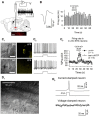
ACh iontophoresis directly stimulated α4β2 neurons in vivo and in vitro. A, Single-unit recordings were performed in STN. A typical 1 s trace is shown. Calibration: 0.5 s, 150 μV. A representative image shows neurobiotin staining in STN. Scale bar, 100 μm. B, ACh (0.5 m) was applied from the recording electrode by iontophoresis (10 s, 30 nA), and stimulated an STN neuron (right). Left, Spike waveform. Calibration: 1 ms, 100 μV. C1, C2, Brain slices of α4-YFP mice were prepared, and whole-cell current-clamp recordings (C2) were made in either α4-YFP-positive (C1, top, C2, top) or α4-YFP-negative (C1, bottom, C2, bottom) neurons. Scale bars: C1, 20 μm. C2, The square waveform under firing traces shows 10 s ACh application by iontophoresis (0.5 m ACh, 30 nA). This increased firing rate in α4-YFP-positive neurons (top), but not in α4-YFP-negative neurons (bottom). C3, ACh increased firing rate by 71 ± 5% (n = 7, p = 0.006) in α4-YFP-positive neurons (filled circle), but not in α4-YFP-negative neurons (open circle, by 8 ± 6%, n = 9, p = 0.50) (α4-YFP-positive vs negative neurons, df = 14; t = 3.9, p < 0.01, unpaired t test). D1, A pair of STN neurons were recorded. Nicotinic responses were detected by puffing ACh (0.1 s, 300 μm, 20 psi) onto neurons. Rec, Recording electrode; ACh, puffer pipette. D2, In a pair of neurons, a single spike was evoked in one neuron in current-clamp mode by injecting a depolarizing current (10 ms, 50 pA), and meanwhile signals were recorded from the other neuron in voltage-clamped mode (VH = −50 mV).
Figure 1.
Figure 1.
Two classes of nicotinic ACh responses in STN neurons. A1, A2, In some STN neurons, ACh puffs (300 μm, 0.1 s, 20 psi) induced slower inward currents (IACh) in the voltage-clamp mode (VH = −65 mV; A1: a typical trace), and accelerated spontaneous firings in the current-clamp mode (A2: a typical trace). B1, B2, In some neurons, ACh puffs induced faster inward currents (VH = −65 mV; B1: a typical trace), and transiently accelerated firings (B2: a typical trace). C, In a typical neuron, on which 0.1 s ACh (300 μm) induced a slower current (left), 3 s ACh evoked a current with slower and incomplete desensitization (right). D, In a typical neuron, on which 0.1 s (300 μm) ACh induced a faster current (left), 3 s ACh evoked a faster current with complete desensitization (right). E, Summary of decay time constant τ, induced by 3 s ACh application, in faster and slower nAChR currents (df = 13, t = 7.2, p < 0.01, unpaired t test). The numbers of recorded neurons are shown in parentheses.
Figure 2.
Figure 2.
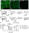
Genetically assisted pharmacological characterization of nAChR subtypes in STN neurons. A1–A3, GFP antibody staining in a PFA-fixed parasagittal brain section from either an α4-YFP (A1, 10×; A2, 60×) or a wild-type mouse (A3, 60×). B1, B2, A slower IACh was abolished by 300 nm DHβE (B1), whereas a faster IACh was eliminated by 10 nm MLA (B2). C1, RJR-2403 (RJR), but not PNU-282987 (PNU), evoked an inward current in an STN neuron showing a slower IACh. C2, PNU-282987, but not RJR-2403, evoked an inward current in an STN neuron showing a faster IACh. Arrows indicate agonist applications (0.1 s, 20 psi). D, Both α4β2 and α7 nAChR currents diminished during nicotine (0.2 and 1 μm) perfusion. The α4β2 nAChR current amplitude sharply decreased by 68 ± 11% (n = 6) and 100% (n = 5) after 10 min perfusion of 0.2 (solid triangle) and 1 μm (solid circle) nicotine, respectively. α7 nAChR currents diminished more slowly and less completely after 10 min nicotine perfusion (0.2 μm nicotine: by 38 ± 12%, n = 5, open triangle; 1 μm nicotine: by 80 ± 7%, n = 5, open circle). Two-way repeated measurements were used to compare nicotine perfusion-induced desensitization of α4β2 and α7 nAChRs. For 1 μm nicotine, both nAChR subtypes (df = 1, F = 38.7, p = 0.00023) and nicotine exposure time (0, 3, and 6 min) (df = 2, F = 249.2, p < 0.00001) significantly affected nicotine effects. For 0.2 μm nicotine, both nAChR subtypes (df = 1, F = 7.2, p = 0.028) and nicotine exposure time (0, 3, and 6 min) (df = 2, F = 26.0, p < 0.00001) affected nicotine effects. However, only α7 nAChR currents fully recovered after 15 min washout (α7: 0.2 μm nicotine: 101 ± 2% of control, n = 5, open triangle; 1 μm: 82 ± 6% of control, n = 5, open circle) (α4β2: 0.2 μm nicotine: 55 ± 19% of control, n = 5, solid triangle; 1 μm: 0% of control, n = 5, solid circle).
Figure 3.
Figure 3.
Glutamatergic and GABAergic synaptic inputs are diverse among STN neurons. A, Inward and outward spontaneous postsynaptic currents, recorded with low Cl (5 mm) intrapipette solution (see Materials and Methods) at VH = −50 mV (left), were blocked by 20 μm CNQX (middle) and 10 μm GABAzine (right), respectively. B, The sIPSC and sEPSC frequency was lower (df = 23, t = 3.24, p < 0.01, unpaired t test) and higher (df = 23, t = 3.71, p < 0.01, unpaired t test), respectively, in α4β2 neurons than in α7 neurons. C, TTX did not change the frequency (left) and amplitude (right) of sEPSCs and sIPSCs in α4β2 (blank dots) and α7 (dark gray dots) neurons (sEPSC frequency: α4β2 vs α7, df = 9, t = 1.41, p > 0.05; sIPSC frequency: α4β2 vs α7, df = 9, t = 0.81, p > 0.05; sEPSC amplitude: α4β2 vs α7, df = 9, t = 0.85, p > 0.05; sIPSC amplitude: α4β2 vs α7, df = 9, t = 0.13, p > 0.05, unpaired t test). Local electrical stimulation evoked larger EPSCs, but smaller IPSCs, in α4β2 neurons than in α7 neurons in STN. D, Typical traces; E, Summary of peak amplitude (eEPSCs: α4β2 vs α7, df = 21, t = 3.3, p < 0.01; eIPSCs: α4β2 vs α7, df = 21, t = 3.7, p < 0.01, unpaired t test). F1, Electrical stimulation evoked a double-peaked outward PSC (VH = −50 mV) in an STN neuron. Bicuculline (BIC) converted this to a monophasic inward current, which was blocked by CNQX. F2, Local electrical stimulation changed interspike intervals in current-clamped neurons. G1, Electrical stimulation evoked an inward PSC (VH = −50 mV) and accelerated firing in an STN neuron (G2). H1, Comparison of the proportions of α4β2 and α7 neurons being stimulated (black) or inhibited (gray) (χ2 = 4.39, p = 0.04). H2, Summary of electrical stimulation-evoked increase and decrease of instantaneous firing rates in α4β2 (blank bars) and α7 (gray bars) neurons. Number of neurons is indicated in parentheses. **p < 0.01; ***p < 0.001.
Figure 4.
Figure 4.
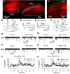
α4β2 and α7 neurons differentially modulate SN neurons. A1, A2, BDA (red), an anterograde tracer, injected in STN, was transported to the SN. Green neurons were TH-positive (A2). B1, A stimulating electrode (Stim) and a recording electrode (Rec) were placed in STN and SNc or SNr, respectively. B2, B3, In the presence of 10 μm bicuculline, electrical stimulation in STN evoked CNQX- sensitive EPSCs in both SNc (B2) and SNr neurons (B3). C, Either RJR-2403 (RJR, 50 μm) or PNU-282987 (PNU, 20 μm) was locally applied (5 s) to alternatively activate α4β2 or α7 nAChRs in STN. Meanwhile, sEPSCs were recorded from either an SNr neuron or an SNc neuron. Either RJR-2403 (D1,E1,F1) or PNU-282987 (D2,E2,F2) in STN increased sEPSC frequency in SNr neurons. PNU-282987 (D2,E2,F2), but not RJR-2403 (D1,E1,F2) in STN, increased sEPSC frequency in SNc neurons. B1, C, The rectangles in SNc and SNr indicate the area containing the recorded neurons.
Figure 6.
Figure 6.
Chronic nicotine upregulates α4β2 nAChRs in STN. α4-YFP was visualized with GFP antibody staining (A, B). More α4-YFP-positive neurons were found in STN of chronic nicotine-treated mice (B) than chronic vehicle-treated mice (A). A, B, Insets, 60× images. C, The number of neurons per STN section was greater in chronic nicotine-treated mice. D1, D2, The integrated YFP density in STN neurons was higher in chronic nicotine-treated mice. E, STN neurons in chronic nicotine-treated (14 d) mice (Nicotine) had larger α4β2 nAChR currents (df = 43; t = 2.77, p = 0.009, unpaired t test; K-S statistics, 0.45, p < 0.05) but similar α7 nAChR currents, compared with those in chronic vehicle-treated mice (Vehicle). F, The 14 d nicotine treatment increased the proportion of α4β2 neurons (black stack) but decreased that of neurons not responding to ACh (dark gray stack) (df = 2, χ2 = 10.7, p < 0.01). G, The 3 μm ACh induced currents in α4β2 neurons of 14 d nicotine-treated (right) mice, but not in those of 14 d vehicle-treated (left) mice. H1, The 14 d nicotine treatment increased spontaneous firing rate in α4β2 neurons (df = 29, t = 3.39, p = 0.02, unpaired t test), but not in α7 neurons (df = 31, t = 1.1, p > 0.05). H2, In chronic vehicle- and nicotine-treated mice, ACh iontophoresis in vivo stimulated 21% (17 of 81) and 44% (38 of 86), respectively, of STN neurons (df = 1, χ2 = 10.1, p < 0.01). Spontaneous firing rate was higher in STN neurons stimulated by ACh iontophoresis in chronic nicotine-treated mice than those in chronic vehicle-treated mice (df = 53, u = 196, p < 0.01, Mann–Whitney test). Numbers of neurons are indicated in each box. *p < 0.05; **p < 0.01.
Figure 7.
Figure 7.
The diversity of synaptic inputs and outputs of STN neurons. α4β2 and α7 nAChRs are the predominant nAChR subtypes in ∼20% and ∼40% of STN neurons, respectively. Compared with α7 nAChR-containing neurons, α4β2 nAChR-containing neurons receive stronger glutamatergic inputs, primarily from the motor cortex, but less GABAergic inputs from the globus pallidus (GP). α4β2 and α7 nAChR-expressing neurons preferentially control SNr and SNc neurons, respectively. SNr GABAergic neurons inhibit SNc neurons. Putative synaptic connections defined in other publications are indicated as dashed lines.

Comment in

Similar articles

Cited by

References

    1. Armstrong DL, Lester HA. The kinetics of tubocurarine action and restricted diffusion within the synaptic cleft. J Physiol. 1979;294:365–386. doi: 10.1113/jphysiol.1979.sp012935. - DOI - PMC - PubMed
    1. Benabid AL, Chabardes S, Mitrofanis J, Pollak P. Deep brain stimulation of the subthalamic nucleus for the treatment of Parkinson's disease. Lancet Neurol. 2009;8:67–81. doi: 10.1016/S1474-4422(08)70291-6. - DOI - PubMed
    1. Benarroch EE. Subthalamic nucleus and its connections: anatomic substrate for the network effects of deep brain stimulation. Neurology. 2008;70:1991–1995. doi: 10.1212/01.wnl.0000313022.39329.65. - DOI - PubMed
    1. Bergman H, Wichmann T, Karmon B, DeLong MR. The primate subthalamic nucleus: II. Neuronal activity in the MPTP model of parkinsonism. J Neurophysiol. 1994;72:507–520. - PubMed
    1. Berry MS, Pentreath VW. Criteria for distinguishing between monosynaptic and polysynaptic transmission. Brain Res. 1976;105:1–20. doi: 10.1016/0006-8993(76)90919-7. - DOI - PubMed

Publication types

LinkOut - more resources