. 2015 Jul;88(1):52-60.
doi: 10.1038/ki.2015.29. Epub 2015 Feb 25.

Correlation of disease activity in proliferative glomerulonephritis with glomerular spleen tyrosine kinase expression

Affiliations

Correlation of disease activity in proliferative glomerulonephritis with glomerular spleen tyrosine kinase expression

Stephen P McAdoo et al. Kidney Int. 2015 Jul.

Abstract

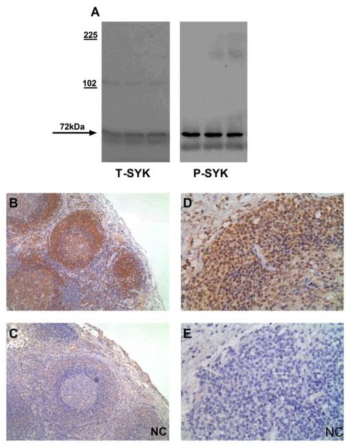
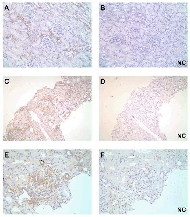
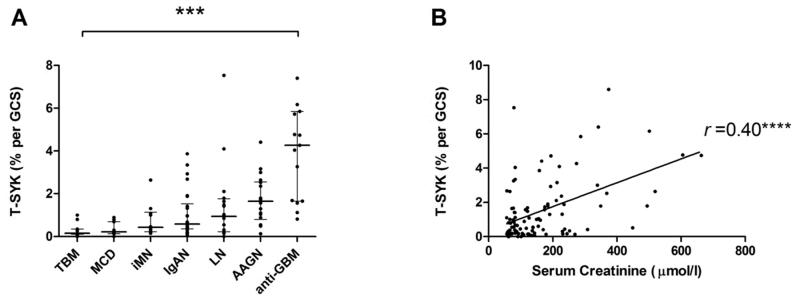
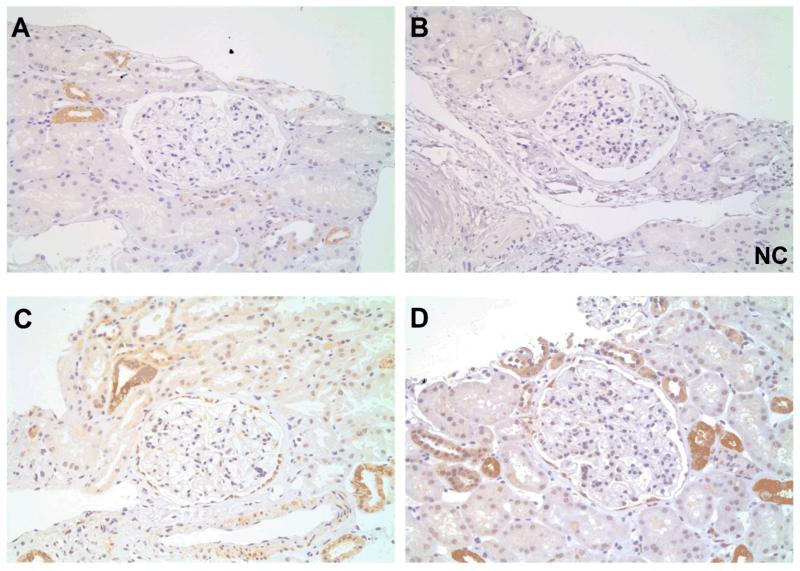
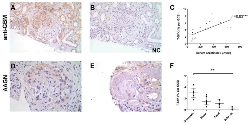
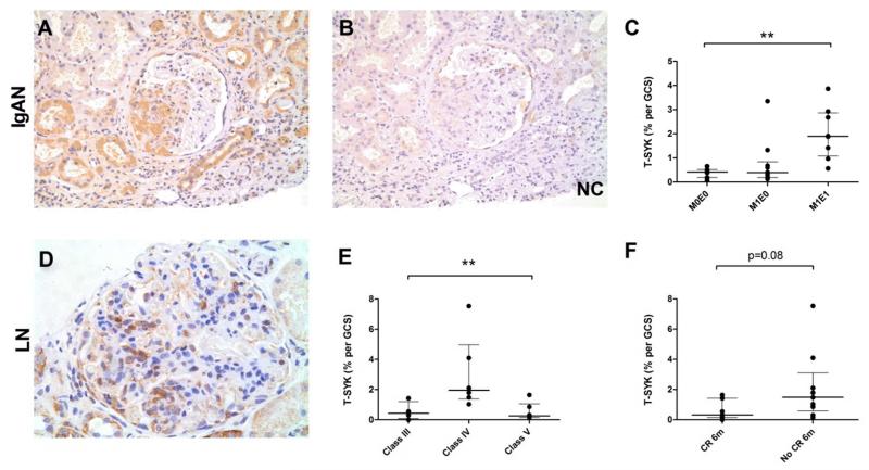
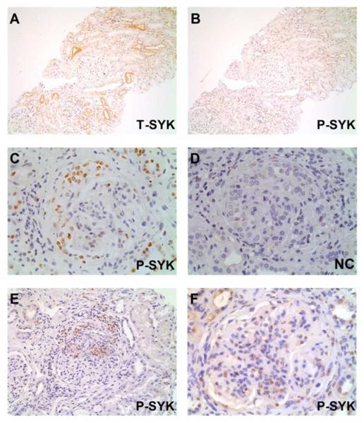
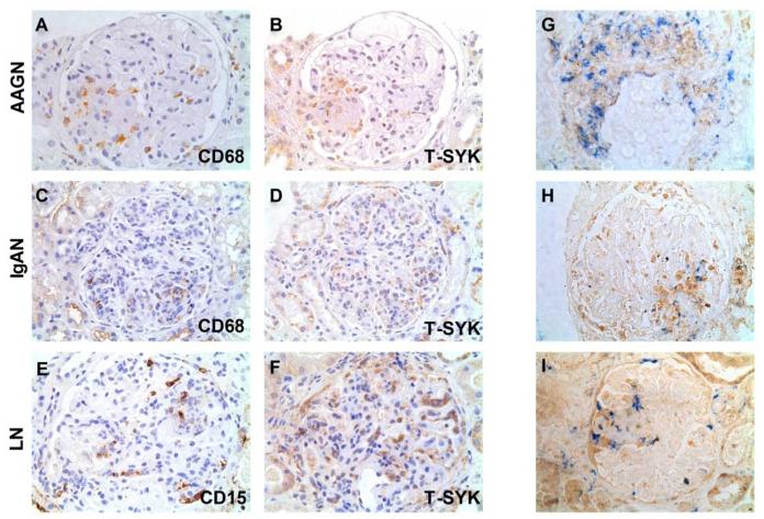
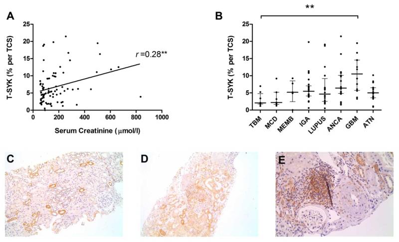
Spleen tyrosine kinase (SYK) is an important component of the intracellular signaling pathway for various immunoreceptors. Inhibition of SYK has shown promise in preclinical models of autoimmune and glomerular disease. However, the description of SYK expression in human renal tissue, which would be desirable ahead of clinical studies, is lacking. Here we conducted immunohistochemical analysis for total and phosphorylated SYK in biopsy specimens from >120 patients with a spectrum of renal pathologies, including thin basement membrane lesion, minimal change disease, membranous nephropathy, IgA nephropathy, lupus nephritis, ANCA-associated glomerulonephritis, antiglomerular basement membrane disease, and acute tubular necrosis. We found significant SYK expression in proliferative glomerulonephritis and that glomerular expression levels correlated with presenting serum creatinine and histological features of disease activity that predict outcome in IgA nephropathy, lupus nephritis, ANCA-associated glomerulonephritis, and antiglomerular basement membrane disease. SYK was phosphorylated within pathological lesions, such as areas of extracapillary and endocapillary proliferation, and appeared to localize to both infiltrating leucocytes and to resident renal cells within diseased glomeruli. Thus SYK is associated with the pathogenesis of proliferative glomerulonephritides, suggesting that these conditions may respond to SYK inhibitor treatment.


Figures

Figure 1
Figure 1. Method development and control immunohistochemical (IHC) stains
(A) Western blot for total (T-) and phophorylated (P-) SYK in lysates of human peripheral blood mononuclear cells, confirming detection of SYK protein at 72kDA. (B & C) Sequential sections showing positive and negative control (NC) staining for T-SYK in human lymph node, with strong staining for SYK localised to lymphoid follicles. (D & E) Sequential sections showing positive and negative control (NC) staining for P-SYK in human lymph node, demonstrating nuclear and cytoplasmic localisation, within cells in lymphoid follicles. All sections are immunoperoxidase stains with haematoxylin counterstain, x200-400 magnification. Negative control (NC) stains were performed by pre-incubating the primary antibody with the relevant immunising peptide.
Figure 2
Figure 2. Total (T-) SYK detection in normal and nephritic renal tissue
(A & B) Paired sections showing positive and negative control (NC) staining for T-SYK in normal renal tissue obtained from nephrectomy specimens, showing intermittent SYK expression in distal tubular epithelial cells (having thin cuboidal epithelium, little brush border and open tubular lumens). (C-F) Two pairs of sequential sections at low and high power, respectively, showing positive and negative control (NC) staining for T-SYK in diffuse proliferative post-infectious glomerulonephritis; SYK is detected within the nephritic glomerular tuft, as well as in some distal tubular epithelial cells, similar to that is seen in normal tissue. All sections are immunoperoxidase stains with haematoxylin counterstain, x100-400 magnification. Negative control (NC) stains were performed by pre-incubating the primary antibody with the relevant immunising peptide.
Figure 3
Figure 3. Quantification of Total (T-) SYK detection in a spectrum of glomerular diseases
(A) Glomerular T-SYK expression in each form of glomerulonephritis, expressed as mean percent stain per glomerular cross section (% per GCS) in each biopsy case; negligible staining was observed in non-proliferative diseases and expression levels were significantly elevated in proliferative and crescentic forms of glomerulonephritis; TBM, thin basement membrane lesion; MCD, minimal change disease; iMN, idiopathic membranous nephropathy; IgAN, immunoglobulin A nephropathy; LN, lupus nephritis; AAGN, anti-neutrophil cytoplasm antibody-associated glomerulonephritis; anti-GBM, anti-glomerular basement membrane disease. (B) Glomerular T-SYK expression correlates with serum creatinine at the time of biopsy (n=107 cases, Spearman r =0.4, p<0.0001). *** p<0.001, ***p<0.0001.
Figure 4
Figure 4. Total (T-) SYK expression non-proliferative glomerulonephritides
(A & B) Sequential sections showing positive and negative control (NC) staining for T-SYK in thin basement membrane lesion. (C) T-SYK staining in minimal change disease. (D) T-SYK staining in idiopathic membranous nephropathy. In all cases, negligible glomerular staining is seen. There is intermittent staining of distal tubular epithelial cells, comparable to that seen in normal kidney tissue. All sections are immunoperoxidase stains with haematoxylin counterstain, x100-400 magnification. Negative control (NC) stains were performed by pre-incubating the primary antibody with the relevant immunising peptide.
Figure 5
Figure 5. Total (T-) SYK expression in anti-glomerular basement membrane (anti-GBM) disease and ANCA-associated glomerulonephritis (AAGN)
(A & B) Sequential sections showing positive and negative control (NC) staining for T-SYK in a crescentic glomerulus in anti-GBM disease. (C) Glomerular T-SYK expression correlates strongly with presenting serum creatinine in anti-GBM disease (n=15 cases; Spearman r = 0.78, p=0.0006). (D) T-SYK staining in AAGN localises to areas of crescent formation. (E) Minimal glomerular staining for T-SYK in a scarred glomerulus in sclerotic-class AAGN. (F) Correlation of glomerular T-SYK staining with histologic class of AAGN, with highest levels seen in crescentic class disease. All sections are immunoperoxidase stains with haematoxylin counterstain, x200-400 magnification. Negative control (NC) stains were performed by pre-incubating the primary antibody with the relevant immunising peptide. GCS, glomerular cross section. ** p<0.01, *** p<0.001.
Figure 6
Figure 6. Total (T-) SYK expression in Immunoglobulin A nephropathy (IgAN) and lupus nephritis (LN)
(A & B) Sequential sections showing positive and negative control (NC) staining for T-SYK in IgAN, localised to a segmental area of endocapillary proliferation. (C) T-SYK detection in IgAN according to Oxford Class of disease; significant T-SYK detection was a feature of IgAN with endocapillary, and not mesangial, proliferation; M0E0, neither mesangial nor endocapillary proliferation; M1E0, mesangial proliferation without endocapillary proliferation; M1E1, both mesangial and endocapillary proliferation. (D) T-SYK detection in class IV lupus nephritis, localised to cells within the glomerular tuft. (E) T-SYK detection in LN according to ISN/RPS class of disease; SYK expression was a feature of class IV (diffuse proliferative) disease only. (F) T-SYK detection was higher in patients who failed to achieve complete remission (CR) at six months (6m) from initiating immunosuppressive treatment after this index renal biopsy. All sections are immunoperoxidase stains with haematoxylin counterstain, x200-400 magnification. Negative control (NC) stains were performed by pre-incubating the primary antibody with the relevant immunising peptide. GCS, glomerular cross section. ** p<0.01
Figure 7
Figure 7. Phophorylated (P-) SYK detection in proliferative glomerulonephritides
(A & B) Sequential sections showing total (T-) SYK and P-SYK detection in minimal change disease; glomerular staining was not observed in either case; T-SYK staining was observed in some distal tubular epithelial cells, although minimal staining for P-SYK was observed within these cells, suggesting that SYK in not significantly activated in this cell type. (C & D) Sequential sections showing positive and negative control (NC) staining for P-SYK in anti-glomerular basement membrane disease; P-SYK is detected within cells of an extracapillary crescent, and within occasional cells in the glomerular tuft. (E) P-SYK detection in ANCA-associated glomerulonephritis, localised to an area of crescent formation and peri-glomerular inflammation; minimal tubular staining for P-SYK is seen (F) P-SYK detection in class IV lupus nephritis, showing P-SYK cells within the glomerular tuft. All sections are immunoperoxidase stains with haematoxylin counterstain, x200-400 magnification. Negative control (NC) stains were performed by pre-incubating the primary antibody with the relevant immunising peptide.
Figure 8
Figure 8. Cellular localisation of SYK in proliferative glomerulonephritides
(A & B) Paired sections showing staining for CD68 and total (T-) SYK, suggesting co-localisation of T-SYK and CD68+ macrophages within a segmental area of inflammation in the glomerulus (at 7 o’clock position) in a case of ANCA-associated glomerulonephritis (AAGN). (C & D) Paired sections suggesting co-localisation of T-SYK and CD68+ macrophages in the glomerulus (at 5 o’clock positition) in a case of IgA nephropathy. (E & F) Paired sections suggesting localisation of T-SYK and CD15+ neutrophils in a case of class IV lupus nephritis (LN). A-F are immunoperoxidase stains with haematoxylin counterstain, x400 magnification. (G,H & I) Two-colour double staining for CD68 (blue) and T-SYK (brown) in three cases of AAGN, showing co-localisation of T-SYK expression with CD68+ macrophage detection, within crescents (G) or segmental areas of glomerular inflammation (H & I). In addition, T-SYK+/CD68-negative cells are observed suggesting that there are additional cell types that may express T-SYK within the inflamed glomerulus. G, H & I are immunoperoxidase stain (brown; T-SYK) and immunophosphatase stain (blue; CD68), without counterstain, to enable identification of blue reporter stain.
Figure 9
Figure 9. Tubulointerstitial staining for total (T-) SYK in glomerulonephritis and in acute tubular necrosis (ATN)
(A) Tubulointerstitial T-SYK detection, expressed as the mean percent stain per tubular cross section (TCS) in each biopsy case, showing a statistically significant correlation with serum creatinine at the time of biopsy (Spearman r =0.28; p=0.009). (B) Tubulointerstitial SYK detection according to diagnosis in a range of glomerulonephritides and in acute tubular necrosis (ATN), demonstrating that tubular SYK expression may be upregulated at times of tubular injury. TBM, thin basement membrane lesion; MCD, minimal change disease; iMN, idiopathic membranous nephropathy; IgAN, immunoglobulin A nephropathy; LN, lupus nephritis; AAGN, anti-neutrophil cytoplasm antibody-associated glomerulonephritis; anti-GBM, anti-glomerular basement membrane disease. ** p<0.01. (C) Tubulointerstitial SYK expression in a case TBM, shown here for comparison. (D) Generalised increased in tubular SYK staining adjacent to a crescentic glomerulus in a severe case of AAGN. (E) Tubulointerstial infiltrate containing SYK positive cells, in a case AAGN with concomitant tubulointerstitial nephritis. (A-C) Immunperoxidase stain with haematoxylin counterstain, x100-200 magnification.

Similar articles

Cited by

References

    1. Geahlen RL. Syk and pTyr’d: Signaling through the B cell antigen receptor. Biochimica et biophysica acta. 2009 Jul;1793(7):1115–1127. - PMC - PubMed
    1. Crowley MT, Costello PS, Fitzer-Attas CJ, et al. A critical role for Syk in signal transduction and phagocytosis mediated by Fcgamma receptors on macrophages. The Journal of experimental medicine. 1997 Oct 6;186(7):1027–1039. - PMC - PubMed
    1. Kiefer F, Brumell J, Al-Alawi N, et al. The Syk protein tyrosine kinase is essential for Fcgamma receptor signaling in macrophages and neutrophils. Molecular and cellular biology. 1998 Jul;18(7):4209–4220. - PMC - PubMed
    1. de Castro RO. Regulation and function of syk tyrosine kinase in mast cell signaling and beyond. Journal of signal transduction. 2011;2011:507291. - PMC - PubMed
    1. Mocsai A, Ruland J, Tybulewicz VL. The SYK tyrosine kinase: a crucial player in diverse biological functions. Nature reviews. Immunology. 2010 Jun;10(6):387–402. - PMC - PubMed

Publication types

MeSH terms