. 2015 Feb 20;347(6224):842-7.
doi: 10.1126/science.1256022.

Photochemistry. Chemiexcitation of melanin derivatives induces DNA photoproducts long after UV exposure

Affiliations

Photochemistry. Chemiexcitation of melanin derivatives induces DNA photoproducts long after UV exposure

Sanjay Premi et al. Science. .

Abstract

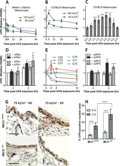
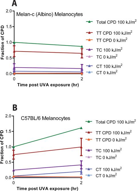
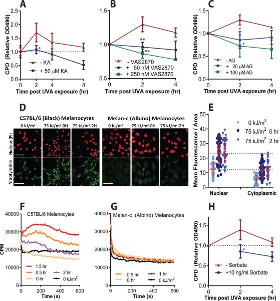
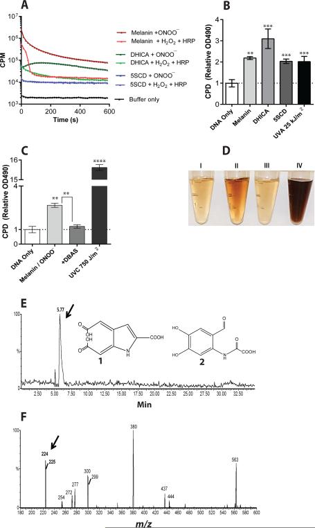
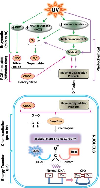
Mutations in sunlight-induced melanoma arise from cyclobutane pyrimidine dimers (CPDs), DNA photoproducts that are typically created picoseconds after an ultraviolet (UV) photon is absorbed at thymine or cytosine. We found that in melanocytes, CPDs are generated for >3 hours after exposure to UVA, a major component of the radiation in sunlight and in tanning beds. These "dark CPDs" constitute the majority of CPDs and include the cytosine-containing CPDs that initiate UV-signature C→T mutations. Dark CPDs arise when UV-induced reactive oxygen and nitrogen species combine to excite an electron in fragments of the pigment melanin. This creates a quantum triplet state that has the energy of a UV photon but induces CPDs by energy transfer to DNA in a radiation-independent manner. Melanin may thus be carcinogenic as well as protective against cancer. These findings also validate the long-standing suggestion that chemically generated excited electronic states are relevant to mammalian biology.


Figures

Fig. 1
Fig. 1. Cyclobutane pyrimidine dimers (CPD) continue to be generated in melanocytes long after UV exposure ends
(A) In albino murine melanocytes CPD induction at time 0 is followed by repair, but in melanin-containing melanocytes (B, C) CPD continue to increase for 4 h after UVA exposure ends. CPD were assayed by DNA ELISA. (D) Additional dark CPD are revealed in melanocytes when excision repair is suppressed by siXpa or siXpc; dark CPD account for half the total CPD. (E) In melanocytes from humans, the production of dark CPD after UVA varies between individuals but as shown in (F) “non-producers” are revealed as producers after DNA repair is suppressed in the cells by siXPA or siXPC. (G) Dark CPD in mouse melanocytes and keratinocytes in vivo. K14-Kitl transgenic mice, which have epidermal melanocytes containing eumelanin, were crossed to mice carrying the Mc1re/e allele which confers epidermal pheomelanin and yellow fur. Scale bar is 50 μm. (H) Quantitation of CPD in epidermal sections. Error bars are SD from 4, 3, or 2 experiments (A-C, D-F, H, respectively). p values by t-test are for the difference between the asterisked timepoint and 0 h or as indicated. *, p ≤ 0.05; ** 0.005; *** 0.0005; **** 0.00005.
Fig. 2
Fig. 2. Mass spectrometry reveals enrichment of dark CPD for CPD that contain cytosine
For energetic reasons, direct absorption of UVA generates primarily thymine-containing (TT) cyclobutane pyrimidine dimers. (A) There is no dark CPD formation in albino melanocytes after UVA exposure. The slope of post-UVA CPD induction in albino cells is indistinguishable from the slope in unirradiated cells. Data represent the average of 3 experiments, expressed as a fraction of the total CPD generated at 0 h. Hence, the total CPD line has no error bar. The total number of CPD at 0 h was 169 CPD per megabase of DNA. (B) Dark CPD are induced in melanin-containing melanocytes by UVA and include a greater number of cytosine-containing TC and CT dimers, capable of causing UV signature C→T mutations. Slopes for post-UVA induction of TT, TC, and CT CPD in melanin-containing cells are greater than those in albino, with p = 0.01, 0.05, and 0.03, respectively. The total number of CPD at 0 h was 87 CPD per megabase of DNA, consistent with the shielding function of eumelanin. Data represent the average of 5 experiments.
Fig. 3
Fig. 3. In the proposed photochemical pathway for production of dark CPD, high energy triplet-state moieties are created by melanin, superoxide, and nitric oxide
Dark CPD are blocked by inhibitors of: (A) melanin synthesis (kojic acid, KA), (B) the superoxide generator NADPH oxidase (VAS2870), and (C) the nitric oxide generator iNOS (aminoguanidine). Inhibitors are not expected to reduce CPD levels below those seen at 0 hr, when CPD are generated primarily by direct UV absorption (dotted line). (D) The product of superoxide and nitric oxide, peroxynitrite, generates 3-nitrotyrosine modified proteins in melanocyte nuclei, except in albino melanocytes, and these increase with UVA exposure. (E) Quantitation of 3-nitrotyrosine in melanin-containing melanocytes. The increase at 0 h arises from protein nitration during the 27 min UVA exposure. (F) Ultra-weak chemiluminescence is generated by permeabilized UVA-irradiated melanized melanocytes that had been incubated with a triplet energy acceptor probe, DBAS. Chemiluminescence was quantified by single-photon counting (cpm, counts per minute). (G) Albino melanocytes do not produce chemiluminescence when irradiated. (H) A triplet-state quencher, ethyl sorbate, inhibits production of dark CPD in pigmented mouse melanocytes after they are exposed to UVA. Data are the average of 3 experiments. (F) and (G) show data from one of 3 similar experiments. p values are for the difference between treated and untreated samples.
Fig. 4
Fig. 4. Excited-state melanin may act as a molecular vector
(A) Synthetic melanin, its eumelanin monomer DHICA, and its pheomelanin monomer 5SCD generate ultra-weak chemiluminescence when oxidized by peroxynitrite or by horseradish peroxidase plus H2O2. DBAS was used as a triplet energy probe, so luminescence indicates the presence of electronically excited triplet states. Data represent one of 6 experiments. (B) CPD are created in the dark when melanin or its monomers are oxidized with peroxynitrite. The level of CPD resulting from direct DNA absorption of UVA is shown for comparison. (C) Diverting triplet energy to luminescence with DBAS blocks production of CPD by oxidized melanin. The UVC dose used for comparison is ~100 fold higher than that lethal to 50% of normal cells. In B and C, data represent the average of 3 experiments. p values are for the difference between treated and “DNA only” samples or as indicated. (D) Synthetic melanin is rapidly solubilized upon exposure to UV or peroxynitrite. Left to right: Supernatants resulting from untreated melanin or from melanin exposed to UVA (200 kJ/m2), 7.8 mM NaOH, or peroxynitrite (1 mM in NaOH stabilizer). (E-F) Identification of a putative triplet-carbonyl carrier. (E) The eumelanin monomer DHICA was oxidized and products were separated by HPLC, monitoring by mass spectrometry in negative ion mode. Monitoring for the 224 m/z ion of 225 dalton compounds 1 and 2, expected for triplet-carbonyl derivatives of dioxetane-adducted DHICA, reveals a single, polar peak. (F) Scanning the polar HPLC peak reveals four principal m/z peaks, one at 224 daltons.
Fig. 5
Fig. 5. A mechanistic model for the generation of dark CPD in melanocytes by chemiexcitation, with melanin as an active participant
Exposing cells to UV radiation is known to upregulate iNOS, NADPH oxidase (NOX), and enzymes of melanin synthesis, presumably causing sustained generation of nitric oxide (NO) and superoxide (O2•–). Cytoplasmic NOS and NOX are indicated on the figure but some isoforms are located in the nucleus. The present experiments show a UV-induced surge in the product of these two radicals, the powerful oxidant peroxynitrite (ONOO), and show that peroxynitrite degrades melanin polymer to fragments. Melanin or melanin fragments then appear in the nucleus. Peroxynitrite is also one of the few biologically-synthesized molecules capable of exciting an electron to a triplet state. The present experiments show that, on a faster time scale, peroxynitrite excites an electron in a melanin fragment to a triplet state that has the high energy of a UV photon. The typical triplet-state reaction intermediate, not demonstrated here (hence indicated in italics), is a cycloaddition of –O–O– to create an unstable dioxetane; dioxetanes undergo spontaneousthermolysis to yield two carbonyls, one of which acquires most of the energy and finishes in a high energy triplet state (*). For the melanin-related triplet, the half-life of the reaction intermediate appeared to be minutes and a carbonyl consistent with a dioxetane precursor was identified by mass spectrometry. Triplet energy then discharges on a microsecond timescale to generate visible luminescence, or discharges in a radiation-independent manner to DBAS to be emitted as fluorescence, to sorbate to be dissipated as isomerization and heat, or evidently toDNA bases where it makes CPD. The presence of melanin, activation of iNOS and NOX, and the triplet state were shown to be required for dark CPD formation.

Comment in

Similar articles

Cited by

References

    1. Kollias N, Sayre RM, Zeise L, Chedekel MR. Photoprotection by melanin. J. Photochem. Photobiol. B. 1991;9:135–160. - PubMed
    1. Williams PF, Olsen CM, Hayward NK, Whiteman DC. Melanocortin 1 receptor and risk of cutaneous melanoma: a meta-analysis and estimates of population burden. Int J Cancer. 2011;129:1730–1740. - PubMed
    1. Noonan FP, et al. Melanoma induction by ultraviolet A but not ultraviolet B radiation requires melanin pigment. Nat Commun. 2012;3:884. - PMC - PubMed
    1. Mitra D, et al. An ultraviolet-radiation-independent pathway to melanoma carcinogenesis in the red hair/fair skin background. Nature. 2012;491:449–453. - PMC - PubMed
    1. Chedekel MR, Smith SK, Post PW, Pokora A, Vessell DL. Photodestruction of pheomelanin: role of oxygen. Proc. Natl. Acad. Sci. U.S.A. 1978;75:5395–5399. - PMC - PubMed

Publication types

MeSH terms