. 2015 Feb 12;11(2):e1004657.
doi: 10.1371/journal.ppat.1004657. eCollection 2015 Feb.

Human adenovirus 52 uses sialic acid-containing glycoproteins and the coxsackie and adenovirus receptor for binding to target cells

Affiliations

Human adenovirus 52 uses sialic acid-containing glycoproteins and the coxsackie and adenovirus receptor for binding to target cells

Annasara Lenman et al. PLoS Pathog. .

Abstract

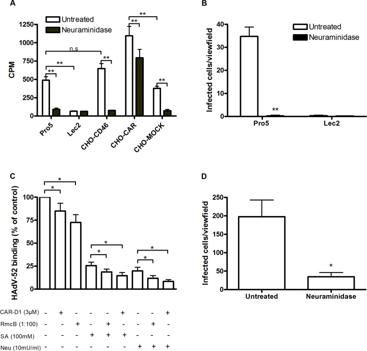
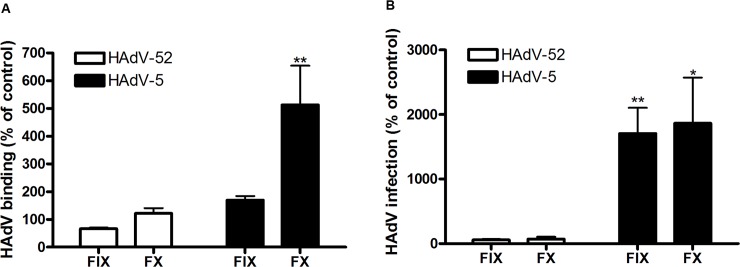
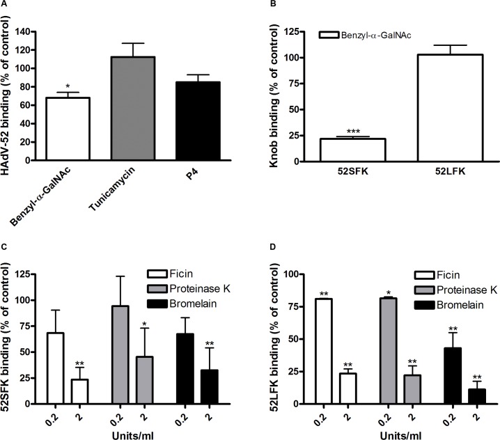
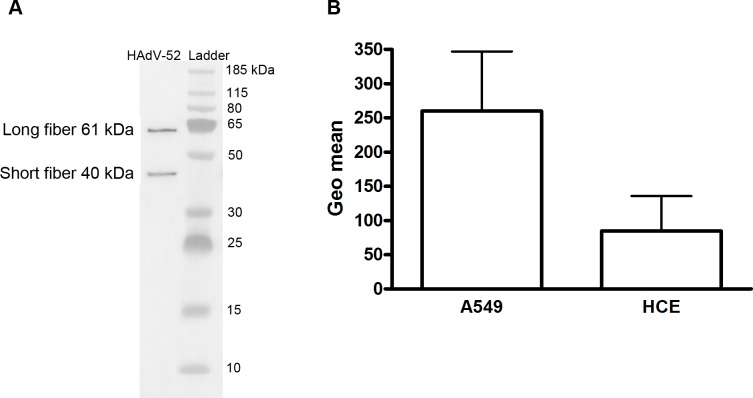
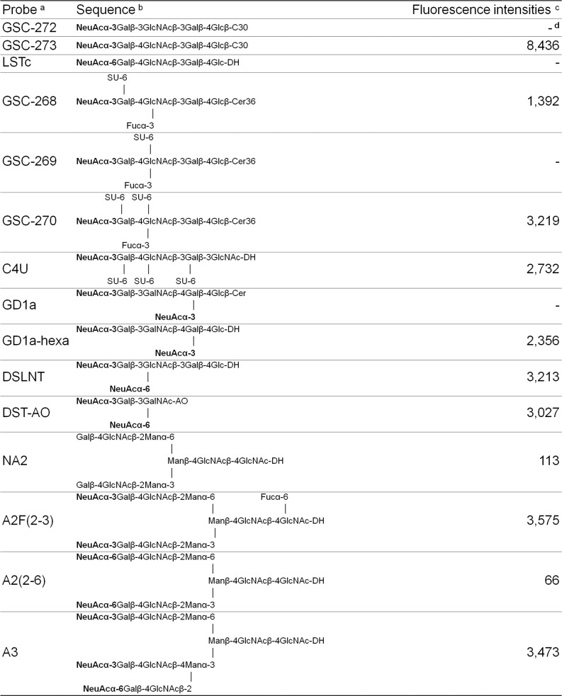
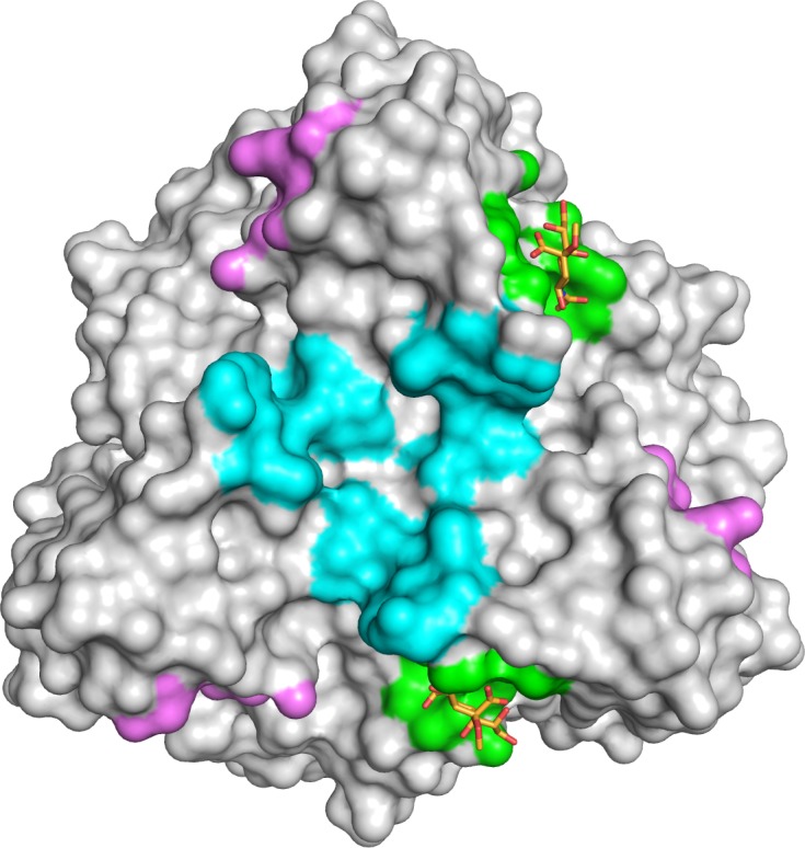
Most adenoviruses attach to host cells by means of the protruding fiber protein that binds to host cells via the coxsackievirus and adenovirus receptor (CAR) protein. Human adenovirus type 52 (HAdV-52) is one of only three gastroenteritis-causing HAdVs that are equipped with two different fiber proteins, one long and one short. Here we show, by means of virion-cell binding and infection experiments, that HAdV-52 can also attach to host cells via CAR, but most of the binding depends on sialylated glycoproteins. Glycan microarray, flow cytometry, surface plasmon resonance and ELISA analyses reveal that the terminal knob domain of the long fiber (52LFK) binds to CAR, and the knob domain of the short fiber (52SFK) binds to sialylated glycoproteins. X-ray crystallographic analysis of 52SFK in complex with 2-O-methylated sialic acid combined with functional studies of knob mutants revealed a new sialic acid binding site compared to other, known adenovirus:glycan interactions. Our findings shed light on adenovirus biology and may help to improve targeting of adenovirus-based vectors for gene therapy.


Conflict of interest statement

The authors have declared that no competing interests exist.

Figures

Fig 1
Fig 1. HAdV-52 uses sialic acid and CAR for binding to and infection of cells.
(A) 35S-labeled HAdV-52 virion binding to CHO cells expressing or lacking known HAdV receptors. Pro-5 is a sialic acid-positive, reference cell line and parental cell line to sialic acid-negative Lec2 cells. CHO-CD46 and CHO-CAR cells express human CD46 and CAR, respectively. CHO-MOCK is mock transfected with respect to CHO-CAR. Black bars show HAdV-52 binding to cells after pretreatment with V. cholerae neuraminidase. Binding was quantified by liquid scintillation counting and shown as counts per minute (CPM). (B) HAdV-52 infection of Pro-5 and Lec2 pretreated with (black bars) or without (white bars) V. cholerae neuraminidase. The number of infected cells was quantified by immunofluorescence. (C) 35S-labeled HAdV-52 virion binding to A549 cells. Virions were preincubated with or without soluble CAR-D1 or sialic acid monosaccharides, and cells were preincubated with or without mouse anti-CAR mab (clone RmcB) or V. cholerae neuraminidase as indicated, prior to binding. (D) HAdV-52 infection of A549 cells pretreated with or without V. cholerae neuraminidase. All experiments were performed three times with duplicate samples in each experiment. Error bars represent mean ± SD. n.s = not significant, * P of < 0.05 and ** P of < 0.01.
Fig 2
Fig 2. HAdV-52 does not use coagulation factors for binding and infection of A549 cells.
(A) 35S-labeled HAdV-52 virion binding to A549 cells after virion preincubation with physiological concentrations of coagulation factor IX and X (FIX: 5μg/ml and FX: 10μg/ml). (B) HAdV-52 infection of A549 cells after virion preincubation with physiological concentrations of coagulation factors. All experiments were performed three times with duplicate samples in each experiment. Error bars represent mean ± SD. * P of < 0.05 and ** P of < 0.01 versus control.
Fig 3
Fig 3. HAdV-52 short fiber knob binds to O-linked glycoproteins on A549 cells.
(A) 35S-labeled HAdV-52 virion binding to A549 cells pretreated with benzyl-α-GalNAc, tunicamycin or P4 (inhibitors of O-linked glycan synthesis, N-linked glycan synthesis, and glycolipid synthesis, respectively). (B) HAdV-52 short fiber knobs (52SFK) and HAdV-52 long fiber knobs (52LFK) binding to A549 cells pretreated with benzyl-α-GalNAc (inhibitor of O-linked glycan synthesis). (C) 52SFK and (D) 52LFK binding to A549 cells pretreated with ficin, proteinase K or bromelain proteases at indicated concentrations. All experiments were performed three times with duplicate samples in each experiment. Error bars represent mean ± SD. * P of < 0.05, ** P of < 0.01 and *** P of < 0.001 versus control.
Fig 4
Fig 4. Virion composition and relative expression of CAR and sialic acid on human epithelial cells.
A) Western blot analysis of HAdV-52 virion fiber content using a mouse mab (clone 4D2) recognizing an epitope (MKRARPSEDTFNPVYPY) conserved in the tail domain of all HAdVs. The experiment was performed three times (with three different virus preparations) and the figure shows one representative set of results. B) Flow cytometry analysis of CAR expression on A549 and human corneal epithelial (HCE) cells using an anti CAR mouse mab (clone E1-1). Data are shown as geometrical mean (geo mean) and the experiment was performed three times with duplicate samples in each experiment. Error bars represent means ± SD.
Fig 5
Fig 5. HAdV-52 short fiber knob binds to sialic acid and long fiber knob binds to CAR.
(A) HAdV-52 long fiber knob (52LFK) and (B) HAdV-52 short fiber knob (52SFK) binding to CHO-cells lacking human CAR (all cells except CHO-CAR), lacking sialic acid (only Lec2) and expressing human CAR (CHO-CAR). (C) 52SFK and 52LFK binding to A549 cells pretreated with V. cholerae neuraminidase. (D) ELISA analysis of 52SFK and 52LFK (in solution) binding to immobilized, sialylated fetuin and asialofetuin type I (chemically prepared) and II (enzymatically prepared). Relative absorbance is shown. (E) Surface plasmon resonance analysis of 52LFK (in solution) binding to CAR (immobilized). A twofold dilution series of 52LFK is shown, ranging from 2 μM to 8 nM. Results are shown as response units (RU). (F) 35S-labeled HAdV-52 virion binding to A549 cells pretreated with V. cholerae neuraminidase (removes α2,3-, α2,6- and α2,8-linked sialic acid) or α2,3-specific neuraminidase at indicated concentrations. All experiments were performed three times with duplicate samples in each experiment. Figure E show one representative set of results. Error bars represent mean ± SD, n.s = not significant, * P of < 0.05, ** P of < 0.01 and *** P of < 0.001 versus control.
Fig 6
Fig 6. Features of 52SFK binding to selected sialyl sequences in the microarray.
a The oligosaccharide probes are all lipid-linked, neoglycolipids (NGLs) or glycosylceramides and are from the collection assembled in the course of research in the Glycosciences Laboratory. b The selected α2-3-linked and α2-6-linked sialyl sequences are marked in bold. For definition of the lipid moieties of the probes, please see https://glycosciences.med.ic.ac.uk/docs/lipids.pdf c Numerical scores for the binding signals are shown as means of duplicate spots at 5 fmol per spot. d-, signal less than 1.
Fig 7
Fig 7. HAdV-52 interaction with sialic acid.
(A) Surface representation of the HAdV-52 short fiber knob structure viewed from the top along the three-fold symmetry axis. The three monomers are shown in green, blue and grey. The 2-O-methyl sialic acid bound to two of three binding sites is shown as orange and red stick model. (B) Detailed view of the interactions in the ligand binding site. The side chain of R316 forms a bidentate salt bridge with the carboxyl group of sialic acid. Residues G303, R316 and G317 form backbone hydrogen bonds with the glycerol, amide and O4 groups of the sialic acid, respectively, while the side chain of N318 engages in a hydrogen bond with the sialic acid carboxylate. The methyl group is not involved in binding contacts. (C) Simulated annealing Fo-Fc omit map for the sialic acid. The map was calculated at 3.0 σ and is displayed with a radius of 1.7 Å around the ligand. 52SFK WT (wild type) and mutant binding to Pro5 (D) and A549 (E) cells. All experiments were performed three times with duplicate samples in each experiment. Error bars represent mean ± SD. * P of < 0.05, ** P of < 0.01 and *** P of < 0.001 versus control.
Fig 8
Fig 8. HAdV-52 has a unique sialic acid binding site compared to other sialic acid binding HAdVs.
Comparison of the sialic acid binding site in HAdV-52 (green with orange sticks) with those of HAdV-37 (cyan) and CAdV-2 (magenta). The protein chains were superposed using the PyMOL (The PyMOL Molecular Graphics System, Version 1.5.0.4 Schrödinger, LLC) align tool, and the contact surface of each ligand was mapped onto HAdV-52 (calculated 4.5 Å around sialic acid).

Similar articles

Cited by

References

    1. Harrach B, Benkö M, Both GW, Brown M, Davison AJ, et al. (2011) Family Adenoviridae In: King AMQ, Adams MJ, Carstens EB, LE J, editors. Virus Taxonomy Ninth Report of the International Committee on Taxonomy of Viruses. San Diego: Elsevier Academic Press; pp. 125–141.
    1. Wold WSM, Horwitz MS (2007) Adenoviruses In: Knipe DM, Howley PM, editors. Fields Virology. 5 ed. Philadelphia: Lippincott Williams & Wilkins; pp. 2395–2436.
    1. Jones MS, Harrach B, Ganac RD, Gozum MM, Dela Cruz WP, et al. (2007) New adenovirus species found in a patient presenting with gastroenteritis. J Virol 81: 5978–5984. - PMC - PubMed
    1. Bergelson JM, Cunningham JA, Droguett G, Kurt-Jones EA, Krithivas A, et al. (1997) Isolation of a common receptor for Coxsackie B viruses and adenoviruses 2 and 5. Science 275: 1320–1323. - PubMed
    1. Tomko RP, Xu R, Philipson L (1997) HCAR and MCAR: the human and mouse cellular receptors for subgroup C adenoviruses and group B coxsackieviruses. Proc Natl Acad Sci U S A 94: 3352–3356. - PMC - PubMed

Publication types

MeSH terms

Substances

Associated data

LinkOut - more resources