. 2015 Feb 6;11(2):e1004659.
doi: 10.1371/journal.ppat.1004659. eCollection 2015 Feb.

Viral and cellular proteins containing FGDF motifs bind G3BP to block stress granule formation

Affiliations

Viral and cellular proteins containing FGDF motifs bind G3BP to block stress granule formation

Marc D Panas et al. PLoS Pathog. .

Abstract

The Ras-GAP SH3 domain-binding proteins (G3BP) are essential regulators of the formation of stress granules (SG), cytosolic aggregates of proteins and RNA that are induced upon cellular stress, such as virus infection. Many viruses, including Semliki Forest virus (SFV), block SG induction by targeting G3BP. In this work, we demonstrate that the G3BP-binding motif of SFV nsP3 consists of two FGDF motifs, in which both phenylalanine and the glycine residue are essential for binding. In addition, we show that binding of the cellular G3BP-binding partner USP10 is also mediated by an FGDF motif. Overexpression of wt USP10, but not a mutant lacking the FGDF-motif, blocks SG assembly. Further, we identified FGDF-mediated G3BP binding site in herpes simplex virus (HSV) protein ICP8, and show that ICP8 binding to G3BP also inhibits SG formation, which is a novel function of HSV ICP8. We present a model of the three-dimensional structure of G3BP bound to an FGDF-containing peptide, likely representing a binding mode shared by many proteins to target G3BP.


Conflict of interest statement

The authors have declared that no competing interests exist.

Figures
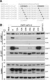
Fig 1
Fig 1. Mutagenesis of the G3BP-binding domain in SFV-nsP3 reveals a core binding motif of FGDF.
(A) C-terminal sequences of pEGFP-C1, pEGFP-nsP3-31-wt, -T2A, -F3A, -G4A, -D5A, -F6A or -D7A. Alanine mutations are shown in bold. (B) BHK cells were mock transfected (M) or transfected with pEGFP-nsP3-31-wt, -T2A, -F3A, -G4A, -D5A, -F6A or -D7A or pEGFP-C1. Cell lysates were prepared 16 h after transfection and immunoprecipitated with anti-GFP or G3BP-1 antisera and separated by SDS–PAGE and transferred to nitrocellulose. Blots of total lysates and IPs were probed for G3BP-1, GFP or actin.
Fig 2
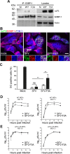
Fig 2. SFV-F3A does not sequester G3BP, induces a stronger SG response and is attenuated in vitro.
(A) BHK cells were infected at MOI 10 with SFV-wt or SFV-F3A. At 8 hpi, cell lysates were prepared and immunoprecipitated with G3BP-1 antisera and separated by SDS–PAGE. Lysates and IPs were probed for nsP3, G3BP-1 or actin. (B) MEF cells were infected with SFV-wt or SFV-F3A at MOI 1. At 8 hpi cells were fixed and stained for nsP3 (green), G3BP-1 (red) and TIA-1 (blue). Bar 20 μm. (C) MEF cells were mock infected or infected with SFV-wt or SFV-F3A at an MOI of 50 for 7 h before 1h treatment with Pat A (100 nM) or mock treatment. Cells were fixed and stained for G3BP-1, TIA-1 and nsP3. Fifty cells per treatment were scored as SG+ or not based on G3BP-1 and TIA-1 colocalization. Data are presented as mean ± SD from five independent experiments. Open bars, mock treated; closed bars, Pat A. Unpaired Student’s t test, **p < 0.01. (D) WT or (E) eIF2α-AA MEFs were infected with SFV-wt or SFV-F3A at an MOI of 10 (left) or an MOI of 0.1 (right). At 4, 8, 12, 24 and 36 hpi, supernatants were collected, and SFV titers were quantified by plaque assay on BHK cells. Data are means of two independent experiments. Error bars indicate SD.
Fig 3
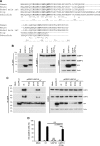
Fig 3. USP10 binds G3BP via a conserved FGDF motif.
(A) Alignment of the N-terminal 80 amino acid residues of higher eukaryote USP10. Human (NM_005153.2), mouse (NP_033488.1), naked mole rat (XP_004842781.1), frog (NP_001080643.1), zebrafish (XP_685621.5). The numbering is based on the human sequence. Conserved phenylalanine residues are indicated in bold. (B) BHK cells were mock-transfected or transfected with pEGFP-C1 or pEGFP- USP101–40-wt. Cell lysates were prepared 16 h after transfection and immunoprecipitated with anti-GFP (left panels) or anti-G3BP-1 (middle panels) antisera. Lysates and IPs were separated by SDS-PAGE and probed for G3BP-1, G3BP-2, GFP or actin. (C) BHK cells were mock-transfected (M) or transfected with pEGFP-C1 or pEGFP-USP101–40-wt, -F10A, -G11A, -D12A or -F13A. Cell lysates were prepared 16 h after transfection and immunoprecipitated with anti-GFP and separated by SDS–PAGE. Lysates and IPs were probed for G3BP-1, G3BP-2, GFP or actin. (D) BHK cells were mock-transfected or transfected with pEGFP-C1, pEGFP-USP10-wt or -F10A. After 23 h, the transfected cells were mock-treated or treated with 0.5 mM sodium arsenite for 1 h, fixed and stained for G3BP-1 and TIA1. Fifty cells per treatment were scored as SG+ based on G3BP-1 and TIA-1 colocalization. Open bars, mock treated; closed bars, sodium arsenite. Data are presented as mean ± SD from three independent experiments. Unpaired Student’s t-test, * p < 0.05, *** p < 0.001.
Fig 4
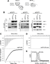
Fig 4. SFV nsP3, containing two FGDF repeats, binds two G3BP molecules, while USP10 binds only one.
(A) Schematic representation of the possible binding of the two FGDF motifs in nsP3-31 (left panel) to two G3BP molecules. The right panel shows the possible binding of one G3BP molecule to one FGDF domain of USP101–40. (B) BHK cells were transfected with pEGFP-C1, pEGFP-USP101–40-wt or pEGFP-nsP3-31-wt. Cell lysates were prepared 16 h after transfection and immunoprecipitated with GFP or G3BP-1 antibody and separated by SDS–PAGE. Lysates and IPs were probed for G3BP-1, G3BP-2, GFP or actin. (C) In isothermal titration calorimetry, injection of nsP3-25 (LTFGDFDEHEVDALASGITFGDFDD) (syringe: 225 μM) to G3BP-NTF2 (cell: 23 μM) resulted in peptide-concentration dependent exothermic heat changes. The binding isotherm of the integrated heat changes reached half-maximum at a molar ratio of approximately 0.5 demonstrating that two molecules of G3BP-NTF2 were bound by one peptide molecule. A one-to-one binding model was used to fit the binding isotherm and gave the following parameters: number of binding sites (n) on nsP3-25 of 2.4 ± 0.1; association constant (KA) of (1.49 ± 0.08)·105 nsP3-25 (dissociation binding constant KD of 6.7 μM); binding enthalpy ΔH of –12.8 ± 0.1 kcal/mol. (D) Titration of the peptide nsP3-25-mut (LTAGDADEHEVDALASGITAGDADD) to G3BP-NTF2 did not produce binding-induced heat changes. (E) In size exclusion chromatography, G3BP-NTF2 alone (dotted line) or pre-incubated with a ten-fold molar excess of nsP3-25 (solid line) eluted at retention volumes of 11.3 and 10 mL, respectively. This shift corresponded to an increase in the apparent molecular weight from 30 to 60 kDa when G3BP was pre-incubated with nsP3-25. The theoretical MW for G3BP-NTF2 and nsP3-25 are 18.4 kDa and 2.7 kDa, respectively.
Fig 5
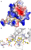
Fig 5. Molecular model of G3BP/FGDF interaction.
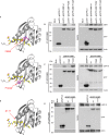
(A) G3BP-NTF2 (PDB ID: 4FCM) with modelled peptide LTFGDFDE (yellow sticks). The protein is shown as the surface colored according to the electrostatic potential (red: acidic, blue: basic). (B) Details of the interaction of the peptide with G3BP-NTF2. G3BP-NTF2 is shown in the cartoon representation. The N-terminal 11 residues as well as several residues lining the peptide-binding groove are displayed as grey sticks. The LTFGDFDE peptide is displayed as yellow sticks, peptide amino acid residues are labeled in red and in bold font. G3BP-1 residues mentioned in the text are labeled in black.
Fig 6
Fig 6. Site-directed mutagenesis in the FGDF peptide binding cleft of G3BP.
(A) Schematic representation of the G3BP-F33W mutant (upper panel) and the G3BP-F124W mutant (lower panel) with the modeled LTFGDFDE peptide. The LTFGDFDE peptide is displayed as sticks with yellow carbon atoms. Mutated tryptophan residues are shown in magenta. (B) HEK293T cells were mock transfected or cotransfected with pEBB/PP-SFV-nsP3 (pnsP3) and either pEGFP-C1, pEGFP-G3BP-wt, -F33W, or -F124W. Cell lysates were prepared 24 h after transfection, immunoprecipitated with anti-GFP and separated by SDS–PAGE. Lysates and IPs were probed using streptavidin-HRP (nsP3), GFP or actin. (C) HEK293T cells were mock transfected or transfected with pEGFP-C1, pEGFP-G3BP-wt, -F33W or -F124W. Cell lysates were prepared 24 h after transfection and immunoprecipitated with GFP antisera and separated by SDS–PAGE. Lysates and IPs were probed for USP10, GFP or actin. (D) Schematic representation of G3BP1-Δ1–11 with the modeled LTFGDFDE peptide. The LTFGDFDE peptide is displayed as sticks with yellow carbon atoms. (E) HEK293T cells were mock transfected or transfected with pEGFP-C1, pEGFP-G3BP1-wt or -Δ1–11. Cell lysates were prepared 24 h after transfection and immunoprecipitated with anti-GFP and separated by SDS–PAGE. Lysates and IPs were probed for USP10, GFP or actin.
Fig 7
Fig 7. An FGDF motif in HSV-1 ICP8 binds G3BP and inhibits SGs.
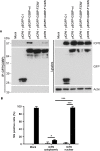
(A) HEK293T cells were mock transfected or cotransfected with pE29 (ICP8) and either pEGFP-C1, pEGFP-G3BP-wt, -F33W or -F124W. Cell lysates were prepared 24 h after transfection and immunoprecipitated with anti-GFP and separated by SDS–PAGE. Lysates and IPs were probed for ICP8, GFP or actin. (B) BHK cells were mock transfected or transfected with pE29 (ICP8). After 23 h the transfected cells were mock treated or treated with sodium arsenite for 1 h fixed and stained for ICP8, G3BP-1 and TIA1. ICP8+ cells were categorized as cytoplasmic or nuclear, and scored as SG+ based on G3BP1 and TIA1 colocalization. Open bars, mock treated; closed bars, sodium arsenite. Data are presented as mean +/- SD from three independent experiments in which 50 cells per treatment were counted. Unpaired Student’s t-test, * p < 0.05, *** p < 0.001.

Similar articles

Cited by

References

    1. Matsuki H, Takahashi M, Higuchi M, Makokha GN, Oie M, et al. (2013) Both G3BP1 and G3BP2 contribute to stress granule formation. Genes Cells 18: 135–146. 10.1111/gtc.12023 - DOI - PubMed
    1. Tourriere H, Chebli K, Zekri L, Courselaud B, Blanchard JM, et al. (2003) The RasGAP-associated endoribonuclease G3BP assembles stress granules. J Cell Biol 160: 823–831. - PMC - PubMed
    1. Kedersha NL, Gupta M, Li W, Miller I, Anderson P (1999) RNA-binding proteins TIA-1 and TIAR link the phosphorylation of eIF-2 alpha to the assembly of mammalian stress granules. J Cell Biol 147: 1431–1442. - PMC - PubMed
    1. Dang Y, Kedersha N, Low WK, Romo D, Gorospe M, et al. (2006) Eukaryotic initiation factor 2alpha-independent pathway of stress granule induction by the natural product pateamine A. J Biol Chem 281: 32870–32878. - PubMed
    1. Suyama M, Doerks T, Braun IC, Sattler M, Izaurralde E, et al. (2000) Prediction of structural domains of TAP reveals details of its interaction with p15 and nucleoporins. EMBO Rep 1: 53–58. - PMC - PubMed

Publication types

MeSH terms