

Deficits in prefrontal cortical and extrastriatal dopamine release in schizophrenia: a positron emission tomographic functional magnetic resonance imaging study
- PMID: 25651194
- PMCID: PMC4768742
- DOI: 10.1001/jamapsychiatry.2014.2414
Deficits in prefrontal cortical and extrastriatal dopamine release in schizophrenia: a positron emission tomographic functional magnetic resonance imaging study
Abstract
Importance: Multiple lines of evidence suggest a deficit in dopamine release in the prefrontal cortex (PFC) in schizophrenia. Despite the prevalence of the concept of prefrontal cortical hypodopaminergia in schizophrenia, in vivo imaging of dopamine release in the PFC has not been possible until now, when the validity of using the positron emission tomographic D2/3 radiotracer carbon 11-labeled FLB457 in combination with the amphetamine paradigm was clearly established.
Objectives: To (1) test amphetamine-induced dopamine release in the dorsolateral PFC (DLPFC) in drug-free or drug-naive patients with schizophrenia (SCZ) and healthy control (HC) individuals matched for age, sex, race/ethnicity, and familial socioeconomic status;(2) test blood oxygenation level-dependent (BOLD) functional magnetic resonance imaging activation during a working memory task in the same participants; and (3) examine the relationship between positron emission tomographic and functional magnetic resonance imaging outcome measures.
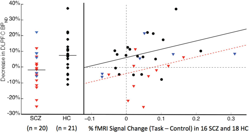
Design, setting and participants: Positron emission tomographic imaging with carbon 11-labeled FLB457 before and following 0.5 mg/kg of amphetamine by mouth. Blood oxygenation level-dependent functional magnetic resonance imaging during the self-ordered working memory task. Twenty patients with schizophrenia recruited from the inpatient and outpatient research facilities at New York State Psychiatric Institute and 21 healthy control individuals participated, and data were acquired between June 16, 2011, and February 25, 2014.
Main outcomes and measure: The percentage change in binding potential (∆BPND) in the DLPFC following amphetamine, BOLD activation during the self-ordered working memory task compared with the control task, and the correlation between these 2 outcome measures.
Results: We observed significant differences in the effect of amphetamine on DLPFC BPND (mean [SD], ∆BPND in HC: -7.5% [11%]; SCZ: +1.8% [11%]; P = .01); a generalized blunting in dopamine release in SCZ involving most extrastriatal regions and the midbrain; and a significant association between ∆BPND and BOLD activation in the DLPFC in the overall sample including patients with SCZ and HC individuals.
Conclusions and relevance: To our knowledge, these results provide the first in vivo evidence for a deficit in the capacity for dopamine release in the DLPFC in SCZ and suggest a more widespread deficit extending to many cortical and extrastriatal regions including the midbrain. This contrasts with the well-replicated excess in dopamine release in the associative striatum in SCZ and suggests a differential regulation of striatal dopamine release in associative striatum vs extrastriatal regions. Furthermore, dopamine release in the DLPFC relates to working memory-related activation of this region, suggesting that blunted release may affect frontal cortical function.
Figures
Similar articles
-
Dynamic Connectivity between Brain Networks Supports Working Memory: Relationships to Dopamine Release and Schizophrenia.J Neurosci. 2016 Apr 13;36(15):4377-88. doi: 10.1523/JNEUROSCI.3296-15.2016. J Neurosci. 2016. PMID: 27076432 Free PMC article.
-
Impaired Prefrontal Cortical Dopamine Release in Schizophrenia During a Cognitive Task: A [11C]FLB 457 Positron Emission Tomography Study.Schizophr Bull. 2019 Apr 25;45(3):670-679. doi: 10.1093/schbul/sby076. Schizophr Bull. 2019. PMID: 29878197 Free PMC article.
-
Dopamine-Related Disruption of Functional Topography of Striatal Connections in Unmedicated Patients With Schizophrenia.JAMA Psychiatry. 2016 Aug 1;73(8):862-70. doi: 10.1001/jamapsychiatry.2016.0178. JAMA Psychiatry. 2016. PMID: 27145361 Free PMC article.
-
Dopaminergic control of working memory and its relevance to schizophrenia: a circuit dynamics perspective.Neuroscience. 2006 Apr 28;139(1):153-71. doi: 10.1016/j.neuroscience.2005.08.070. Epub 2005 Dec 1. Neuroscience. 2006. PMID: 16324800 Review.
-
Specific relationship between prefrontal neuronal N-acetylaspartate and activation of the working memory cortical network in schizophrenia.Am J Psychiatry. 2000 Jan;157(1):26-33. doi: 10.1176/ajp.157.1.26. Am J Psychiatry. 2000. PMID: 10618009 Review.
Cited by
-
Increased dopamine D2 receptor activity in the striatum alters the firing pattern of dopamine neurons in the ventral tegmental area.Proc Natl Acad Sci U S A. 2015 Mar 24;112(12):E1498-506. doi: 10.1073/pnas.1500450112. Epub 2015 Feb 9. Proc Natl Acad Sci U S A. 2015. PMID: 25675529 Free PMC article.
-
Dynamic Connectivity between Brain Networks Supports Working Memory: Relationships to Dopamine Release and Schizophrenia.J Neurosci. 2016 Apr 13;36(15):4377-88. doi: 10.1523/JNEUROSCI.3296-15.2016. J Neurosci. 2016. PMID: 27076432 Free PMC article.
-
Activation of brainstem and midbrain nuclei during cognitive control in medicated patients with schizophrenia.Hum Brain Mapp. 2019 Jan;40(1):202-213. doi: 10.1002/hbm.24365. Epub 2018 Sep 5. Hum Brain Mapp. 2019. PMID: 30184301 Free PMC article.
-
Defining the Locus of Dopaminergic Dysfunction in Schizophrenia: A Meta-analysis and Test of the Mesolimbic Hypothesis.Schizophr Bull. 2018 Oct 17;44(6):1301-1311. doi: 10.1093/schbul/sbx180. Schizophr Bull. 2018. PMID: 29301039 Free PMC article.
-
Effects of Amisulpride Adjunctive Therapy on Working Memory and Brain Metabolism in the Frontal Cortex of Patients with Schizophrenia: A Preliminary Positron Emission Tomography/Computerized Tomography Investigation.Clin Psychopharmacol Neurosci. 2019 May 31;17(2):250-260. doi: 10.9758/cpn.2019.17.2.250. Clin Psychopharmacol Neurosci. 2019. PMID: 30905125 Free PMC article.
References
-
- Weinberger DR, Berman KF, Daniel DG. Mesoprefrontal cortical dopaminergic activity and prefrontal hypofunction in schizophrenia. Clin Neuropharmacol. 1992;15(Suppl 1 Pt A):568A–569A. - PubMed
-
- Green MF. What are the functional consequences of neurocognitive deficits in schizophrenia? Am J Psychiatry. 1996 Mar;153(3):321–330. - PubMed
-
- Arnsten AF, Goldman-Rakic PS. Noise stress impairs prefrontal cortical cognitive function in monkeys: evidence for a hyperdopaminergic mechanism. Arch Gen Psychiatry. 1998;55(4):362–368. - PubMed
-
- Arnsten AF, Cai JX, Murphy BL, Goldman-Rakic PS. Dopamine D1 receptor mechanisms in the cognitive performance of young adult and aged monkeys. Psychopharmacology (Berl) 1994 Oct;116(2):143–151. - PubMed
-
- Sawaguchi T, Goldman-Rakic PS. D1 dopamine receptors in prefrontal cortex: involvement in working memory. Science. 1991;251(4996):947–950. - PubMed
Publication types
MeSH terms
Substances
Grants and funding
LinkOut - more resources
Full Text Sources
Other Literature Sources
Medical
Miscellaneous
