. 1989 Mar 24;56(6):957-68.
doi: 10.1016/0092-8674(89)90629-6.

Expression and function of Drosophila cyclin A during embryonic cell cycle progression

Affiliations

Expression and function of Drosophila cyclin A during embryonic cell cycle progression

C F Lehner et al. Cell. .

Abstract

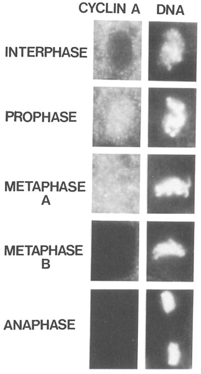
Cyclin proteins are thought to trigger entry into mitosis. During mitosis they are rapidly degraded. Therefore, mitosis and consequently cyclin degradation might be triggered at a time when cyclins have reaccumulated to a critical level. We cloned and sequenced a Drosophila cyclin A homolog and identified mutations in the corresponding gene. Immunofluorescent staining revealed that cyclin A accumulates in the interphase cytoplasm of cellularized embryos, but relocates to the nuclear region early in prophase and is completely degraded within metaphase. Cyclin A was expressed in dividing cells throughout development, and a functional cyclin A gene was required for continued division after exhaustion of maternally contributed cyclin A. Importantly, the timing of post cellularization divisions was not governed by the rate of accumulation or level of cyclin A.


Figures

Figure 1
Figure 1. Nucleotide Sequence and Predicted Amino Acid Sequence of the Drosophila Cyclin A cDNA
The nucleotide sequence of a Drosophila cyclin A cDNA (2385 bp) has a long open reading frame encoding a protein of 491 amino acids. The predicted amino acid sequence is shown in three letter code above the nucleotide sequence. The putative initiating ATG is underlined along with flanking bases matching the consensus sequences for translational start sites in Drosophila (Cavener, 1987). Five additional upstream ATG codons and a putative polyadenylation signal are also indicated by underlining.
Figure 2
Figure 2. Amino Acid Sequence Comparison of the Drosophila Cyclin A with Other Known Cyclin Proteins
The amino acid sequences of the domain conserved in Drosophila cyclin A (D.A), clam cyclin A (C.A), clam cyclin B (C.B), a sea urchin cyclin (SU), and the S. pombe cdc13+ gene product (SP) are aligned. Residues identical in all the sequences are marked with asterisks (*). Bold letters indicate residues that are conserved in at least three sequences. The arrows delineate an especially highly conserved region (see text). Gaps, introduced for optimal alignment, are represented by dashes. Regions are boxed where the two A type sequences (D.A and C.A) and at least two of the B type sequences (C.B, SU, and SP) are similar (identical or conserved replacements using the following grouping: A,L,V,I,M; K,R; D,E; S,T; N,Q; Y,F). Black dots mark regions where distinctive differences distinguish A and B type cyclins.
Figure 3
Figure 3. Identification of Mutations in the Cyclin A Gene
(A) Cytogenetic map of the region on chromosome arm 3L containing the cyclin A gene. The regions deleted in the chromosomal deficiencies vin2 and vin3 (Akam et al., 1978) are indicated by black bars. In situ hybridization localized the cyclin A sequence within the two proximal breakpoints of vin2 and vin3 (arrow). Five lethal complementation groups (rsg11–rsg15) have been localized between these breakpoints (Hoogwerf et al., 1988). The EMS-induced allele I(3)183 of the complementation group rsg11 complements neither the EMS-induced allele I(3)v4-4 nor the allele neo114 isolated after P element insertion mutagenesis (Cooley et al., 1988) (B) Mapping the P element insertion neo114. Southern analysis with a cyclin A cDNA probe was used to compare genomic restriction fragments from an rsg11 allele (neo114), a deletion of the region (vin3), and a control chromosome. The chromosome carrying the allele neo116 was chosen as control because it is derived from the same parental chromosome as neo114 and it also has a P element insert but in a different location. Since the chromosomes being compared carry recessive lethals, DNA was prepared from heterozygous flies. Novel fragments absent from the control DNA (neo116/TM3, lanes 1, 3, and 7) but apparent in neo114/TM3 DNA are marked with arrowheads. Lanes 1 and 2: Sall; lanes 3 and 4: EcoRI; lanes 5–8: HindIII. Despite the appearance of novel fragments, no loss of bands is observed in Sall and EcoRI digests of neo114/TM3 DNA because of the contribution of the balancer chromosome. However, one band present in the control (lane 7) cannot be detected in HindIII digests of neo114/TM3 DNA (lane 8, arrow). This disappearance indicates the presence of a restriction site polymorphism. In digests of DNA from flies carrying a deficiency deleting the cyclin A gene (vin3), the hybridizing fragments are uniquely derived from the balancer chromosome, and a comparison of the banding pattern derived from two different balancer chromosomes, In(3L)P (lane 5) and TM3 (lane 6), reveals a HindIII restriction site polymorphism. This polymorphism allows the unambiguos assignment of the hybridizing fragments to the different chromosomes present in neo116/TM3 flies or neo114/TM3 flies, respectively. Position and size (kbp) of molecular weight markers are indicated on the left side.
Figure 4
Figure 4. Cyclin A mRNA Levels during Embryonic Cell Cycle Progression
Total RNA from an equal number of early embryos, engaged in the rapid cleavage divisions (lane 1), early cycle 14 embryos in interphase (lane 2), embryos engaged in embryonic cell divisions 15 and 16 (lane 3), and older embryos where cell divisions are restricted to the nervous system (lane 4) were probed on Northern blots with the cyclin A cDNA. The estimated size (kb) of the cyclin mRNAs are indicated on the left side. Embryos were collected and aged at 25°C for the times indicated and staged under the microscope before isolation of RNA. In order to visualize the signals from cyclin A mRNAs in older embryos, a longer exposure is shown in the lower panel.
Figure 5
Figure 5. Cyclin A Abundance during Embryonic Development
Total protein extracts from embryos collected and aged at 25°C for the times indicated were resolved on SDS gels, transferred to nitrocellulose, and probed with affinity purified antibodies against cyclin A followed by 125I iodinated protein A. The position and size (kd) of molecular weight markers are indicated on the right side.
Figure 6
Figure 6. Intracellular Distribution of Cyclin A during Cell Division
Embryos were double labeled with affinity purified antibodies against cyclin A followed by rhodamine conjugated secondary antibodies (left column) and with the DNA stain Hoechst 33258 (right column). Cells in interphase, prophase, metaphase, and anaphase are shown. Cyclin A is degraded within metaphase and therefore metaphases with cyclin A staining (Metaphase A) as well as without (Metaphase B) can be detected.
Figure 7
Figure 7. Accumulation and Degradation of Cyclin A during Embryonic Cell Cycle Progression
Embryos were stained with affinity purified antibodies against cyclin A and rhodamine conjugated secondary antibodies. The following developmental stages are shown (the age of the embryos is given as the time after onset of mitosis 14, which is about 200 min after egg deposition): (a) immediately before onset of mitosis 14. (b) 5 min after onset of mitosis 14. The cells in the unstained domains in the head have already completed mitosis. Arrows point to a domain where mitosis is initiated but not yet completed. (c) 20 min divisions in the lateral regions posterior to the cephalic furrow have started. (d) 30 min most cells have completed mitosis 14 except cells in the amnioserosa (A), the neurogenic region (N), and a few cells in the head region. (e) 70 min onset of mitosis 15. Cyclin A staining is again present in cells unstained in the 330 min embryo (d), and a few cells (arrow) in the thoracic region have already completed mitosis 15 and are unstained. (f) 7 hr cell divisions are restricted to the peripheral and central nervous system. Assignment of the developmental ages was according to Foe (1989). Embryos are oriented anterior to the left, posterior to the right, dorsal up and ventral down, except the embryo in (b), which shows a dorsal view. The bar in (f) corresponds to 50 μm.
Figure 8
Figure 8. Cyclin A Distribution and Nuclear Density in Mutant Embryos
Embryos were stained with affinity purified antibodies against cyclin A followed by rhodamine conjugated goat anti-rabbit antibodies (a–d) and with a monoclonal antibody against a nuclear envelope component followed by fluorescein conjugated goat anti-mouse antibodies (e and f). (a, c, and e) Normal embryos, (b, d, and f) Mutant embryos. Embryos are at the time of mitosis 14 (a and b), mitosis 15 (c and d), or after mitosis 16 (9 hr, e and f). The level of cyclin A staining in the progeny of the cross neo114/TM3 × vin3/TM3 is wild type (a and c) in 25%, intermediate (data not shown) in 50%, and clearly lower (b and d) in 25% of the embryos. Double labeling with anti–cyclin A antibodies (data not shown) and antinuclear envelope antibodies (e and f) revealed that embryos, which have no detectable cyclin A (f), have less than half the number of nuclei than in embryos where cyclin A was readily detected (e). The bar in (d) corresponds to 50 μm. Only the dorsolateral epidermis of three embryonic segments is shown in (e) and (f). The bar in (f) corresponds to 5 μm.

Similar articles

Cited by

References

    1. Akam ME, Roberts DB, Richards GP, Ashburner M. Drosophila: the genetics of two major larval proteins. Cell. 1978;13:215–225. - PubMed
    1. Blumenthal AB, Kriegstein HJ, Hogness DS. The units of DNA replication in Drosophila melanogaster chromosomes. Cold Spring Harbor Symp Quant Biol. 1973;38:205–233. - PubMed
    1. Booher R, Beach D. Interaction between cdc13+ and cdc2+ in the control of mitosis in fission yeast; dissociation of the G1 and G2 roles of the cdc2+ protein kinase. EMBO J. 1987;6:3441–3447. - PMC - PubMed
    1. Booher R, Beach D. Involvement of cdc13+ in mitotic control in Schizosaccharomyces pombe: possible interaction of the gene product with microtubules. EMBO J. 1988;7:2321–2327. - PMC - PubMed
    1. Carroll SB, Laughon A. Production and purification of polyclonal antibodies to the foreign segment of (β-galactosidase fusion proteins. In: Glover D, editor. DNA Cloning: A Practical Approach. Oxford: IRL Press; 1987. pp. 89–111.

Publication types

MeSH terms

Associated data