. 2015 Mar 13;290(11):7291-303.
doi: 10.1074/jbc.M114.616730. Epub 2015 Jan 30.

A novel mechanism of regulating the ATPase VPS4 by its cofactor LIP5 and the endosomal sorting complex required for transport (ESCRT)-III protein CHMP5

Affiliations

A novel mechanism of regulating the ATPase VPS4 by its cofactor LIP5 and the endosomal sorting complex required for transport (ESCRT)-III protein CHMP5

Cody J Vild et al. J Biol Chem. .

Abstract

Disassembly of the endosomal sorting complex required for transport (ESCRT) machinery from biological membranes is a critical final step in cellular processes that require the ESCRT function. This reaction is catalyzed by VPS4, an AAA-ATPase whose activity is tightly regulated by a host of proteins, including LIP5 and the ESCRT-III proteins. Here, we present structural and functional analyses of molecular interactions between human VPS4, LIP5, and the ESCRT-III proteins. The N-terminal domain of LIP5 (LIP5NTD) is required for LIP5-mediated stimulation of VPS4, and the ESCRT-III protein CHMP5 strongly inhibits the stimulation. Both of these observations are distinct from what was previously described for homologous yeast proteins. The crystal structure of LIP5NTD in complex with the MIT (microtubule-interacting and transport)-interacting motifs of CHMP5 and a second ESCRT-III protein, CHMP1B, was determined at 1 Å resolution. It reveals an ESCRT-III binding induced moderate conformational change in LIP5NTD, which results from insertion of a conserved CHMP5 tyrosine residue (Tyr(182)) at the core of LIP5NTD structure. Mutation of Tyr(182) partially relieves the inhibition displayed by CHMP5. Together, these results suggest a novel mechanism of VPS4 regulation in metazoans, where CHMP5 functions as a negative allosteric switch to control LIP5-mediated stimulation of VPS4.

Keywords: ATPase; CHMP5; Crystal Structure; Endosomal Sorting Complexes Required for Transport (ESCRT); LIP5; MIT-MIM; Membrane Trafficking; Multivesicular Body; Protein-Protein Interaction; VPS4.


Figures

FIGURE 1.
FIGURE 1.
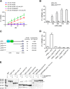
LIP5 stimulates the ATPase activity of VPS4. A, time course of ATP hydrolysis by VPS4B. Reactions with different combinations of VPS4B and LIP5 were allowed to proceed for 1 h. Production of inorganic phosphate was monitored at 10-min intervals. B, concentration-dependent stimulation of VPS4B by LIP5. The ATPase activity of VPS4B (125 nm) was measured in the presence of increasing concentrations of either LIP5 or LIP5253–307. Reactions were allowed to proceed for 1 h. C, schematic diagram showing the C-terminal fragments of LIP5 used in the assays and their corresponding Vta1 fragments. D, the C-terminal domain of LIP5 alone does not stimulate VPS4B. The ATPase activity of VPS4B (125 nm) was measured in the presence of various C-terminal fragments of LIP5 (375 nm). Reactions were allowed to proceed for 1 h. E, the C-terminal domain of LIP5 is responsible for binding VPS4B. Various C-terminal fragments of LIP5 were assayed for their binding affinity for VPS4B. The GST-tagged or GST-SUMO-tagged protein-immobilized glutathione beads were incubated with 1 ml of VPS4BE233Q (1 μm) for 1 h at 4 °C in the presence or absence of ATP. GST-SUMO was used as a tag for LIP5163–307 and LIP51–162/215–307, because the GST-tagged proteins run at a position similar to that of VPS4BE233Q. E233Q is an ATPase-deficient mutant of VPS4B. LIP51–162 lacks the VPS4B binding C-terminal domain and was intended as a negative control for binding. The arrow indicates the running position of VPS4BE233Q. Error bars, S.E.
FIGURE 2.
FIGURE 2.
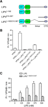
Both the N- and C-terminal domains of LIP5 are required for its VPS4 stimulatory activity. A, schematic diagram showing the LIP5 fragments used in the assays. B, linking the N- and C-terminal domains together (LIP51–162/215–307) provides partial VPS4B stimulation. The ATPase activity of VPS4B (125 nm) was measured in the presence of different LIP5 fragments (375 nm). Reactions were allowed to proceed for 1 h. Note that adding the N- and C-terminal domains in trans does not provide VPS4B stimulation. C, concentration-dependent stimulation of VPS4B by LIP51–162/215–307. The ATPase activity of VPS4B (125 nm) was measured in the presence of increasing concentrations of either LIP5 or LIP51–162/215–307. Reactions were allowed to proceed for 1 h. Error bars, S.E.
FIGURE 3.
FIGURE 3.
The ESCRT-III proteins regulate LIP5-mediated VPS4 stimulation. A, effect of CHMP5 or CHMP1B on LIP5-mediated VPS4B stimulation. The ATPase activity of VPS4B (125 nm) was measured in the presence of 375 nm LIP5 and increasing concentrations of CHMP5151–190, CHMP1B176–199, CHMP5151–190,L4D, or BSA. Reactions were allowed to proceed for 1 h. B, concentration-dependent stimulation of VPS4B79–444 by LIP5 or LIP51–162/215–307. The ATPase activity of VPS4B79–444 (375 nm) was measured in the presence of increasing concentrations of either LIP5 or LIP51–162/215–307. Reactions were allowed to proceed for 1 h (LIP5) or 15 min (LIP51–162/215–307). C, effect of CHMP5 or CHMP1B on LIP5-mediated VPS4B79–444 stimulation. The ATPase activity of VPS4B79–444 (375 nm) was measured in the presence of 375 nm LIP5 and increasing concentrations of CHMP5151–190, CHMP1B176–199, CHMP5151–190,L4D, or BSA. Reactions were allowed to proceed for 1 h. D, the inhibitory activity of CHMP5 does not depend on the linker region of LIP5. LIP5, LIP51–162/215–307, and LIP5165–307 (375 nm) were assayed for their abilities to stimulate VPS4B79–444 (375 nm) ATPase activity in the presence of increasing concentrations of CHMP5. Reactions were allowed to proceed for 1 h. Error bars, S.E.
FIGURE 4.
FIGURE 4.
The LIP5NTD-CHMP1B complex structure. A, an overview of the LIP5NTD-CHMP1B complex structure. Shown is a schematic representation of LIP5NTD in complex with CHMP1B176–199 in two orthogonal views. CHMP1B is colored in yellow. MIT1, MIT2, and the short linker between the two MIT domains of LIP5NTD are colored cyan, green, and white, respectively. Helices of LIP5NTD are labeled α1–α7. B, the CHMP1B-binding surface of LIP5NTD is hydrophobic. LIP5NTD is shown as a surface representation and colored based on the underlying atoms: hydrophobic side chain atoms (orange), polar and charged side chain atoms (blue), and main chain atoms (white). CHMP1B is shown as a yellow coil. C and D, detailed interactions at the LIP5NTD-CHMP1B interface. Shown is an enlarged schematic representation showing residues involved in hydrophobic (C) and polar (D) interactions between LIP5NTD and CHMP1B. Residues in contact are shown as stick models. Hydrogen bonds are denoted as dashed lines. Color schemes are the same as in A except for the following atoms: oxygen (red), nitrogen (blue), and sulfur (orange).
FIGURE 5.
FIGURE 5.
Critical residues at the LIP5NTD-CHMP1B interface. A, critical residues on LIP5NTD. GST-CHMP1B176–199 was used to analyze its interaction with LIP5 and various LIP5 mutants. B, critical residues on CHMP1B. GST-CHMP1B176–199, GST-CHMP1B176–199,L195D, or GST-CHMP1B176–199,R196A was used to analyze its interaction with LIP5. The GST-tagged protein-immobilized glutathione beads were incubated with 1 ml of LIP5 or LIP5 mutant (375 nm) for 1 h at 4 °C. The arrow indicates the running position of LIP5 or LIP5 mutants.
FIGURE 6.
FIGURE 6.
The LIP5NTD-CHMP1B-CHMP5 complex structure. A, overview of the LIP5NTD-CHMP1B-CHMP5 complex structure. Shown is a schematic representation of LIP5NTD in complex with CHMP1B176–199 and CHMP5151–190 in front (left) and back (right) views. Color schemes and labeling schemes are the same as in Fig. 4A except that CHMP5 is colored in magenta. B, CHMP5 makes extensive contacts with LIP5NTD. The interface is divided and shown in three separate panels based on three sequence segments of CHMP5: 151–159 (left), 160–176 (center) and 176–190 (right). CHMP5 is shown as a magenta coil. LIP5NTD is shown as a semitransparent surface along with a stick representation of side chains that contribute to van der Waals interactions. Surface and sticks are colored using the following scheme: carbon atoms of MIT1 (cyan), carbon atoms of MIT2 (green), oxygen (red), and nitrogen (blue). C, Trp147 of LIP5 flips upon binding CHMP5. An enlarged schematic representation shows the conformation of Trp147 before (pale green) and after (green) binding CHMP5 (magenta).
FIGURE 7.
FIGURE 7.
Conformational change at the LIP5 MIT1-MIT2 domain interface. A, superimposition of the LIP5NTD-CHMP1B complex structure with the LIP5NTD-CHMP1B-CHMP5 complex structure. The color scheme used is the same as in Figs. 3A and 5A (the binary complex has a pale color tone). The view in the right panel is nearly orthogonal to that of the left panel and highlights the movement of the MIT domains. Helices are shown as cylinders, and loops in the right panel are omitted for clarity. B, Glu26-Lys116 salt bridge is broken in the ternary complex structure. Side chains of Glu26 and Lys116 in each of the three crystal structures as indicated are shown in stick representations. Carbon atoms are colored using the same scheme as in A. C, Glu26 and Lys116 are engaged in new interactions in the ternary complex structure. The left panel shows an enlarged view of interactions involving Glu26 and Lys116 before and after CHMP5 binding. Residues involved are shown as sticks and labeled. New hydrogen bonds are denoted as dashed lines with distances indicated. The right panel omits the binary complex structure but shows 2FoFc electron density (2.0σ) associated with interacting residues in the ternary complex structure. D, insertion of Tyr182 at the MIT domain interface. Left, superimposition of the LIP5NTD-CHMP1B-CHMP5 structure with the LIP5NTD-CHMP5 structure (only one of the 10 NMR structure assemblies is shown) (52). Right, conformation of Tyr182 in the two structures. Tyr182 in the ternary complex structure adopts a “flipped in” conformation. Tyr182 in the binary complex structure can adopt either a “flipped in” or “flipped out” conformation.
FIGURE 8.
FIGURE 8.
Tyr182 of CHMP5 is important for its inhibitory activity. A, Y182A partially reverses the inhibitory activity of CHMP5 toward LIP5-mediated VPS4B stimulation. The ATPase activity of VPS4B79–444 (375 nm) was measured in the presence of 375 nm LIP5 and increasing concentrations of CHMP5, CHMP5Y182A, or BSA. Reactions were allowed to proceed for 1 h. B, CHMP5Y182A binds to LIP5. GST-SUMO, GST-SUMO-CHMP5151–190, or GST-SUMO-CHMP5151–190,Y182A was used to analyze its interaction with LIP5. The GST-SUMO-tagged protein-immobilized glutathione beads were incubated with 1 ml of LIP5 (375 nm) for 1 h at 4 °C. GST-SUMO was used as a tag because GST-CHMP5151–190 runs at a position similar to that of LIP5. Arrow, running position of LIP5. Error bars, S.E.
FIGURE 9.
FIGURE 9.
A model of VPS4 regulation by the LIP5-CHMP5 complex. The C-terminal domain of LIP5 makes an initial contact with VPS4. This interaction brings its N-terminal domain close to VPS4, which enables an additional interaction between the N-terminal domain and VPS4. The combined actions of the two domains lead to full activity of VPS4. Binding of CHMP5 to LIP5 weakens the interaction between the N-terminal domain and VPS4 and leads to an inhibition of LIP5-mediated VPS4 stimulation.

Similar articles

Cited by

References

    1. Hurley J. H. (2010) The ESCRT complexes. Crit. Rev. Biochem. Mol. Biol. 45, 463–487 - PMC - PubMed
    1. Henne W. M., Buchkovich N. J., Emr S. D. (2011) The ESCRT pathway. Dev. Cell 21, 77–91 - PubMed
    1. McCullough J., Colf L. A., Sundquist W. I. (2013) Membrane fission reactions of the mammalian ESCRT pathway. Annu. Rev. Biochem. 82, 663–692 - PMC - PubMed
    1. Hanson P. I., Cashikar A. (2012) Multivesicular body morphogenesis. Annu. Rev. Cell Dev. Biol. 28, 337–362 - PubMed
    1. Katzmann D. J., Babst M., Emr S. D. (2001) Ubiquitin-dependent sorting into the multivesicular body pathway requires the function of a conserved endosomal protein sorting complex, ESCRT-I. Cell 106, 145–155 - PubMed

Publication types

MeSH terms

Substances

LinkOut - more resources