. 2015 Feb 18;43(3):1498-512.
doi: 10.1093/nar/gku1387. Epub 2015 Jan 20.

ARGONAUTE2 cooperates with SWI/SNF complex to determine nucleosome occupancy at human Transcription Start Sites

Affiliations

ARGONAUTE2 cooperates with SWI/SNF complex to determine nucleosome occupancy at human Transcription Start Sites

Claudia Carissimi et al. Nucleic Acids Res. .

Abstract

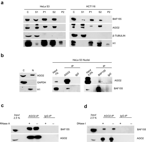
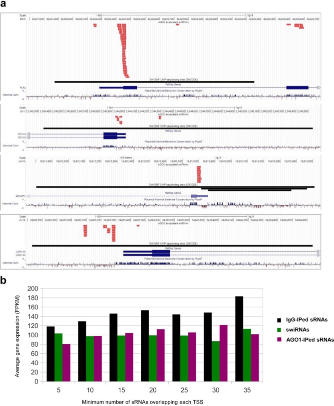
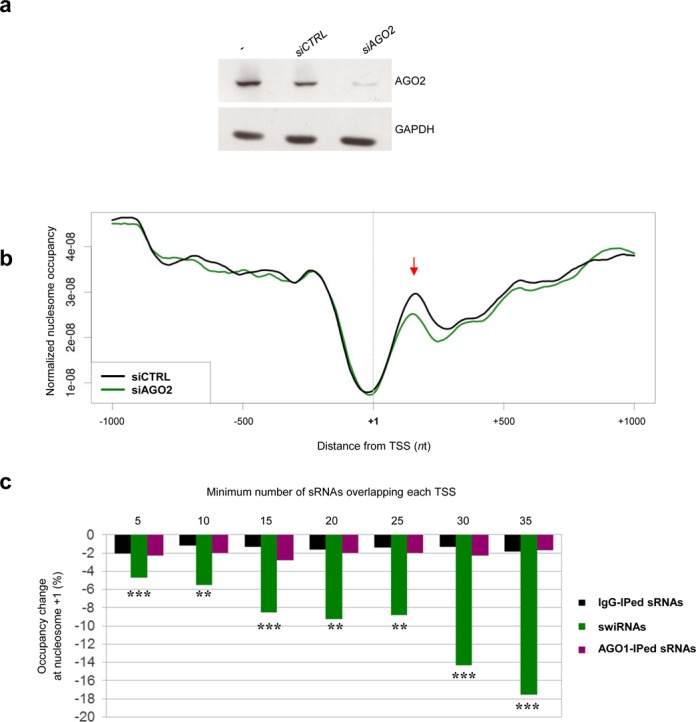
Argonaute (AGO) proteins have a well-established role in post-transcriptional regulation of gene expression as key component of the RNA silencing pathways. Recent evidence involves AGO proteins in mammalian nuclear processes such as transcription and splicing, though the mechanistic aspects of AGO nuclear functions remain largely elusive. Here, by SILAC-based interaction proteomics, we identify the chromatin-remodelling complex SWI/SNF as a novel AGO2 interactor in human cells. Moreover, we show that nuclear AGO2 is loaded with a novel class of Dicer-dependent short RNAs (sRNAs), that we called swiRNAs, which map nearby the Transcription Start Sites (TSSs) bound by SWI/SNF. The knock-down of AGO2 decreases nucleosome occupancy at the first nucleosome located downstream of TSSs in a swiRNA-dependent manner. Our findings indicate that in human cells AGO2 binds SWI/SNF and a novel class of sRNAs to establish nucleosome occupancy on target TSSs.


Figures

Figure 1.
Figure 1.
AGO2 interacts with components of SWI/SNF complex in human cell lines. (a) Schematic view of SILAC-based analysis of the AGO2 interactome, carried out in forward (for) and reverse (rev) setup (upper panel). The scatter plot based on protein SILAC-ratios (Heavy/Light) in forward and reverse experiments (x and y axes, respectively), shows that the outliers (true binders; blue) are in the top right quadrant, while background proteins (unspecific binders; green) cluster around protein ratio 1:1 (0 on a logarithmic scale). (b) Experimental scatterplot of H/L protein ratios obtained in the forward and reverse AGO2-IP SILAC experiment with HeLaS3 lysates. Components of SWI/SNF and TNRC6 complex are highlighted in dark and light blue respectively, while the bait is in red. (c) HeLaS3 and HCT116 cell extracts were immunoprecipitated (IP) with AGO2 antibody or isotype-matched immunoglobulins (IgG) as a negative control. (d) HeLaS3 and HCT116 cell extracts were immunoprecipitated with BAF155 antibody or IgG as a negative control. (e) AGO1 does not interact with SWI/SNF complex in human cell lines. HeLaS3 cell extract was immunoprecipitated with AGO1 or BAF155 antibody (with IgG as a negative control). Input and IPs were immunoblotted with the indicated antibodies.
Figure 2.
Figure 2.
AGO2 and SWI/SNF complex are associated with chromatin and interact in the nucleus in a RNA- and DNA-independent manner. (a) Whole cell extract (C), triton-soluble protein (S1), chromatin-bound, nuclear-matrix-bound and insoluble protein (P1), DNase-released chromatin associated protein (S2) and insoluble, cytoskeletal and nuclear matrix protein (P2) fractions corresponding to the same amount of HeLaS3 or HCT116 cells were analysed by western blot for the presence of indicated proteins. β-tubulin serves as a chromatin-unbound marker whereas histone H1 as chromatin-bound marker. (b) Cytosolic and nuclear fractions of HeLaS3 cells were prepared. Fractionation efficiency was verified by western blot for the presence of GAPDH (cytosolic marker) and H1 (nuclear marker) (Left panel). Nuclear lysates were immunoprecipitated with indicated antibodies or IgG as a negative control (right panel). (c) Total cell extract pre-treated or not with RNase A (left panel) or DNase I (right panel) were immunoprecipitated with AGO2 antibody or IgG as a negative control. Input and IPs were immunoblotted with the indicated antibodies.
Figure 3.
Figure 3.
Nuclear AGO2 is associated with a novel class of sRNAs arising from TSSs bound by SWI/SNF complex (swiRNAs). (a) Genomic annotation of sRNA-Seq data (hg19). The histograms illustrate the sRNA classes identified in AGO2- or IgG-IP samples. The main RNA classes in the AGO2-IP sample are represented by miRNAs followed by ‘other sRNAs’. (b) Size distribution of AGO2- and IgG-IP ‘other sRNAs’. The fraction of ‘other sRNAs’ in AGO2- or IgG-IP samples and corresponding length (nt) are plotted. (c and d) Percentage of ‘other sRNAs’ in AGO2- or IgG-IP samples mapping within SWI/SNF biding sites (ENCODE ChIP-seq data) (c) or within 1 kb of TSSs of expressed genes (TSSs were defined by mRNA-seq) (d). (e and f) Average per gene coverage of AGO2-associated ‘other sRNAs’ around TSSs with a SWI/SNF binding site (ENCODE ChIP-seq data) within ±1 kb (e) and around TSSs without SWI/SNF binding sites (f). Average per gene coverage is defined as the sum of sRNAs mapping to a given position (relative to TSS) divided by number of TSSs analysed. Only ‘other sRNAs’ lying within clusters of less than 50 molecules were considered. Red and blue represent sRNAs in the sense and anti-sense orientation with respect to gene transcription, respectively.
Figure 4.
Figure 4.
The number of AGO2-associated swiRNAs overlapping each TSS does not correlates with gene expression level. (a) Genome browser (USCS) view of representative clusters of AGO2-associated swiRNAs in HelaS3 cells. swiRNAs map to TSSs overlapped by SWI/SNF binding sites. Reads corresponding to AGO2-associated swiRNAs are red-coloured and orientation respect to reference genome is indicated for each read. SWI/SNF binding sites are black-coloured (BAF155, BAF170, BRG1, BAF47. Data from ENCODE). (b) Bar height represents average gene expression of loci with TSS (±150 nt) overlapped by at least the indicated number of AGO2-bound swiRNAs (green), IgG-IPed ‘other sRNAs’ (black) and AGO1-associated ‘other sRNAs’ (purple) in HeLaS3 cells.
Figure 5.
Figure 5.
swiRNAs are processed in a Dicer-dependent manner.Coverage of swiRNAs in parental HCT116 cell line (a) and in HCT116 DicerEx5 cell line (b.) Coverage at each position represents the number of swiRNAs mapping at the indicated distance from a TSS, normalized by the total number of ‘other sRNAs’. Only ‘other sRNAs’ lying within clusters of <50 molecules were considered. Red and blue represent sRNAs in the sense and anti-sense orientation with respect to gene transcription, respectively. (c) swiRNAs display a 2 nt 3′ overhang typical of Dicer-processed sRNAs. The length (nt) of 3′ overhang for pairs of complementary swiRNAs was plotted. Negative values represent a 5′ overhang. Most pairs display a 2 nt 3′ overhang, suggestive of Dicer processing.
Figure 6.
Figure 6.
AGO2 knock-down affects nucleosome occupancy at TSSs bound by SWI/SNF. (a) HeLaS3 cells were transfected with a control siRNA (siCTRL) or a pool of AGO2 siRNA (siAGO2). Down-regulation of AGO2 protein was verified by western blot. GAPDH was used as loading control. (b) Chromatin from siCTRL- or siAGO2-treated HeLaS3 cells was digested by MNase and recovered DNA fragments were sequenced. Nucleosome occupancy profile for siCTRL and siAGO2 cells was plotted for TSSs with at least 30 swiRNAs (siCTRL, black line; siAGO2, green line). The occupancy at the nucleosome +1 (arrow) is reduced in AGO2 knock-down cells. (c) Bars height represents percent reduction of nucleosome occupancy (siAGO2 versus siCTRL) at TSS ±150 nt overlapped by at least the indicated number of swiRNAs (green), IgG-IP ‘other sRNAs’ (black) and AGO1-associated ‘other sRNAs’ (purple). **P value < 0.01, ***P value < 10−3, paired Wilcoxon test (see 'Materials and Methods' section for details).

Similar articles

Cited by

References

    1. Napoli C., Lemieux C., Jorgensen R. Introduction of a chimeric chalcone synthase gene into petunia results in reversible co-suppression of homologous genes in trans. Plant Cell. 1990;2:279–289. - PMC - PubMed
    1. Romano N., Macino G. Quelling: transient inactivation of gene expression in Neurospora crassa by transformation with homologous sequences. Mol. Microbiol. 1992;6:3343–3353. - PubMed
    1. Tabara H., Sarkissian M., Kelly W.G., Fleenor J., Grishok A., Timmons L., Fire A., Mello C.C. The rde-1 gene, RNA interference, and transposon silencing in C. elegans. Cell. 1999;99:123–132. - PubMed
    1. Fagard M., Boutet S., Morel J.B., Bellini C., Vaucheret H. AGO1, QDE-2, and RDE-1 are related proteins required for post-transcriptional gene silencing in plants, quelling in fungi, and RNA interference in animals. Proc. Natl. Acad. Sci. U.S.A. 2000;97:11650–11654. - PMC - PubMed
    1. Meister G. Argonaute proteins: functional insights and emerging roles. Nat. Rev. Genet. 2013;14:447–459. - PubMed

Publication types

LinkOut - more resources