. 2015 May;65(1):1-16.
doi: 10.1016/j.molimm.2014.12.010. Epub 2015 Jan 16.

Desialylation of airway epithelial cells during influenza virus infection enhances pneumococcal adhesion via galectin binding

Affiliations

Desialylation of airway epithelial cells during influenza virus infection enhances pneumococcal adhesion via galectin binding

Mihai Nita-Lazar et al. Mol Immunol. 2015 May.

Abstract

The continued threat of worldwide influenza pandemics, together with the yearly emergence of antigenically drifted influenza A virus (IAV) strains, underscore the urgent need to elucidate not only the mechanisms of influenza virulence, but also those mechanisms that predispose influenza patients to increased susceptibility to subsequent infection with Streptococcus pneumoniae. Glycans displayed on the surface of epithelia that are exposed to the external environment play important roles in microbial recognition, adhesion, and invasion. It is well established that the IAV hemagglutinin and pneumococcal adhesins enable their attachment to the host epithelia. Reciprocally, the recognition of microbial glycans by host carbohydrate-binding proteins (lectins) can initiate innate immune responses, but their relevance in influenza or pneumococcal infections is poorly understood. Galectins are evolutionarily conserved lectins characterized by affinity for β-galactosides and a unique sequence motif, with critical regulatory roles in development and immune homeostasis. In this study, we examined the possibility that galectins expressed in the airway epithelial cells might play a significant role in viral or pneumococcal adhesion to airway epithelial cells. Our results in a mouse model for influenza and pneumococcal infection revealed that the murine lung expresses a diverse galectin repertoire, from which selected galectins, including galectin 1 (Gal1) and galectin 3 (Gal3), are released to the bronchoalveolar space. Further, the results showed that influenza and subsequent S. pneumoniae infections significantly alter the glycosylation patterns of the airway epithelial surface and modulate galectin expression. In vitro studies on the human airway epithelial cell line A549 were consistent with the observations made in the mouse model, and further revealed that both Gal1 and Gal3 bind strongly to IAV and S. pneumoniae, and that exposure of the cells to viral neuraminidase or influenza infection increased galectin-mediated S. pneumoniae adhesion to the cell surface. Our results suggest that upon influenza infection, pneumococcal adhesion to the airway epithelial surface is enhanced by an interplay among the host galectins and viral and pneumococcal neuraminidases. The observed enhancement of pneumococcal adhesion may be a contributing factor to the observed hypersusceptibility to pneumonia of influenza patients.

Keywords: Airway A549 cells; Galectin; Influenza; Neuraminidase; Pneumococcus pneumoniae.


Figures

Fig. 1
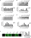
Fig. 1. Galectin expression and release, correlated with changes in glycosylation in the mouse lung during the progression of influenza (PR8) and pneumococcal (Sp3) infections
The mice were infected with influenza strain PR8 followed by a secondary bacterial infection with Streptococcus pneumoniae (Sp3) after 14 days. Each group of mice, ranging from two to four animals, was euthanized according to the following schedule: 3 days post-PR8 (PR8 3d), 7 days post-PR8 (PR8 7d), 10 days post PR8 (PR8 10d), 14 days post PR8 without Sp3 challenge (PR8 14d), 14 days post PR8 and 1 hour post Sp3 challenge (PR8 14d + Sp3 1h), 14 days post PR8 and 18 hours post Sp3 (PR8 14d + Sp3 18h), ctrl: non-challenged mice. Transcript levels of galectin-1 (Gal1) and galectin-3 (Gal3) were assessed by RT-PCR in lung tissues (A) or bronchoalveolar lavage (BAL) cells (B). Protein levels of selected galectins (Gal1 and Gal3) were assessed by Western blot in lungs (C) or BAL fluid (D) collected from control (non-challenged) or challenged groups of mice. (E) Sialylation status of lung tissues was assessed by immunofluorescence with FITC-PNA, which revealed the exposed galactosyl moieties. Bar graphs represent fold change in galectin transcript or protein levels from challenged mice in comparison with unchallenged mice, normalized to β-actin Images and the bar graphs are representative data from at least two independent experiments. *p <0.05; **p<0.001, non-paired Student’s t test.
Fig. 2
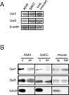
Fig. 2. Expression of galectins in epithelial cell cultures and mouse lung tissue
Transcript (A) or protein (B) levels of selected galectins (Gal1 and Gal3) were assessed with RT-PCR or Western blot, respectively, in lung carcinoma epithelial cells (A549), small airway epithelial cells (SAEC), or mouse lung tissues (lg). RT-PCR was performed with the total RNA extracted from A549, SAEC, or mouse lungs. Western blot was performed from cell lysate (c), culture supernatant (sn), lung tissue lysates (lg), or bronchoalveolar lavage (bal).
Fig. 3
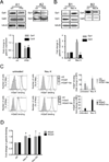
Fig. 3. In vitro effect of PR8 and neuraminidase exposure of A549 cells on expression and binding of galectins to A549 cells
Exposure of A549 cells to PR8 (A) or neuraminidase (B) modulates galectin expression (A1 and B1) and secretion (A2 and B2). A549 cells were infected with PR8 (MOI 5) for 72h (A), or treated with a bacterial neuraminidase cocktail (Arthrobacter ureafaciens and Clostridium perfringens; Neu K) (B). Galectin (Gal1 and Gal3) expression or secretion was assessed from cell lysates (A1 and B1) or culture supernatant (A2 and B2) with Western blot. Bar graphs show fold changes of galectin expression from PR8-infected or neuraminidase-treated cells (Neu K) in comparison with untreated cells (ctrl) normalized to tubulin. (C) Neuraminidase-treated (Neu K) or untreated (ctrl) cells were incubated with 15 µg/ml of biotinylated rhGal1, rhGal3, or galectins in the presence of lactose (0.1M) (Gal1+lac, Gal3+lac), followed by streptavidin-APC for flow cytometry analysis. The exogenous galectin binding to A549 cells was normalized to A549 cells without exogenous galectin (ctrl). (D) Effects of influenza viral neuraminidase (Neu N2) and bacterial neuraminidases (Arthrobacter ureafaciens and Clostridium perfringens; Neu K) on galectin binding to A549 cells. A549 cells grown in ELISA plates were subject to neuraminidase treatment (Neu K or Neu N2), followed by incubation with 15 µg/ml of exogenous rhGal1 or rhGal3. Galectin binding to the cells was assessed using primary antibodies against Gal1 or Gal3 followed by an HRP-conjugated secondary antibody. The galectin binding levels to neuraminidase-treated cells were normalized to the binding to the untreated cells (ctrl). Data shown and the bar graphs are representative data from at least three independent experiments. *p <0.05; **p<0.001, non-paired Student’s t test.
Fig. 4
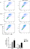
Fig. 4. Effect of neuraminidase treatment on the pro-apoptotic activity of galectin-1 and galectin-3
A549 cells treated with bacterial neuraminidases (Arthrobacter ureafaciens and Clostridium perfringens; Neu K) and untreated controls were incubated with 15 µg/ml of exogenous rhGal1 or rhGal3 and analyzed by TUNEL assay in flow cytometry for apoptosis. The apoptosis percentage in A549 cells in the presence of rhGal1 or rhGal3 was normalized to A549 cells without exogenous galectin (ctrl). Data shown and the bar graphs are representative data from at least three independent experiments. *p <0.05; **p<0.001, non-paired Student’s t test.
Fig. 5
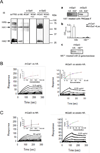
Fig. 5. Binding of galectins to influenza PR8 virion components and effect of neuraminidase exposure
(A) Binding of rhGal1 and rhGal3 to influenza hemagglutinin (HA0, precursor HA containing a hydrophobic signal sequence; HA1 and HA2, subunits of HA). Lysates from PR8 virus were prepared and subjected to Western blot. The total viral proteins were revealed by an α-PR8 antibody, and the viral HA by an α-HA antibody. Galectin binding was performed by overlaying membrane with rhGal1, rhGal3, and the specificity of the binding assessed by preincubation of the galectins with lactose (0.2 M) (rhGal1+lac, rhGal3+lac), followed by anti-Gal1 or anti-Gal3 antibodies, followed by HRP-conjugated secondary antibodies. To confirm the carbohydrate specificity of the galectin-HA interactions, HA was treated with PNGase F (HA* in a) or β-galactosidase (HA** in b or c) and subjected to lectin blot (a and c) as described above, or ELISA (b). (B) SPR sensogram of binding rhGal1 to HA. SPR was measured with immobilized rhGal1 and using HA (left panel) or desialylated HA (right panel) as analytes. HA starts at 12.5µg/ml, with 2-fold serial dilution; asialo-HA starts at 6.25 µg/ml. (C) SPR sensogram of binding of rhGal3 to HA. SPR was measured with immobilized rhGal3 and using HA (left panel) or desialylated HA (right panel) as analytes. HA starts at 50 µg/ml, with 2-fold serial dilution; asialo-HA starts at 6.25 µg/ml.
Fig. 6
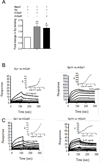
Fig. 6. Galectin-mediated adhesion of S. pneumoniae to airway epithelial cells and binding to capsular polysaccharides
A549 cells were grown in ELISA plates and treated with bacterial neuraminidase (Arthrobacter ureafaciens and Clostridium perfringens; Neu K). The cells were exposed for 1h to Sp3 (MOI 10) alone or Sp3 previously incubated with 15 µg/ml of rhGal1 or rhGal3. Bacterial binding to the cells was assessed using a primary anti-Streptococcus pneumoniae antibody, followed by an HRP-conjugated secondary antibody. The bacterial adhesion to cells in the presence of galectins was normalized to the bacterial adhesion in the absence of exogenous galectin. Data shown and the bar graphs are representative data from at least three independent experiments. *p <0.05; **p<0.001, non-paired Student’s t test. (B) SPR sensogram of binding rhGal1 and rhGal3 to S. pneumoniae capsular polysaccharides type I and type XIV: rhGal1 and type I polysaccharide (Sp1) at starting concentration of 12.5 mg/ml, with 2-fold serial dilutions (left panel); rhGal1 and type XIV polysaccharide (Sp14) at starting concentration of 6.25 µg/ml with 2-fold serial dilutions (right panel). (C) rhGal3 and type I polysaccharide (Sp1) at starting concentration of 12.5 mg/ml with 2-fold serial dilutions (left panel); rhGal3 and type XIV polysaccharide (Sp14) at starting concentration of 6.25 µg/ml, with 2-fold serial dilutions (right panel).
Fig. 7
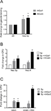
Fig. 7. Effect of S. pneumoniae neuraminidase and PR8 infection on galectin-mediated adhesion of S pneumoniae to A549 cells
(A) A549 cells grown in ELISA plates were treated with S. pneumoniae neuraminidase (Neu Sp) or untreated (ctrl) followed by incubation with 15 µg/ml of exogenous rhGal1 or rhGal3. Galectin binding to the cells was assessed using primary antibodies against Gal1 or Gal3 followed by and HRP-conjugated secondary antibody. The galectin binding levels to the neuraminidase-treated cells were normalized to the untreated cells (ctrl). (B) A549 cells grown on the 10 cm plates were treated with bacterial neuraminidases (Arthrobacter ureafaciens and Clostridium perfringens; Neu K) or S. pneumoniae neuraminidase (Neu Sp), and subsequently incubated with S. pneumoniae (MOI 10) in the absence (Sp) or presence of 15 µg/ml of exogenous rhGal1 (Sp + rhGal1) or rhGal3 (Sp + rhGal3). The bound bacteria were released in water then quantified after 24h incubation on 5% sheep’s blood agar plates by counting colony forming units (CFU). The bar graphs represent the fold change of CFU from galectin-mediated Sp3 adhesion compared to the Sp3 adhesion in the absence of exogenous galectin. (C) A549 cells grown on the 10 cm plates were exposed to PR8 (MOI 5) for 72 h, then incubated with Sp3 (MOI 10) without (Sp) or with 15 ug/ml of exogenous rhGal1 (Sp + rhGal1) or rhGal3 (Sp + rhGal3) for bacterial adhesion. The colony forming units (CFU) released from bound bacteria were counted as described above. The bar graphs represent the fold change of CFU from galectin-mediated Sp adhesion compared to the Sp adhesion in the absence of exogenous galectin. Data shown and the bar graphs are representative data from at least three independent experiments. *p <0.05; **p<0.001.

Similar articles

Cited by

References

    1. Ahmad N, Gabius HJ, et al. Galectin-3 precipitates as a pentamer with synthetic multivalent carbohydrates and forms heterogeneous cross-linked complexes. J Biol Chem. 2004;279(12):10841–10847. - PubMed
    1. Air GM. Influenza virus-glycan interactions. Curr Opin Virol. 2014;7C:128–133. - PMC - PubMed
    1. Barker WH, Mullooly JP. Impact of epidemic type A influenza in a defined adult population. Am J Epidemiol. 1980;112(6):798–811. - PubMed
    1. Barker WH, Mullooly JP. Pneumonia and influenza deaths during epidemics: implications for prevention. Arch Intern Med. 1982;142(1):85–89. - PubMed
    1. Barrionuevo P, Beigier-Bompadre M, et al. A novel function for galectin-1 at the crossroad of innate and adaptive immunity: galectin-1 regulates monocyte/macrophage physiology through a nonapoptotic ERK-dependent pathway. J Immunol. 2007;178(1):436–445. - PubMed

Publication types

MeSH terms