. 2015 Jan 15;11(1):e1004834.
doi: 10.1371/journal.pgen.1004834. eCollection 2015 Jan.

Inactivation of PNKP by mutant ATXN3 triggers apoptosis by activating the DNA damage-response pathway in SCA3

Affiliations

Inactivation of PNKP by mutant ATXN3 triggers apoptosis by activating the DNA damage-response pathway in SCA3

Rui Gao et al. PLoS Genet. .

Abstract

Spinocerebellar ataxia type 3 (SCA3), also known as Machado-Joseph disease (MJD), is an untreatable autosomal dominant neurodegenerative disease, and the most common such inherited ataxia worldwide. The mutation in SCA3 is the expansion of a polymorphic CAG tri-nucleotide repeat sequence in the C-terminal coding region of the ATXN3 gene at chromosomal locus 14q32.1. The mutant ATXN3 protein encoding expanded glutamine (polyQ) sequences interacts with multiple proteins in vivo, and is deposited as aggregates in the SCA3 brain. A large body of literature suggests that the loss of function of the native ATNX3-interacting proteins that are deposited in the polyQ aggregates contributes to cellular toxicity, systemic neurodegeneration and the pathogenic mechanism in SCA3. Nonetheless, a significant understanding of the disease etiology of SCA3, the molecular mechanism by which the polyQ expansions in the mutant ATXN3 induce neurodegeneration in SCA3 has remained elusive. In the present study, we show that the essential DNA strand break repair enzyme PNKP (polynucleotide kinase 3'-phosphatase) interacts with, and is inactivated by, the mutant ATXN3, resulting in inefficient DNA repair, persistent accumulation of DNA damage/strand breaks, and subsequent chronic activation of the DNA damage-response ataxia telangiectasia-mutated (ATM) signaling pathway in SCA3. We report that persistent accumulation of DNA damage/strand breaks and chronic activation of the serine/threonine kinase ATM and the downstream p53 and protein kinase C-δ pro-apoptotic pathways trigger neuronal dysfunction and eventually neuronal death in SCA3. Either PNKP overexpression or pharmacological inhibition of ATM dramatically blocked mutant ATXN3-mediated cell death. Discovery of the mechanism by which mutant ATXN3 induces DNA damage and amplifies the pro-death signaling pathways provides a molecular basis for neurodegeneration due to PNKP inactivation in SCA3, and for the first time offers a possible approach to treatment.


Conflict of interest statement

The authors have declared that no competing interests exist.

Figures
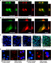
Figure 1
Figure 1. PNKP interacts with both wild-type and mutant ATXN3 in cells and human brain sections.
(A) Plasmids pGFP-ATXN3-Q28 and pCherry-PNKP were co-transfected into SH-SY5Y cells and co-localization of PNKP and ATXN3-Q28 was assessed by confocal microscopy; the merge of green and red fluorescence from ATXN2-Q28 and PNKP, respectively, appears as yellow/orange fluorescence. Nuclei were stained with DAPI in C-D. (B) Plasmids pGFP-ATXN3-Q84 and pCherry-PNKP were co-transfected into SH-SY5Y cells and co-localization of PNKP and ATXN3-Q84 was assessed as in A. (C) Bimolecular fluorescence complementation assay. SH-SY5Y cells were transfected with plasmid: Panel 1) pVN173-PNKP; Panel 2) pVC155-ATXN3-Q28; Panel 3) pVC155-ATXN3-Q84; Panel 4) co-transfected with pVN173-PNKP and pVC155-ATXN3-Q28; or Panel 5) pVN173-PNKP and pVC155-ATXN3-Q84. Reconstitution of green/yellow fluorescence was assessed 48 hours after transfection using fluorescence microscopy (20X). Nuclei were stained with DAPI in Figs. 1C and 1D. (D) Proximity ligation assays (PLAs) were performed on control and SCA3 human brain sections, using mouse anti-PNKP and rabbit anti-ATXN3 primary antibodies, and anti-DNA ligase 3 (DNA LIG3) (rabbit) with anti-ATXN3 (mouse) antibodies as control. The generation of red fluorescence was monitored under a fluorescence microscope.
Figure 2
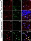
Figure 2. PNKP co-localizes with wild-type and mutant ATXN3 in human brain sections.
(A) Normal human brain sections were analyzed by immunostaining with anti-PNKP (red) and anti-ATXN3 (green) antibodies to assess in vivo interaction of PNKP and ATXN3; merge of red and green fluorescence appears as yellow/orange fluorescence, Nuclei were stained with DAPI in Figs. 1A to 1D; (B) SCA3 (expressing mutant ATXN3-Q72) brain sections were analyzed by immunostaining with anti-PNKP (red), and anti-ATXN3 (green) antibodies to assess in vivo interaction of PNKP and ATXN3; merge of red and green fluorescence appears as yellow/orange fluorescence; (C) Normal human brain sections were analyzed by immunostaining with anti-PNKP (red) and anti-polyQ antibody (green) antibodies to assess the presence of PNKP in the polyQ aggregates; (D) SCA3 (expressing ATXN3-Q72) brain sections were analyzed by immunostaining with anti-PNKP (red), and anti-polyQ (green) antibodies to determine the presence of PNKP in the polyQ aggregates; merge of red and green fluorescence appears as yellow/orange fluorescence.
Figure 3
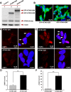
Figure 3. Expression of mutant ATXN3 in cells induces DNA strand breaks.
(A) Expression of GFP-ATXN3-Q84 and GFP-ATXN3-Q28 was induced in SH-SY5Y cells; 48 hours after induction, the cells were harvested and their lysates analyzed by Western blotting with anti-ATXN3 monoclonal antibody to detect endogenous ATXN3 and exogenous GFP-ATXN3-Q28 and GFP-ATXN3-Q84 levels (shown by arrows). Lane 1, control SH-SY5Y cells; lane 2, SH-SY5Y cells expressing GFP-ATXN3-Q28; lane 3, SH-SY5Y cells expressing ATXN3-Q84; β-actin was used as loading control. (B) Confocal images showing expression of GFP-tagged ATXN3-Q28 and ATXN3-Q84 in SH-SY5Y cells. Nuclei were stained with DAPI in B, C and E. (C) Expression of either GFP-ATXN3-Q28 or GFP-ATXN3-Q84 was induced and the cells analyzed by immunostaining with anti-p-53BP1-S1778 antibody (red) to assess DNA strand breaks; 53BP1 foci are shown by arrows. (D) Relative number of 53BP1 foci in the SH-SY5Y cells expressing ATXN3-Q28 or ATXN3-Q84 (n = 100, data represent mean ± SD, *** = p < 0.001). (E) SH-SY5Y cells expressing either ATXN3-Q84 or ATXN3-Q28 analyzed by immunostaining with anti-γH2AX-S139 antibody (red); γH2AX foci are shown by arrows. (F) Relative number of γH2AX foci in SH-SY5Y cells expressing ATXN3-Q28 or ATXN3-Q84 (n = 100, data represents mean ± SD, *** = p < 0.001).
Figure 4
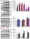
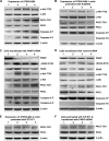
Figure 4. Mutant ATXN3 activates DNA damage-response in vitro and in vivo.
(A) Expression of ATXN3-Q84 was induced in differentiated SH-SY5Y cells; cells were harvested 0, 3, 6 and 12 days post-induction (lanes 1 to 4) and their lysates analyzed by Western blotting to determine the levels of ATM-S1981, total ATM, γH2AX-S139, total H2AX, Chk2-T68, total Chk2, p53-S15, p53-S20, p53-S46 and total p53; β-actin was used as the loading control in A, C and E. (B) Levels of ATM-S1981, γH2AX-S139, Chk2-T68, p53-S15, p53-S20 and p53-S46 relative to their respective total protein levels in cells expressing ATXN3-Q84. Cells were harvested 0 (Grey), 3 (black), 6 (blue) and 12 (red) days post ATXN3-Q84 expression in Figs. B and D (n = 4, data represent mean ± SD, *** = p < 0.001 for B and F) (C) Expression of ATXN3-Q28 was induced in differentiated SH-SY5Y cells; cells were harvested 0, 3, 6 and 12 days post-induction (lanes 1 to 4) and their lysates analyzed by Western blotting to determine ATM-S1981, total ATM, γH2AX-S139, total H2AX, p53-S15 and total p53 levels. (D) Cells expressing ATXN3-Q28 were analyzed as in B and relative levels of ATM-S1981, γH2AX and p53-S15 vs. the respective total protein levels are shown; NS denotes non-significant. (E) Total protein was isolated from the deep cerebellar nuclei (DCN) of SCA3 transgenic mice (24 weeks old) constitutively expressing human mutant ATXN3 (lanes 3 and 4) and age-matched control mice (lanes 1 and 2) and analyzed by Western blotting to determine ATM-S1981, total ATM, γH2AX-S139, total H2AX, p53-S15 and total p53 levels. Each lane represents total protein from a pool of DCN tissue from 4–5 wild-type or an equal number of transgenic littermates. (F) Relative levels of ATM-S1981, γH2AX, p53-S15 with respect to total protein in SCA3 transgenic mouse DCN (black bars) vs. age-matched control DCN (grey bars); each bar represents a pool of DCN tissue collected from 4 to 5 littermate mice (either wild-type or transgenic). Data represent mean ± SD; *** = p < 0.001.
Figure 5
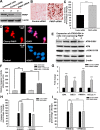
Figure 5. The mutant ATXN3-induced apoptotic pathway is rescued by PNKP-overexpression or inhibition of ATM and p53.
(A) Total protein was isolated from control untreated SH-SY5Y cells (lane 1), SH-SY5Y cells transfected with control-siRNA (lane 2) and SH-SY5Y cells transfected with PNKP-siRNA (lane 3), the cells harvested 48 hours after transfection, and their lysates analyzed by Western blotting to determine the PNKP levels; β-actin was used as the loading control. (B) Light microscopic images showing TUNEL staining of SH-SY5Y cells transfected with control-siRNA or PNKP-siRNA. TUNEL staining was performed 48 hours post-transfection. (C) Caspase-3 activities in SH-SY5Y cells treated with control-siRNA or PNKP-siRNA were measured 48 hours after transfecting the cells with the respective siRNA. Data represent means ± SD; (n = 3, *** = p < 0.001) in C, and F-I. (D) Fluorescence microscopic images showing SH-SY5Y cells overexpressing Cherry-tagged PNKP and control untreated SH-SY5Y cells. (E) Expression of ATXN3-Q84 was induced in SH-SY5Y cells overexpressing PNKP, the cells harvested 0, 3, 6 and 12 days post-induction (lanes 1 to 4), and their lysates analyzed by Western blotting to determine ATM-S1981, total ATM, γ-H2AX-S139 and total H2AX levels; β-actin was used as the loading control. (F) Caspase-3 activities in cells expressing ATXN3-Q28 and ATXN3-Q84, co-expressing exogenous PNKP and ATXN3-Q28, and co-expressing PNKP and ATXN3-Q84. (G) SH-SY5Y cells were transfected with PNKP- or control-siRNA; 48 hours post-transfection, total RNA was isolated, and the expression of BAX, BBC3, Bcl2L11 and PMAIP1 determined by qRT-PCR. (H) Caspase-3 activities in SH-SY5Y cells transfected with PNKP-siRNA, control-siRNA; and in SH-SY5Y cells pre-treated with the ATM inhibitor Ku55933 and transfected with control-siRNA and PNKP-siRNA. Caspase-3 activities are expressed as percentage change normalized to the control in H and I. (I) Caspase-3 activities in SH-SY5Y cells transfected with control-siRNA and PNKP-siRNA, and in SH-SY5Y cells pre-treated with the p53 inhibitor Pifithrin-α and transfected with control-siRNA and PNKP-siRNA.
Figure 6
Figure 6. ATXN3-Q84 expression or PNKP deficiency activates the c-Abl→PKCδ signaling pathway.
(A) SH-SY5Y cells were differentiated, the expression of ATXN3-Q84 was induced, and cells were harvested 0, 3, 6 and 12 days post-induction (lanes 1 to 4). Cell lysates were analyzed by Western blotting to determine the levels of c-Abl-T735, total c-Abl, PKCδ-T311, total PKCδ, full-length caspase-3 (-F), and cleaved caspase-3 (-C); β-actin was used as a loading control in A to F. (B) SH-SY5Y cells were differentiated, and treated with the ATM inhibitor Ku55933, expression of ATXN3-Q84 induced, and the cell lysates analyzed as in A. (C) SH-SY5Y cells were differentiated, transfected with 0, 50, 100 and 200 pmoles of PNKP-siRNA (lanes 1 to 4), harvested 48 hours post-transfection, and the cell lysates analyzed as in A. (D) SH-SY5Y cells were differentiated, transfected with 0, 50, 100 and 200 pmoles of control-siRNA (lanes 1 to 4), harvested 48 hours post-transfection, and the cell lysates analyzed as in C. (E) SH-SY5Y cells were differentiated, incubated with the c-Abl inhibitor STI-571, and expression of ATXN3-Q84 induced. The cells were harvested 0, 3, 6 and 12 days post-induction (lanes 1 to 4) and their lysates analyzed by Western blotting to determine PKCδ-T311 and total PKCδ levels. (F) SH-SY5Y cells were differentiated, and incubated with STI-571, and transfected with 0, 50, 100 and 200 pmoles (lanes 1 to 4) of PNKP-siRNA. Cells were harvested 48 hours post transfection and the cell lysates analyzed as in E.
Figure 7
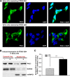
Figure 7. Expression of ATXN3-Q84 in SH-SY5Y cells facilitates nuclear translocation of PKCδ.
(A) SH-SY5Y cells (2 × 10⁴ cells) were plated on chamber slides, expression of ATXN3-Q84 or ATXN3-Q28 was induced, and cells were analyzed by immunostaining with anti-PKCδ antibody (green). The subcellular distribution of PKCδ was assessed by confocal microscopy; nuclei were stained with DAPI. (B) Nuclear and cytosolic protein fractions were isolated from the SH-SY5Y cells expressing ATXN3-Q84 and control un-induced SH-SY5Y cells and the protein fractions analyzed by Western blotting with anti-PKCδ antibody to determine the relative nuclear vs. cytosolic abundance of PKCδ in induced (+) vs. uninduced (-) cells; GAPDH and hnRNPC1 were used as cytosolic and nuclear markers, respectively. (C) Relative levels of PKCδ in nuclear protein fractions of SH-SY5Y cells expressing ATXN3-Q84 (Dox-treated) vs. control cells (Dox-untreated). Data represent mean ± SD (n = 4; *** = p < 0.001).
Figure 8
Figure 8. Proposed mechanism where interaction of PNKP with mutant expanded polyQ-containing ATXN3 abrogates PNKP’s 3’-phosphatase activity.
This atypical interaction perturbs the efficacy of DNA repair, leading to the persistent accumulation of DNA damage/strand breaks in SCA3. The wild-type ATXN3 binds with and stimulates PNKP’s enzymatic activity to repair damaged DNA, contributing to genomic DNA sequence integrity and neuronal survival. By contrast, mutant ATXN3 interacts with and decreases PNKP’s 3’-phosphatase activity, leading to the persistent accumulation of DNA damage in the post-mitotic neurons and resulting in chronic activation of the DNA damage-response ATM→p53 signaling pathway in SCA3. This triggers apoptosis by increasing expression of such p53 target genes as BAX, PUMA and NOXA in SCA3. In parallel, activated ATM phosphorylates c-Abl kinase, which in turn phosphorylates PKCδ, facilitating nuclear translocation of PKCδ and thus further amplifying the pro-apoptotic output in SCA3 ultimately leading to neuronal death in SCA3.

Comment in

Similar articles

Cited by

References

    1. Kawaguchi Y, Okamoto T, Taniwaki M, Aizawa M, Inoue M et al., (1994) CAG expansion in a novel gene for Machado-Joseph disease at chromosome 14q32.1, Nat Genet, 8, 221–228. 10.1038/ng1194-221 - DOI - PubMed
    1. Zoghbi HY, Orr HT (2000) Glutamine repeats and neurodegeneration. Ann Rev Neurosci, 23, 213–247. 10.1146/annurev.neuro.23.1.217 - DOI - PubMed
    1. Kobayashi T, Kakizuka A (2003) Molecular analysis of Machado-Joseph disease, Cytogenetic and Genome Res, 100, 261–275. 10.1159/000072862 - DOI - PubMed
    1. Rosenberg RN (1992) Machado-Joseph disease: an autosomal dominant system degeneration. Mov Disord, 3, 193–203. 10.1002/mds.870070302 - DOI - PubMed
    1. Takiyama Y, Oyanagi S, Kawashima S, Sakamoto H, Saito K et al., (1994) A clinical and pathologic study of a large Japanese family with Machado-Joseph disease tightly linked to the DNA markers on chromosome 14q, Neurology, 44, 1302–1308. 10.1212/WNL.44.7.1302 - DOI - PubMed

Publication types

MeSH terms