. 2015 Jan 1;29(1):1-6.
doi: 10.1101/gad.253682.114.

A laminin 511 matrix is regulated by TAZ and functions as the ligand for the α6Bβ1 integrin to sustain breast cancer stem cells

Affiliations

A laminin 511 matrix is regulated by TAZ and functions as the ligand for the α6Bβ1 integrin to sustain breast cancer stem cells

Cheng Chang et al. Genes Dev. .

Abstract

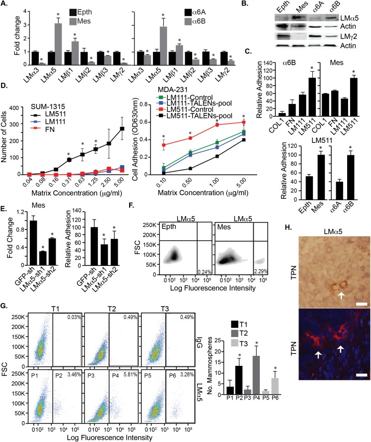
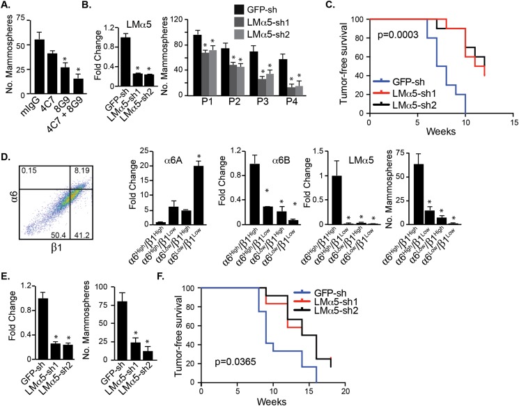
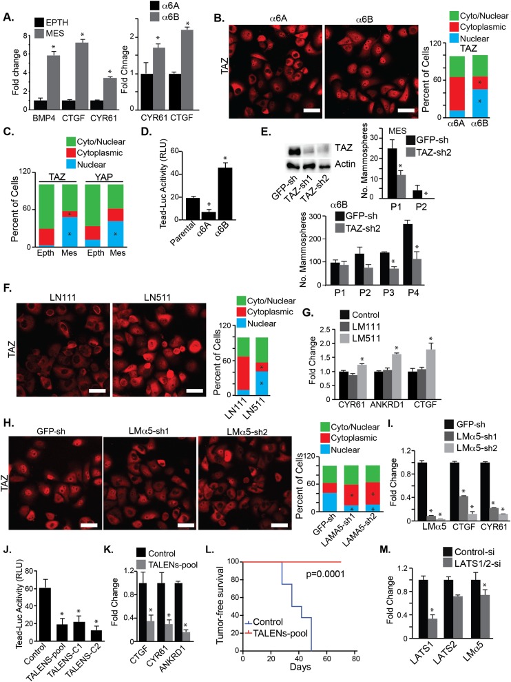
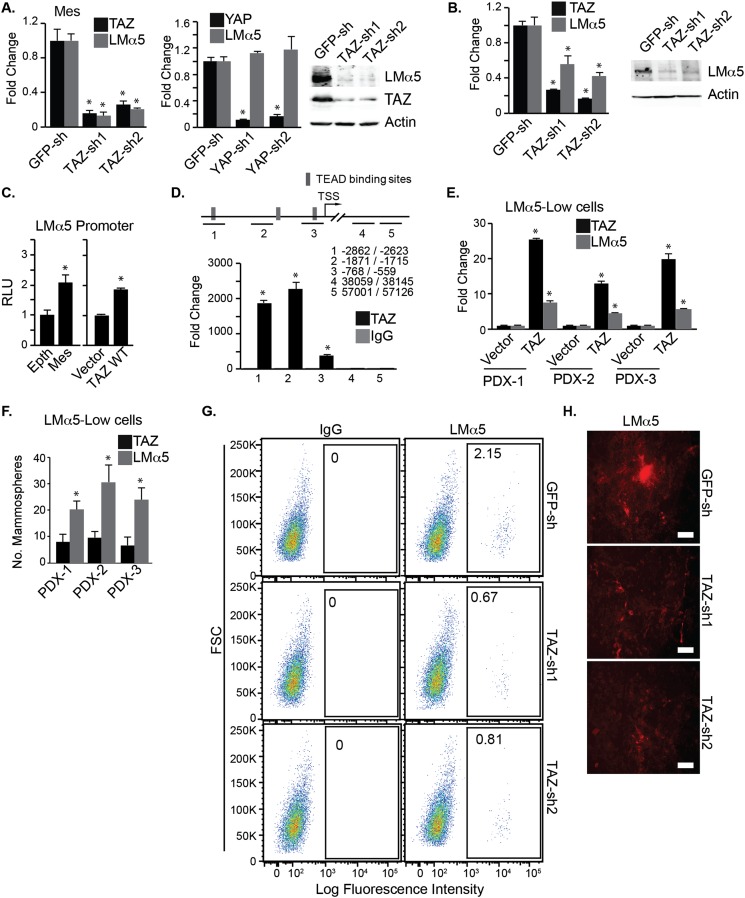
Understanding how the extracellular matrix impacts the function of cancer stem cells (CSCs) is a significant but poorly understood problem. We report that breast CSCs produce a laminin (LM) 511 matrix that promotes self-renewal and tumor initiation by engaging the α6Bβ1 integrin and activating the Hippo transducer TAZ. Although TAZ is important for the function of breast CSCs, the mechanism is unknown. We observed that TAZ regulates the transcription of the α5 subunit of LM511 and the formation of a LM511 matrix. These data establish a positive feedback loop involving TAZ and LM511 that contributes to stemness in breast cancer.

Keywords: TAZ; cancer stem cell; extracellular matrix; integrin; laminin.


Figures

Figure 1.
Figure 1.
LM511 is the preferred ligand for integrin α6Bβ1. (A) Relative mRNA expression of LMα5, LMβ2, LMα3, LMβ3, LMβ1, and LMγ2 in the MES and EPTH populations of CD44+/CD24 Src-transformed MCF10A cells and α6Aβ1- and α6Bβ1-expressing SUM1315 cells was quantified by qPCR. (B) The expression of LMα5, LMγ2, and actin was assessed by immunoblotting in these cells. (C) The cells described in A were assayed for their ability to adhere to COL 1, FN, LM111, and LM511 (1 μg/mL). (D, left) The ability of α6Bβ1-expressing SUM1315 cells to adhere within 30 min to increasing concentrations of FN, LM111, and LM511 was determined. (Right) Control and α6B-depleted MDA-MB-231 cells were compared for their ability to adhere to increasing concentrations of LM111 and LM511. α6B expression was depleted using TALENs as described (Goel et al. 2014). (E) LMα5 expression was diminished in the MES population of CD44+/CD24 Src-transformed MCF10A cells using shRNAs, and the ability of these cells to adhere to glass was assayed. (F) Flow cytometric analysis of surface-bound LMα5 expression in EPTH and MES cells. (G) Three primary human breast tumors (T1, T2, and T3) were dissociated and sorted by FACS using a LMα5 Ab. Cells with low surface-bound LMα5 (P1, P3, and P4) were compared with cells with high surface-bound LMα5 (P2, P4, and P6) for their ability to form mammospheres (bar graph). (H) Frozen sections of human triple-negative breast cancers were stained with a LMα5 Ab, 4C7, using either immunohistochemistry (top) or immunofluorescence (bottom). Arrows depict individual cells with intense staining. Bar, 100 μm.
Figure 2.
Figure 2.
Autocrine LM511 is necessary for self-renewal and tumor initiation. (A) Mammosphere cultures of MES cells were treated with LMα5-blocking antibodies (4C7 and 8G9) daily for 1 wk and quantified. (B) LMα5 expression was diminished in α6Bβ1-expressing SUM1315 cells using shRNAs, and these cells were used for serial passaging of mammospheres. (C) Control (shGFP) and LMα5-diminished MES cells were injected into the mammary fat pads of NSG mice, and tumor formation was assessed by palpation. The curve comparison was done using a log rank test (P < 0.05). (D) TBP mammary tumor cells were sorted by FACS using α6 and β1 integrin Abs. The four populations generated were analyzed for α6B and LMα5 expression and mammosphere formation. Subsequently, LMα5 expression was diminished in the α6high/β1high population using shRNAs, and the impact on mammosphere formation and tumor-free survival (P < 0.05) was determined. (E,F) LMα5 expression was diminished in the α6high/β1high population using shRNAs, and the impact on mammosphere formation (E) and tumor-free survival (F) (P < 0.05) was determined.
Figure 3.
Figure 3.
LM511/α6Bβ1 promotes TAZ activation. (A) Relative mRNA expression of TAZ target genes in the MES and EPTH populations of CD44+/CD24 Src-transformed MCF10A cells and α6Aβ1- and α6Bβ1-expressing SUM1315 cells was quantified by qPCR. (B) α6Aβ1- and α6Bβ1-expressing SUM1315 cells were plated on a LM511 matrix, and the localization of TAZ was assessed by immunofluorescence. (C) The same assay as in B was performed using the MES and EPTH populations of CD44+/CD24 Src-transformed MCF10A cells, and both YAP localization and TAZ localization were assessed. (D) TEAD transcriptional activity was assayed in α6Aβ1- and α6Bβ1-expressing SUM1315 cells and parental cells by transient expression of the 8XGTIIC-luciferase reporter construct. (E) TAZ expression was diminished using shRNA in the MES and α6Bβ1-expressing SUM1315 cells, and the impact on serial mammosphere passage was evaluated. (F) TAZ localization was assessed by immunofluorescence in confluent cultures of SUM1315 cells on either a LM111 or LM511 matrix. (G) Expression of TAZ target genes was quantified by qPCR in SUM1315 cells plated on either LM111 or LM511. (H,I) LMα5 expression was diminished in SUM1315 cells, and the impact on the expression of TAZ localization (H) and TAZ target genes (I) was determined. (J,K) Expression of integrin α6B was depleted in MDA-MB-231 cells using α6B-specific TALENs (see Fig. 1E). These cells (Puro alone, TALENs-pool, TALENs-C1, and TALENs-C2) were used to assay TEAD transcriptional activity (J) and expression of TAZ target genes (K). (L) Control (Puro alone) and TALENs-pool cells were injected into the mammary fat pads of NSG mice, and tumor formation was assessed by palpation. The curve comparison was done using a log rank test (P < 0.05). (M) Expression of Lats1/2 was diminished in EPTH cells using siRNAs, and mRNA expression of Lats1/2 and LMα5 was quantified by qPCR. Bar graphs in this figure represent the average of three independent experiments, and the P-value was determined using Student’s t-test. Bars, 100 μm.
Figure 4.
Figure 4.
TAZ regulates LMα5 expression. (A) Expression of either TAZ or YAP was diminished in MES cells, and the impact on LMα5 mRNA expression was quantified by qPCR. LMα5 protein expression in TAZ-depleted cells was also evaluated by immunoblotting. (B) TAZ expression was diminished in SUM1315 cells, and the impact on LMα5 mRNA and protein expression was determined. (C, left) A luciferase construct containing the LMα5 promoter was generated and used to assay LMα5 transcriptional activity in the MES and EPTH cells. (Right) LMα5 promoter activity was assayed using the same reporter construct in control and TAZ-expressing HEK293 cells. (D, top) Schematic of the LMα5 promoter indicating the location of the putative TEAD-binding sites. (Bottom) Binding of TAZ to the LMα5 promoter was assayed by ChIP. (E,F) Cells with low surface-bound LMα5 (from PDX breast tumors) were isolated and infected with lentiviral particles expressing TAZ. (E) Expression of TAZ and LMα5 mRNA expression was quantified by qPCR. (F) These transfected cells were also assayed for their ability to form mammospheres. (G) Surface-bound LMα5 was quantified by flow cytometry in the EPTH cells transfected with empty vector or TAZ shRNAs. (H) MES cells were cultured for 4 d and detached using EDTA (25 mM), and the exposed matrix was analyzed for LMα5 expression by immunofluorescence.

Similar articles

Cited by

References

    1. Al-Hajj M, Wicha MS, Benito-Hernandez A, Morrison SJ, Clarke MF. 2003. Prospective identification of tumorigenic breast cancer cells. Proc Natl Acad Sci 100: 3983–3988. - PMC - PubMed
    1. Aumailley M, Bruckner-Tuderman L, Carter WG, Deutzmann R, Edgar D, Ekblom P, Engel J, Engvall E, Hohenester E, Jones JC, et al. . 2005. A simplified laminin nomenclature. Matrix Biol 24: 326–332. - PubMed
    1. Baccelli I, Trumpp A. 2012. The evolving concept of cancer and metastasis stem cells. J Cell Biol 198: 281–293. - PMC - PubMed
    1. Beck B, Blanpain C. 2013. Unravelling cancer stem cell potential. Nat Rev Cancer 13: 727–738. - PubMed
    1. Calcagno AM, Salcido CD, Gillet JP, Wu CP, Fostel JM, Mumau MD, Gottesman MM, Varticovski L, Ambudkar SV. 2010. Prolonged drug selection of breast cancer cells and enrichment of cancer stem cell characteristics. J Natl Cancer Inst 102: 1637–1652. - PMC - PubMed

Publication types

MeSH terms