

Lymphatic system: an active pathway for immune protection
- PMID: 25534659
- PMCID: PMC4397130
- DOI: 10.1016/j.semcdb.2014.11.012
Lymphatic system: an active pathway for immune protection
Abstract
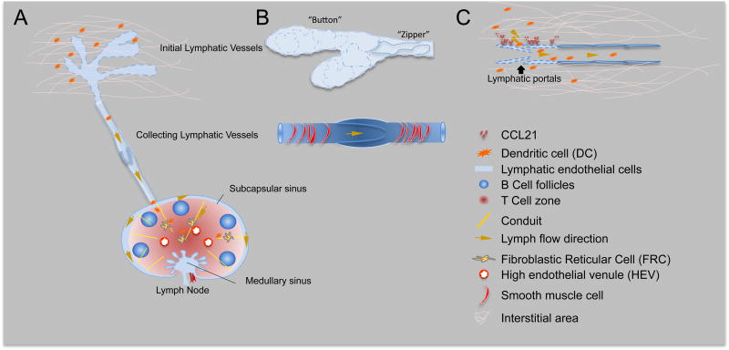
Lymphatic vessels are well known to participate in the immune response by providing the structural and functional support for the delivery of antigens and antigen presenting cells to draining lymph nodes. Recent advances have improved our understanding of how the lymphatic system works and how it participates to the development of immune responses. New findings suggest that the lymphatic system may control the ultimate immune response through a number of ways which may include guiding antigen/dendritic cells (DC) entry into initial lymphatics at the periphery; promoting antigen/DC trafficking through afferent lymphatic vessels by actively facilitating lymph and cell movement; enabling antigen presentation in lymph nodes via a network of lymphatic endothelial cells and lymph node stroma cell and finally by direct lymphocytes exit from lymph nodes. The same mechanisms are likely also important to maintain peripheral tolerance. In this review we will discuss how the morphology and gene expression profile of the lymphatic endothelial cells in lymphatic vessels and lymph nodes provides a highly efficient pathway to initiate immune responses. The fundamental understanding of how lymphatic system participates in immune regulation will guide the research on lymphatic function in various diseases.
Keywords: Antigen delivery; Dendritic cell trafficking; Immune regulation; Lymph node; Lymphatic function.
Copyright © 2014 Elsevier Ltd. All rights reserved.
Conflict of interest statement
Figures
Similar articles
-
Inflammation, lymphatic function, and dendritic cell migration.Lymphat Res Biol. 2006;4(4):217-28. doi: 10.1089/lrb.2006.4406. Lymphat Res Biol. 2006. PMID: 17394405 Review.
-
Dendritic-cell trafficking to lymph nodes through lymphatic vessels.Nat Rev Immunol. 2005 Aug;5(8):617-28. doi: 10.1038/nri1670. Nat Rev Immunol. 2005. PMID: 16056255 Review.
-
Neutrophil Interactions with the Lymphatic System.Cells. 2021 Aug 17;10(8):2106. doi: 10.3390/cells10082106. Cells. 2021. PMID: 34440875 Free PMC article. Review.
-
In Sickness and in Health: The Immunological Roles of the Lymphatic System.Int J Mol Sci. 2021 Apr 24;22(9):4458. doi: 10.3390/ijms22094458. Int J Mol Sci. 2021. PMID: 33923289 Free PMC article. Review.
-
Dendritic cell migration through the lymphatic vasculature to lymph nodes.Adv Immunol. 2013;120:51-68. doi: 10.1016/B978-0-12-417028-5.00002-8. Adv Immunol. 2013. PMID: 24070380 Review.
Cited by
-
Specialized Pro-Resolving Mediators and the Lymphatic System.Int J Mol Sci. 2021 Mar 9;22(5):2750. doi: 10.3390/ijms22052750. Int J Mol Sci. 2021. PMID: 33803130 Free PMC article. Review.
-
Much More than M1 and M2 Macrophages, There are also CD169(+) and TCR(+) Macrophages.Front Immunol. 2015 May 26;6:263. doi: 10.3389/fimmu.2015.00263. eCollection 2015. Front Immunol. 2015. PMID: 26074923 Free PMC article. Review.
-
The anatomy and metabolome of the lymphatic system in the brain in health and disease.Brain Pathol. 2020 Mar;30(2):392-404. doi: 10.1111/bpa.12805. Epub 2019 Dec 5. Brain Pathol. 2020. PMID: 31747475 Free PMC article.
-
Blood⁻Brain Barrier, Lymphatic Clearance, and Recovery: Ariadne's Thread in Labyrinths of Hypotheses.Int J Mol Sci. 2018 Nov 30;19(12):3818. doi: 10.3390/ijms19123818. Int J Mol Sci. 2018. PMID: 30513598 Free PMC article. Review.
-
Immune heterogeneity of head and tail pancreatic lymph nodes in non-obese diabetic mice.Sci Rep. 2019 Jul 5;9(1):9778. doi: 10.1038/s41598-019-45899-1. Sci Rep. 2019. PMID: 31278331 Free PMC article.
References
-
- Liao S, Ruddle NH. Synchrony of high endothelial venules and lymphatic vessels revealed by immunization. J Immunol. 2006;177:3369–79. - PubMed
-
- Kuka M, Iannacone M. The role of lymph node sinus macrophages in host defense. Ann N Y Acad Sci. 2014 - PubMed
-
- Randolph GJ, Angeli V, Swartz MA. Dendritic-cell trafficking to lymph nodes through lymphatic vessels. Nature reviews Immunology. 2005;5:617–28. - PubMed
-
- Reynoso ED, Lee JW, Turley SJ. Peripheral tolerance induction by lymph node stroma. Advances in experimental medicine and biology. 2009;633:113–27. - PubMed
Publication types
MeSH terms
Grants and funding
LinkOut - more resources
Full Text Sources
Other Literature Sources
