. 2015 Jan 22;57(2):207-18.
doi: 10.1016/j.molcel.2014.11.013. Epub 2014 Dec 18.

mTORC1 phosphorylates UVRAG to negatively regulate autophagosome and endosome maturation

Affiliations

mTORC1 phosphorylates UVRAG to negatively regulate autophagosome and endosome maturation

Young-Mi Kim et al. Mol Cell. .

Abstract

mTORC1 plays a key role in autophagy as a negative regulator. The currently known targets of mTORC1 in the autophagy pathway mainly function at early stages of autophagosome formation. Here, we identify that mTORC1 inhibits later stages of autophagy by phosphorylating UVRAG. Under nutrient-enriched conditions, mTORC1 binds and phosphorylates UVRAG. The phosphorylation positively regulates the association of UVRAG with RUBICON, thereby enhancing the antagonizing effect of RUBICON on UVRAG-mediated autophagosome maturation. Upon dephosphorylation, UVRAG is released from RUBICON to interact with the HOPS complex, a component for the late endosome and lysosome fusion machinery, and enhances autophagosome and endosome maturation. Consequently, the dephosphorylation of UVRAG facilitates the lysosomal degradation of epidermal growth factor receptor (EGFR), reduces EGFR signaling, and suppresses cancer cell proliferation and tumor growth. These results demonstrate that mTORC1 engages in late stages of autophagy and endosome maturation, defining a broader range of mTORC1 functions in the membrane-associated processes.


Figures

Figure 1
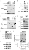
Figure 1. mTORC1 binds to UVRAG and induces UVRAG phosphorylation
(A) mTOR binds to the UVRAG-containing Vps34 complex. Myc-tagged proteins were transiently expressed in HEK293T cells. Endogenous mTOR recovered with myc immunoprecipitate (IP) was analyzed by western blotting (WB). (B) mTORC1 interacts with UVRAG. Myc-tagged proteins were transiently expressed with HA-UVRAG in HEK293T cells. UVRAG recovered with myc IP was analyzed by WB. An irrelevant lane in the middle was eliminated. (C) Endogenous UVRAG recovered with RUBICON, raptor, and control IgG IP was analyzed by WB. (D) The UVRAG-mTORC1 interaction is negatively regulated by Torin1 or HBSS, but not by rapamycin. HEK293T cells were transiently transduced to express myc-UVRAG, and incubated in HBSS for 2 h or treated with Torin1 or rapamycin for 1 h. Endogenous mTOR or raptor isolated with myc-UVRAG was analyzed by WB. (E) mTOR inhibition induces a mobility shift of UVRAG band on SDS-PAGE. HEK293T cells were incubated in DMEM or HBSS for 2 h, or treated with Torin1 for 1 h. Cells were also incubated in leucine-deprived medium (−) then supplemented with leucine (+). (F) Deficiency of either TSC1 or TSC2 in MEFs induces a mobility shift of UVRAG. (G) mTOR phosphorylates UVRAG in vitro. GST-tagged UVRAG fragments were prepared from bacteria and incubated with the active mTOR fragment in the presence of 32P-ATP. Incorporation of 32P into UVRAG fragments was analyzed by autoradiography. See also Figure S1.
Figure 2
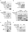
Figure 2. UVRAG Ser498 is an mTORC1-dependent phosphorylation site
(A) Alignment of the sequence surrounding UVRAG S498 with those of the known mTORC1 target sites. (B) Rheb induces UVRAG S498 phosphorylation. HA-Rheb was expressed with myc-UVRAG in HEK293T cells. UVRAG S498 phosphorylation was analyzed by WB. (C) UVRAG phosphorylation in MEFs is inhibited by Torin1, Akt inhibitor VIII or HBSS, but not by rapamycin. Immunoprecipitated UVRAG was analyzed for the phosphorylation by WB. (D) The S498 phosphorylation is enhanced by Akt and Rheb. HA-UVRAG was transiently expressed in HEK293T cells alone (−) or together with myc-tagged constitutively active Akt mutant (cAkt) or Rheb. Two days post-transfection, cells were treated with vehicle (−) or Torin1 (+) for 1 h. (E) Deficiency of AMPK, PTEN, or TSC1 in MEFs enhances UVRAG phosphorylation. MEFs were treated with vehicle (−) or Torin1 (+) for 1 h. The UVRAG phosphorylation was analyzed by immunoprecipitation followed by WB. See also Figure S2.
Figure 3
Figure 3. mTORC1 phosphorylates UVRAG Ser498
(A) mTOR phosphorylates UVRAG S498 in vitro. The active form of mTOR was incubated with puririfed UVRAG in the presence of ATP. Akt1 was used as a negative control. UVRAG S498 phosphorylation was analyzed by WB. (B) Myc-mTOR WT or its kinase dead mutant (KD), transiently expressed in HEK293T cells, was isolated by immunoprecipitation using anti-myc antibody and incubated with UVRAG and ATP. (C) Endogenous mTOR IP was isolated from HEK293T cells, and its kinase activity was analyzed using UVRAG 271–699 fragment as substrate. (D) mTOR, raptor, or rictor IPs were obtained from HEK293T or HeLa cells, and the kinase reaction was analyzed as in (C). (E) The phosphorylation state of S498 was analyzed for endogenous UVRAG isolated from shRNA-transduced HEK293T cells. (F) Torin1 inhibits UVRAG S498 phosphorylation in vitro. The active fragment of mTOR was incubated with UVRAG WT or S498A purified from bacteria. The kinase reaction was performed in the presence or absence of Torin1. See also Figure S3.
Figure 4
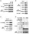
Figure 4. UVRAG Ser498 phosphorylation positively regulates the UVRAG-RUBICON interaction
(A) S498 phosphorylation positively regulates the UVRAG-RUBICON interaction. Myc-UVRAG WT or mutant was transiently expressed in UVRAG-silenced HEK293T cells. Endogenous RUBICON recovered with myc-UVRAG IP was analyzed by WB. (B) Torin1 negatively regulates the UVRAG-RUBICON interaction. HEK293T cells were treated with Torin1 for 4 h. The interaction between endogenous UVRAG and RUBICON was analyzed by immunoprecipitation and WB. (C) Leucine deprivation reduces the UVRAG-RUBICON interaction. HEK293T cells were incubated in medium with (+) or without leucine (−) for 2 h. Endogenous RUBICON recovered with UVRAG IP was analyzed by WB. (D) S498A mutation has a negative effect on the interaction between RUBICON and the UVRAG-containing Vps34 complex. The indicated proteins were expressed in UVRAG-silenced HEK293T cells. Two days post-transfection, the cells were treated with Torin1 as in (B). See also Figure S4.
Figure 5
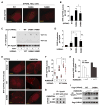
Figure 5. UVRAG Ser498 phosphorylation is important for the inhibitory effect of RUBICON on Vps34
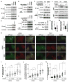
(A) Prevention of S498 phosphorylation increased the accumulation of PI3P in HeLa cells. RFP-tagged 2xFYVE, transiently expressed in WT or mutant UVRAG-reconstituted HeLa cells, was monitored by fluorescence microscope. Scale bar, 10 μm. (B) Quantitative analysis of FYVE puncta formation. Results are represented as means ± standard deviation (SD) (*, p<0.05 vs WT; **, p<0.01 vs WT; #, p<0.01; n≥17). (C) Prevention of S498 phosphorylation increased the kinase activity of Vps34. HA-Vps34 was expressed alone or together with myc-UVRAG in UVRAG-depleted HEK293T cells. HA-Vps34 IP was obtained and incubated with phosphatidylinositol (PI) and ATP in the presence or absence of wortmannin (200 nM). The amount of PI3P was analyzed as described in Experimental Procedures. (D) Quantitative analysis of Vps34 kinase activity. Results are represented as means ± SD (*, p<0.01 vs empty vector; #, p<0.01; NS, Non-Significant; n=3). (E) RUBICON depends on S498 phosphorylation to suppress UVRAG-induced production of PI3P. RFP-2xFYVE expressed alone (−) or together with RUBICON in empty vector- or UVRAG-transduced HCT116 cells was analyzed as described in (A). Scale bar, 5 μm. (F) Quantitative analysis of PI3P puncta formation. The error bars represent means ± SD (#, p<0.01; n=20). (G and H) RUBICON depends on S498 phosphorylation to suppress UVRAG-mediated stimulation of Vps34 kinase activity. UVRAG-reconstituted HEK293T cells were transiently transfected with empty vector or RUBICON construct. The in vitro kinase assay was conducted as in (C). The graph is representative of two independent experiments. (I) Vps34 recovered with Vps34 IP was analyzed by WB. See also Figure S5.
Figure 6
Figure 6. Prevention of UVRAG Ser498 phosphorylation facilitates autophagosome maturation
(A) S498 phosphorylation negatively regulates the UVRAG-Vps39 interaction. HA-Vps39 was transiently expressed in UVRAG-reconstituted HEK293T cells, and their interaction was analyzed by immunoprecipitation and WB. (B) S498 phosphorylation negatively regulates the UVRAG-Vps16 interaction. Myc-Vps16 recovered with GFP-UVRAG IP from HEK293T cells was analyzed by WB. (C) Torin1 enhances the UVRAG-Vps39 interaction. HEK293T cells transiently expressing HA-Vps39 and myc-UVRAG were treated with Torin1 or vehicle (−). HA-Vps39 recovered with myc-UVRAG IP was analyzed by WB. (D) RUBICON knockdown enhances the UVRAG-Vps16 interaction in WT cells but not in S498A cells. GFP-Vps16 recovered with myc-UVRAG IP from shRNA-transduced HEK293T cells was analyzed by WB. (E) S498 phosphorylation negatively regulates the Rab7-RILP interaction. Rab7 recovered with HA-RILP from WT or mutant UVRAG-reconstituted HEK293T cells was analyzed by WB. (F) S498 phosphorylation negatively regulates Rab7. The GTP-bound Rab7 was detected as described in Experimental Procedures. (G) Quantitative analysis of (F). Data are means ± SD (n=2). (H) S498 phosphorylation negatively regulates autophagosome maturation. mCherry-GFP-LC3 was expressed in empty vector- or UVRAG-transduced HCT116 cells. Cells were treated with Torin1 or vehicle. LC3 was monitored by fluorescence microscope. Scale bar, 5 μm. (I and J) Quantitative analysis of GFP puncta (I) and mCherry puncta (J) (*, p<0.01 vs empty vector; #, p<0.01; NS; n≥35). Mean value is shown as a horizontal bar. (K) Quantitative analysis of mCherry only puncta (*, p<0.01 vs empty vector; #, p<0.01; n≥35). Mean and SD are shown as horizontal bars. (L) S498A mutation enhances autophagy flux. HEK293T cells reconstituted with UVRAG constructs were treated with Torin1 in the presence or absence of Bafilomycin A1 (exp1) or E-64/Pepstatin A (exp2). (M) Quantitative analysis of the fold increase of LC3-II level induced by the lysosomal inhibitors in Torin1-treated cells. Data are means ± SD (*, p<0.05; n=5). See also Figure S6.
Figure 7
Figure 7. Prevention of UVRAG Ser498 phosphorylation enhances EGFR degradation, reduces EGFR signaling, and suppresses cancer cell proliferation and tumor growth
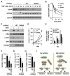
(A) S498A mutation enhances EGFR degradation. HEK293T cells stably transduced by empty vector (−) or UVRAG construct were starved of serum overnight, and treated EGF. EGFR level in cell lysate was analyzed by WB. (B) EGFR level was presented relative to that at time zero. Data are means ± SD (n=4). (C) S498A mutation suppresses EGFR signaling. HEK293T cells, transduced as in (A), were serum-starved overnight and treated with EGF. pERK1/2 (Thr202/Tyr204) or pAKT (Thr308) were analyzed by WB. (D) S498A mutation suppresses cancer cell proliferation. HCT116 cells stably transduced by empty vector (−) or UVRAG construct were cultured in DMEM. The number of viable cells was counted by hemacytometer. Data are means ± SD (n=3). (E) Viable cells were assayed using MTT. Data are means ± SD (n=3). (F) S498A mutation significantly inhibits anchorage-independent growth of cancer cells. HCT116 cells prepared as in (D) were used. Data are shown as mean ± SD (*, p<0.01 vs empty vector; #, p<0.01; n=3). (G) S498A mutation significantly inhibits tumor growth in mice. Values represent means ± SD (*, p=0.012; n=5). (H) Tumor weight relative to the whole body weight was analyzed. Values represent mean ± SD (*, p<0.01; n=5). (I) Regulatory function of UVRAG S498 phosphorylation in autophagosome and endosome maturation. See also Figure S7.

Comment in

Similar articles

Cited by

References

    1. Chang YY, Neufeld TP. An Atg1/Atg13 complex with multiple roles in TOR-mediated autophagy regulation. Mol Biol Cell. 2009;20:2004–2014. - PMC - PubMed
    1. Cui Y, Zhao Q, Gao C, Ding Y, Zeng Y, Ueda T, Nakano A, Jiang L. Activation of the Rab7 GTPase by the MON1-CCZ1 Complex Is Essential for PVC-to-Vacuole Trafficking and Plant Growth in Arabidopsis. Plant Cell. 2014;26:2080–2097. - PMC - PubMed
    1. Dunlop EA, Hunt DK, Acosta-Jaquez HA, Fingar DC, Tee AR. ULK1 inhibits mTORC1 signaling, promotes multisite Raptor phosphorylation and hinders substrate binding. Autophagy. 2011;7:737–747. - PMC - PubMed
    1. Egan DF, Shackelford DB, Mihaylova MM, Gelino S, Kohnz RA, Mair W, Vasquez DS, Joshi A, Gwinn DM, Taylor R, et al. Phosphorylation of ULK1 (hATG1) by AMP-activated protein kinase connects energy sensing to mitophagy. Science. 2011;331:456–461. - PMC - PubMed
    1. Farkas T, Daugaard M, Jaattela M. Identification of small molecule inhibitors of phosphatidylinositol 3-kinase and autophagy. J Biol Chem. 2011;286:38904–38912. - PMC - PubMed

Publication types

MeSH terms

Substances