Review
. 2015 Jan;16(1):18-29.
doi: 10.1038/nrm3920.

Intrinsically disordered proteins in cellular signalling and regulation

Affiliations
Review

Intrinsically disordered proteins in cellular signalling and regulation

Peter E Wright et al. Nat Rev Mol Cell Biol. 2015 Jan.

Abstract

Intrinsically disordered proteins (IDPs) are important components of the cellular signalling machinery, allowing the same polypeptide to undertake different interactions with different consequences. IDPs are subject to combinatorial post-translational modifications and alternative splicing, adding complexity to regulatory networks and providing a mechanism for tissue-specific signalling. These proteins participate in the assembly of signalling complexes and in the dynamic self-assembly of membrane-less nuclear and cytoplasmic organelles. Experimental, computational and bioinformatic analyses combine to identify and characterize disordered regions of proteins, leading to a greater appreciation of their widespread roles in biological processes.


Figures
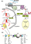
Figure 1
Figure 1. Intrinsic Disorder in Signaling
(a) The metabolic hormone glucagon (an early example of intrinsic disorder in a functional molecule ) binds to a structured, membrane-bound cell surface receptor, a G-protein-coupled receptor (GPCR) causing the translocation of the α subunit of the coupled G protein to the membrane-bound adenylyl cyclase, with concomitant formation of GTP from GDP. Cyclic AMP is generated, and activates protein kinase A (PKA), which has two downstream effects, firstly initiating the phosphorylation cascade that results in the phosphorylation of glycogen and the mobilization of stored glucose. The second effect is that activated PKA is translocated to the nucleus, where it phosphorylates the transcription factor cyclic-AMP response element binding protein (CREB), an intrinsically disordered protein. It appears that CREB is constitutively bound to the CRE DNA sequence through dimerization of the C-terminal basic leucine zipper domain (bzip, red cross). Phosphorylation of the kinase-inducible (KID) domain causes this domain to fold into a helical structure on the KIX domain of the transcriptional coactivator CREB-binding protein (CBP) (yellow), recruiting it to the promoter and promoting the transcription of downstream signal-response genes (reviewed in ). In this case, intrinsically disordered proteins function both in the original reception of the signal and in the promotion of gene transcription in response to the signal. (b) Domain organization of CBP, showing a subset of the IDPs that bind to each of the four main interaction domains, the folded domains TAZ1, KIX and TAZ2, and the disordered (probably molten globular) NCBD.
Figure 2
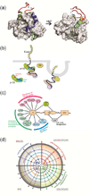
Figure 2. Variable Binding Affinities of IDPs
(a) One member of an NMR-derived family of the structure of the complex between the folded CBP TAZ1 domain (grey surface) and the transactivation domain of RelA (NFκB p65). Backbone dynamics of RelA in the complex were estimated using 1H-15N NOE measurements, and are mapped onto the RelA backbone in red (most flexible), yellow (less flexible), green (less flexible again) and blue (least flexible). The regions of RelA colored blue coincide with the hydrophobic docking interactions that dominate association with the TAZ1 domain. The N-terminal helix (green) is only transiently populated, and is dynamically disordered on a nanosecond timescale, yet contributes to binding affinity. The figure was made from coordinates 2LWW and data in . (b) Schematic illustration of pathway cross talk mediated by differential binding of an IDP. The disordered cytoplasmic tail of E-cadherin binds to the armadillo repeat region of the p120 catenin through interaction of conserved sequence motifs containing phosphorylated tyrosines (Y) and glycines (G) at a high-affinity static binding site (blue), while the LL motif binds at the dynamic site (red), effectively chaperones this region. The adaptor protein (AP) recognizes the exposed LL motif, leading to clathrin-mediated endocytosis of E-cadherin. Figure adapted from . (c) Interactions between adenovirus E1A and cellular proteins. The intrinsically disordered N-terminal region of E1A binds numerous cellular proteins to disrupt cellular regulation. Interactions that function in the repression of cellular genes are shown in pink, in chromatin remodeling and transcriptional activation in green and in the cytoplasm in blue. Interactions with pRb (gray) are essential for deregulation of the cell cycle, while binding to CBP/p300 (green) disrupts cellular transcriptional programs. (d) Schematic summary of the allosteric modulation of the E1A signaling network through complex formation with CBP and pRb. Signaling pathways are modulated allosterically by interactions with various binding partners, as represented by a central phase diagram of the hub, with four states of E1A: free (gray), E1A–pRb (blue), E1A–CBP/p300 (green) and ternary complex (red). Circles outside the hub show additional protein partner interactions that influence regulatory pathways within the cell. Figure reproduced from with permission.
Figure 3
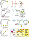
Figure 3. Response to multisite phosphorylation in IDRs
Multisite phosphorylation of disordered proteins can give rise to a range of signaling responses. (a) For phosphorylation at a single site, the response takes the form of a simple hyperbolic saturation curve, as would be the case for single-site phosphorylation of CREB at Ser133 and its interaction with the KIX domain of CBP,. (b) In the Sic1-SCF ubiquitin ligase system, the response to phosphorylation at multiple sites takes the form of sigmoidal threshold response curves, with cooperativity increasing as an increasing number of sites (1–6) are phosphorylated. (c) The interaction of Eco1 with the Cdc4 subunit of the SCF ubiquitin ligase shows coincidence detection, where a certain threshold level of phosphorylation must be achieved before response is initiated. (d) Under conditions of genotoxic stress the affinity of the p53 transactivation domain for CBP/p300 increases with each successive phosphorylation event, relative to the affinities of other transcription factors (denoted TF1 and TF2). This is an example of a rheostat response. Figure adapted from ,, with permission.
Figure 4
Figure 4. Autoinhibition through interactions with IDRs
A disordered acidic domain inhibits the Vav1 nucleotide exchange factor by interacting in cis with the catalytic Dbl homology (DH) and calponin (CH) domains, forming localized elements of helical structure (red rectangles) that incorporate tyrosine residues. The acidic domain undergoes dynamic fluctuations, exposing the tyrosines to phosphorylation which results in dissociation of the bound inhibitory domain and activation of Vav1,. Figure adapted from with permission.
Figure 5
Figure 5. Disorder mediates stress-induced translational silencing
Stress granules, which contain RNA and protein in a membraneless condensed particle, form and coalesce in response to cellular stress, sequestering proteins, including the dual-specificity kinase DYRK3 and mTORC1, the cellular factor that activates translation. Translational silencing is mediated by DYRK3 under stress conditions in two ways, through stabilization of stress granules by the interaction of the disordered N-terminal tail, thus prolonging the sequestration of mTORC1. When cellular stress is relieved, DYRK3 and mTORC1 are released from the stress granules. Active DYRK3 acts as a kinase to phosphorylate PRAS40, relieving its inhibition of mTORC1 and allowing the resumption of protein synthesis. Figure adapted from with permission.

Similar articles

Cited by

References

    1. Wright PE, Dyson HJ. Intrinsically unstructured proteins: Re-assessing the protein structure-function paradigm. Journal of Molecular Biology. 1999;293:321–331. - PubMed
    1. Dunker AK, Brown CJ, Lawson JD, Iakoucheva LM, Obradovic Z. Intrinsic disorder and protein function. Biochemistry. 2002;41:6573–6582. - PubMed
    1. Dyson HJ, Wright PE. Intrinsically unstructured proteins and their functions. Nature Reviews Molecular Cell Biology. 2005;6:197–208. - PubMed
    1. van der Lee R, et al. Classification of intrinsically disordered regions and proteins. Chemical Reviews. 2014;114:6589–6631. - PMC - PubMed
    1. Dunker AK, Cortese MS, Romero P, Iakoucheva LM, Uversky VN. Flexible nets. The roles of intrinsic disorder in protein interaction networks. FEBSJournal. 2005;272:5129–5148. - PubMed

Publication types

MeSH terms

Substances