. 2015 Mar;89(5):2628-42.
doi: 10.1128/JVI.02458-14. Epub 2014 Dec 17.

Ataxia telangiectasia mutated kinase mediates NF-κB serine 276 phosphorylation and interferon expression via the IRF7-RIG-I amplification loop in paramyxovirus infection

Affiliations

Ataxia telangiectasia mutated kinase mediates NF-κB serine 276 phosphorylation and interferon expression via the IRF7-RIG-I amplification loop in paramyxovirus infection

Ling Fang et al. J Virol. 2015 Mar.

Abstract

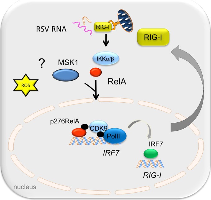
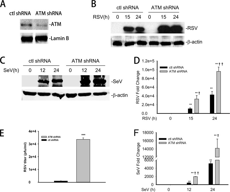
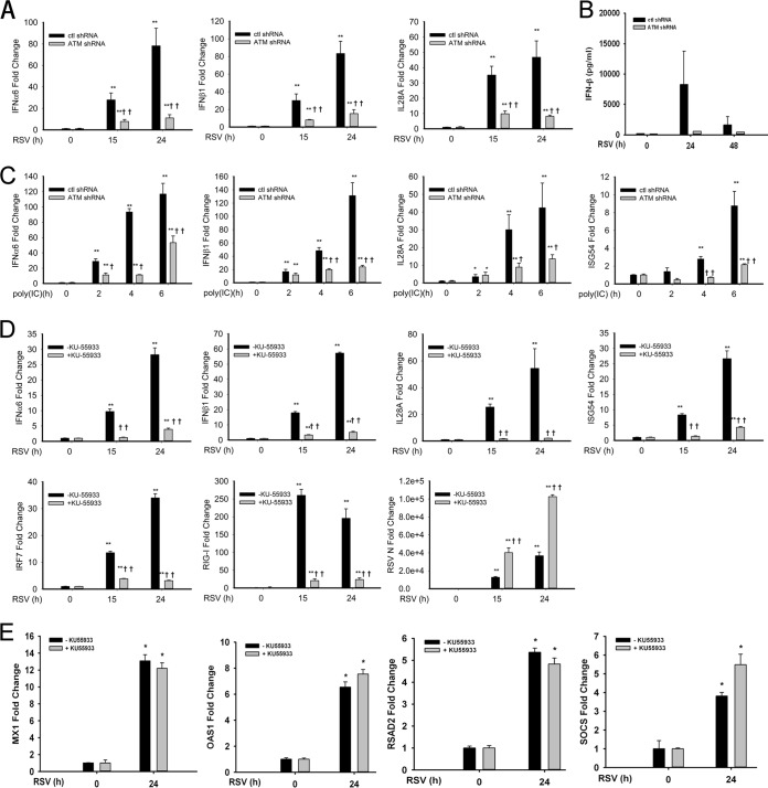
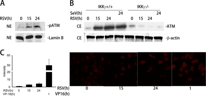
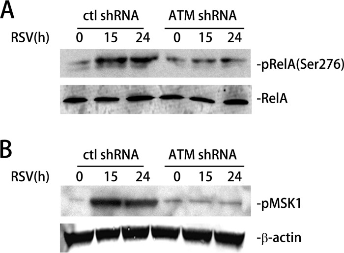
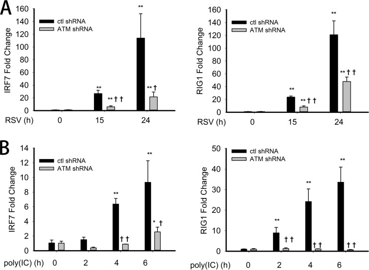
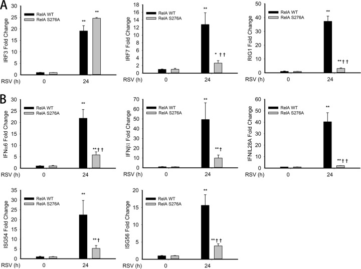
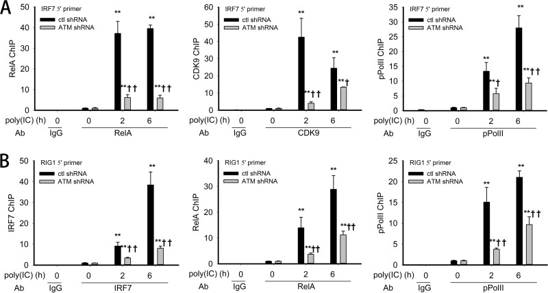
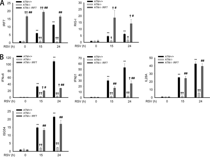
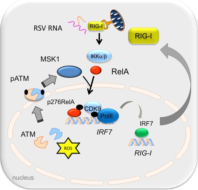
Respiratory syncytial virus (RSV) is a primary etiological agent of childhood lower respiratory tract disease. Molecular patterns induced by active infection trigger a coordinated retinoic acid-inducible gene I (RIG-I)-Toll-like receptor (TLR) signaling response to induce inflammatory cytokines and antiviral mucosal interferons. Recently, we discovered a nuclear oxidative stress-sensitive pathway mediated by the DNA damage response protein, ataxia telangiectasia mutated (ATM), in cytokine-induced NF-κB/RelA Ser 276 phosphorylation. Here we observe that ATM silencing results in enhanced single-strand RNA (ssRNA) replication of RSVand Sendai virus, due to decreased expression and secretion of type I and III interferons (IFNs), despite maintenance of IFN regulatory factor 3 (IRF3)-dependent IFN-stimulated genes (ISGs). In addition to enhanced oxidative stress, RSV replication enhances foci of phosphorylated histone 2AX variant (γH2AX), Ser 1981 phosphorylation of ATM, and IKKγ/NEMO-dependent ATM nuclear export, indicating activation of the DNA damage response. ATM-deficient cells show defective RSV-induced mitogen and stress-activated kinase 1 (MSK-1) Ser 376 phosphorylation and reduced RelA Ser 276 phosphorylation, whose formation is required for IRF7 expression. We observe that RelA inducibly binds the native IFN regulatory factor 7 (IRF7) promoter in an ATM-dependent manner, and IRF7 inducibly binds to the endogenous retinoic acid-inducible gene I (RIG-I) promoter. Ectopic IRF7 expression restores RIG-I expression and type I/III IFN expression in ATM-silenced cells. We conclude that paramyxoviruses trigger the DNA damage response, a pathway required for MSK1 activation of phospho Ser 276 RelA formation to trigger the IRF7-RIG-I amplification loop necessary for mucosal IFN production. These data provide the molecular pathogenesis for defects in the cellular innate immunity of patients with homozygous ATM mutations.

Importance: RNA virus infections trigger cellular response pathways to limit spread to adjacent tissues. This "innate immune response" is mediated by germ line-encoded pattern recognition receptors that trigger activation of two, largely independent, intracellular NF-κB and IRF3 transcription factors. Downstream, expression of protective antiviral interferons is amplified by positive-feedback loops mediated by inducible interferon regulatory factors (IRFs) and retinoic acid inducible gene (RIG-I). Our results indicate that a nuclear oxidative stress- and DNA damage-sensing factor, ATM, is required to mediate a cross talk pathway between NF-κB and IRF7 through mediating phosphorylation of NF-κB. Our studies provide further information about the defects in cellular and innate immunity in patients with inherited ATM mutations.


Figures

FIG 1
FIG 1
Working model for the ROS-dependent formation of Ser 276-phosphorylated RelA in response to RSV infection. RSV dsRNA is sensed by RIG-I, triggering NF-κB release from cytoplasmic stores. In parallel, oxidative stress is required for RelA Ser 276 phosphorylation, mediated by the R6SK MSK1. Downstream, activated phospho-Ser 276 RelA triggers IRF7 gene expression and RIG-I upregulation. The resynthesized RIG-I sustains IFN expression.
FIG 2
FIG 2
Enhanced RSV and Sendai virus replication in ATM knockdown cells. (A) A549 cells were transfected with ATM shRNA; 72 h later, equal amounts of nuclear extract (NE) were analyzed by Western blotting to detect ATM. Lamin B was used as internal control. (B) Control-shRNA-A549 or ATM-shRNA-A549 cells were infected with RSV (MOI, 1.0) for 0, 15, or 24 h. Equal amounts of whole-cell extract (WCE) were analyzed by WB to determine the level of RSV protein expression. β-Actin was used as internal control. (C) Control-shRNA-A549 or ATM-shRNA-A549 cells were mock infected or infected with SeV (MOI, 1.0) for 15 or 24 h. Equal amounts of WCE were used to determine the SeV protein expression by WB. Shown is SeV nucleoprotein (NP). β-Actin was used as internal control. (D) Control-shRNA-A549 or ATM-shRNA-A549 cells were mock or RSV infected (MOI, 1.0) for 15 or 24 h. Total RNA was extracted. Fold changes in the mRNA level of RSV N protein were measured. (E) Control-shRNA-A549 or ATM-shRNA-A549 cells were RSV infected (MOI, 1.0) for 48 h. The RSV titer (PFU/ml) was determined by methylcellulose plaque assay. (F) Control-shRNA-A549 or ATM-shRNA-A549 cells were mock or SeV infected (MOI, 1.0) for 15 or 24 h. The fold changes in SeV mRNA were measured. *, significantly different from RSV- or SeV-treated (0 h) samples, P < 0.05; **, significantly different from RSV- or SeV-treated (0 h) samples, P < 0.01; †, significantly different from ATM+/+ samples, P < 0.05; ††, significantly different from ATM+/+ samples, P < 0.01.
FIG 3
FIG 3
ATM disruption suppresses IFN and ISG expression after RSV infection or poly(I·C) treatment. (A) Control-shRNA-A549 and ATM-shRNA-A549 cells were mock or RSV infected (MOI, 1.0) for 15 or 24 h. Fold changes of mRNA for IFN-α6, IFN-β1, IL-28A, and ISG54 were measured by Q-RT-PCR. (B) Conditioned medium from control-shRNA-A549 and ATM-shRNA-A549 cells taken 0, 24, or 48 h p.i. was assayed for IFN-β protein by ELISA. (C) Control-shRNA-A549 and ATM-shRNA-A549 cells were electroporated with 10 μg poly(I·C) for 2, 4, or 6 h. Fold changes of mRNA for IFN-α6, IFN-β1, IL-28A, and ISG54 were measured. (D) hSAECs were infected with RSV (MOI, 1.0) for 0, 15, or 24 h with or without KU-55933 pretreatment (10 μM, 1 h). Fold changes in mRNA of IFN-α6, IFN-β1, IL-28A, ISG54, IRF7, and RIG-I were measured. (E) hSAECs were infected with RSV (MOI, 1.0) for 0 or 24 h with or without KU-55933 treatment as above. Fold changes in mRNA of MX1, OAS1, RSAD2/CIG5, and SOCS were determined by Q-RT-PCR. *, significantly different from RSV- or poly(I·C)-treated (0 h) samples, P < 0.05; **, significantly different from RSV- or poly(I·C)-treated (0 h) samples, P < 0.01; †, significantly different from ATM+/+ samples, P < 0.05; ††, significantly different from ATM+/+ samples, P < 0.01.
FIG 4
FIG 4
ATM is activated by RSV-induced DSB and requires IKKγ for nuclear export. (A) A549 cells were mock or RSV infected (MOI, 1.0) for 15 or 24 h. Equal amounts of nuclear extract (NE) were assayed to detect the level of phospho-Ser 1981 ATM (pATM) by Western blotting (WB). Lamin B was used as internal control. (B) IKKγ+/+ and IKKγ−/− MEFs were mock or RSV infected (MOI, 1.0) for 15 or 24 h or with SeV (MOI, 1.0) for 24 h as indicated. ATM was detected in equal amounts of cell extract (CE) by WB. β-Actin was used as internal control. (C) Right panel, A549 cells were mock or RSV infected (MOI, 1.0) for 15 or 24 h or treated with VP16 (10 μM) for 1 h as indicated. The cells were fixed, incubated with anti-γH2AX Ab, and then stained with fluorescein isothiocyanate-conjugated anti-rabbit secondary Ab. The nuclei were stained with DAPI (4′,6-diamidino-2-phenylindole). The slides were imaged using confocal microscopy. The expression of γH2AX is shown in red. Left panel, quantification of the signal intensity.
FIG 5
FIG 5
RSV-induced phospho-Ser 376 MSK1 and phospho-Ser 276 RelA formation is ATM dependent. (A) Control-shRNA-A549 and ATM-shRNA-A549 cells were mock or RSV infected (MOI, 1.0) for 15 or 24 h. Phospho-Ser 276 RelA (top) and total RelA (bottom) were detected in whole-cell extract (WCE) using Western blotting (WB). (B) Control-shRNA-A549 or ATM-shRNA-A549 cells were mock or RSV infected (MOI, 1.0) for 15 or 24 h. Phospho-Ser 375 MSK1 (pMSK1) was detected by WB. β-Actin was used as internal control.
FIG 6
FIG 6
Suppressed IRF7 and RIG-I expression upon RSV infection and poly(I·C) treatment in ATM knockdown cells. (A) Control-shRNA-A549 or ATM-shRNA-A549 cells were mock or RSV infected (MOI, 1.0) for 15 or 24 h. Fold changes in IRF7 and RIG-I mRNA abundance were measured by Q-RT-PCR. (B) Control-shRNA-A549 or ATM-shRNA-A549 cells were electroporated with 10 μg poly(I·C) for 0, 2, 4, or 6 h. Fold changes in IRF7 and RIG-I were measured. *, significantly different from RSV- or poly(I·C)-treated (0 h) samples, P < 0.05; **, significantly different from RSV- or poly(I·C)-treated (0 h) samples, P < 0.01; †, significantly different from ATM+/+ samples, P < 0.05; ††, significantly different from ATM+/+ samples, P < 0.01.
FIG 7
FIG 7
RSV-induced IRF7-RIG-I expression is RelA Ser276A dependent. (A) RelA-deficient MEFs stably expressing enhanced green fluorescent protein (EGFP)-tagged RelA WT (RelA WT) or EGFP-tagged nonphosphorylatable Rel Ser 276 Ala mutant (RelA S276A) was mock or RSV infected (MOI, 1.0) for 24 h. Fold changes in IRF3, IRF7, and RIG-I mRNA expression were measured by Q-RT-PCR. (B) RelA WT and Rel Ser276A MEFs were mock infected or infected as described for Fig. 6A. IFN-α6, IFN-β1, IL-28A, ISG54, and ISG56 mRNA abundance was measured. *, significantly different from RSV- or poly(I·C)-treated (0 h) samples, P < 0.05; **, significantly different from RSV or poly(I·C)-treated (0 h) samples, P < 0.01; †, significantly different from RelA WT samples, P < 0.05; ††, significantly different from RelA WT samples, P < 0.01.
FIG 8
FIG 8
Requirement of ATM in RelA/coactivator recruitment to IRF7 and IRF7 recruitment to RIG-I gene promoters. (A) Control-shRNA-A549 or ATM-shRNA-A549 cells were electroporated with 10 μg poly(I·C) for 0, 2, or 6 h. The corresponding chromatin was dually cross-linked and immunoprecipitated with anti-RelA, anti-CDK9, and anti-phospho Ser 2 Pol II (pPol II) Abs. IgG was the negative control. The fold change in IRF7 gene enrichment was quantified by Q-gPCR using the IRF7 5′ primer set. (B) XChIP was performed as described for Fig. 7A. The corresponding chromatin was immunoprecipitated with anti-IRF7, anti-RelA, and anti-pPol II Abs. IgG was the negative control. Fold change in RIG-I gene enrichment was quantified by Q-gPCR using the RIG-I 5′ primer set. *, significantly different from TNF-treated (0 h) samples, P < 0.05; **, significantly different from TNF-treated (0 h) samples, P < 0.01; †, significantly different from ATM+/+ samples, P < 0.05; ††, significantly different from ATM+/+ samples, P < 0.01.
FIG 9
FIG 9
Exogenous IRF7 rescues antiviral gene expression in ATM-shRNA-A549 cells. (A) Expression vectors encoding empty vector or IRF7 were transfected into ATM-shRNA-A549 cells; 72 h later, cells were mock or RSV infected (MOI, 1.0) for 15 or 24 h. mRNA levels of IRF7 and RIG-I were measured by Q-RT-PCR. (B) The same total RNA as in panel A were extracted. mRNA levels of IFN-α6, IFN-β1, IL-28A, and ISG54 were measured. †, significantly different from ATM+/+ samples, P < 0.05; ††, significantly different from ATM+/+ samples, P < 0.01; #, significantly different from ATM-shRNA-A549 samples, P < 0.05; ##, significantly different from ATM-shRNA-A549 samples, P < 0.01.
FIG 10
FIG 10
Working model for the role of ATM in the phospho-RelA-mediated antiviral response. RSV-induced oxidative stress and/or DSB activates ATM to autophosphorylate and induces its IKKγ-dependent export. In the cytosol, ATM is required for RelA Ser 276 phosphorylation by MSK1. RelA Ser 276 phosphorylation is required for IRF7 gene expression. RIG-I gene expression is mediated by IRF7, and resynthesized RIG-I plays an essential role in the antiviral response.

Similar articles

Cited by

References

    1. Glezen WP, Taber LH, Frank AL, Kasel JA. 1986. Risk of primary infection and reinfection with respiratory syncytial virus. Am J Dis Child 140:543–546. - PubMed
    1. Welliver RC. 2004. Respiratory syncytial virus infection: therapy and prevention. Paediatr Respir Rev 5(Suppl A):S127–S133. doi:10.1016/S1526-0542(04)90024-3. - DOI - PubMed
    1. Hall CB, Weinberg GA, Iwane MK, Blumkin AK, Edwards KM, Staat MA, Auinger P, Griffin MR, Poehling KA, Erdman D, Grijalva CG, Zhu Y, Szilagyi P. 2009. The burden of respiratory syncytial virus infection in young children. N Engl J Med 360:588–598. doi:10.1056/NEJMoa0804877. - DOI - PMC - PubMed
    1. Nair H, Nokes DJ, Gessner BD, Dherani M, Madhi SA, Singleton RJ, O'Brien KL, Roca A, Wright PF, Bruce N, Chandran A, Theodoratou E, Sutanto A, Sedyaningsih ER, Ngama M, Munywoki PK, Kartasasmita C, Simoes EA, Rudan I, Weber MW, Campbell H. 2010. Global burden of acute lower respiratory infections due to respiratory syncytial virus in young children: a systematic review and meta-analysis. Lancet 375:1545–1555. doi:10.1016/S0140-6736(10)60206-1. - DOI - PMC - PubMed
    1. Hall CB, Douglas RG Jr, Schnabel KC, Geiman JM. 1981. Infectivity of respiratory syncytial virus by various routes of inoculation. Infect Immun 33:779–783. - PMC - PubMed

Publication types

MeSH terms

LinkOut - more resources