. 2014 Nov 6;15(5):589-604.
doi: 10.1016/j.stem.2014.10.003. Epub 2014 Nov 6.

In vivo activation of a conserved microRNA program induces mammalian heart regeneration

Affiliations

In vivo activation of a conserved microRNA program induces mammalian heart regeneration

Aitor Aguirre et al. Cell Stem Cell. .

Erratum in

  • Cell Stem Cell. 2014 Dec 4;15(6):805

Abstract

Heart failure is a leading cause of mortality and morbidity in the developed world, partly because mammals lack the ability to regenerate heart tissue. Whether this is due to evolutionary loss of regenerative mechanisms present in other organisms or to an inability to activate such mechanisms is currently unclear. Here we decipher mechanisms underlying heart regeneration in adult zebrafish and show that the molecular regulators of this response are conserved in mammals. We identified miR-99/100 and Let-7a/c and their protein targets smarca5 and fntb as critical regulators of cardiomyocyte dedifferentiation and heart regeneration in zebrafish. Although human and murine adult cardiomyocytes fail to elicit an endogenous regenerative response after myocardial infarction, we show that in vivo manipulation of this molecular machinery in mice results in cardiomyocyte dedifferentiation and improved heart functionality after injury. These data provide a proof of concept for identifying and activating conserved molecular programs to regenerate the damaged heart.


Figures

Figure 1
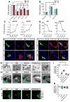
Figure 1. miR-99/100 and its direct targets contribute to zebrafishheartregeneration and development
(A, B) Real time RT-PCR for microRNA candidates miR-99/100 and Let-7a/c (A) and downstream targets fntb and smarca5 (B) in regenerating zebrafish hearts 3 and 7 days post-amputation (dpa) (n=8). (C) Real time RT-PCR revealed that expression of miR-99/100 is very low during the first stages of development and dramatically increased at 3 days post-fertilization (dpf) in zebrafish (n=10). (D) fntb and smarca5 expression inversely correlates with miR-99/100 in developing embryos (n=10). Note that in C and D, analyzes were done in whole embryos at 1, 3, and 5dpf, and whole heart at the adult stage. (E, F) Representative pictures demonstrating that both Fntβ and Smarca5 are present at high levels in the ventricles of developing hearts at 2 and 5dpf (n=10). V: ventricle; A: atrium; Eb: erythroblasts. (G–I) Knock-down of Fntβ and/or Smarca5 in zebrafishembryos resulted in abnormally small animals (G) and reduced ventricle size in cmlc2:GFP animals (H–I). The same phenotypes were observed upon injection of miR-99/100 mimics (G–I) (n>50). Rescue experiments for the conditions tested in G were performed by co-injecting in vitro transcribed mRNAs with modified 5′UTRs with corresponding morpholinos to determine the specificity of the observed phenotypes (J) (n=30). (K) Quantification of the ventricle size for the rescue experiments. Data are represented as mean +/− s.e.m. *p<0.05. See also Figure S1 and Table S1.
Figure 2
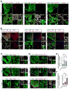
Figure 2. miR-99/100 and its downstream targets are differentially regulated in dedifferentiated cardiomyocytes during heart regeneration
(A, B) FISH/immunofluorescence were used to determine cardiomyocyte specific expression (MyHC) of miR-99/100, Fntβ and Smarca5 in uninjured and regenerating zebrafish hearts (3 and 7dpa). Cardiomyocytes in regenerating hearts exhibited low levels of miR-99/100, and inversely correlating high levels of Fntβ and Smarca5 (n=8). (C–D) Immunofluorescence analysis demonstrating high levels of Fntβ (C) and Smarca5 (D) alongside markers indicative of proliferation, PCNA (C) and H3P (D), in dedifferentiating cardiomyocytes (n=5). (E–F) Quantitative analysis confirming the significantly higher number of MyHC+ cardiomyocytes co-expressing Fntβ/PCNA (E) and Smarca5/H3P (F) in the regenerating zebrafish heart (n=5 animals, three different sections per animal). Dashed line: amputation plane. Boxed area: magnified field. Arrows indicate cells of interest. Data are represented as mean ± s.e.m. *p<0.05. See also Figure S2 and S3.
Figure 3
Figure 3. Heart regeneration is controlled by miR-99/100
(A–C) Following adult zebrafish heart amputation, exogenous intra-cardiac administration of 0.2 μg miR-99/100 mimics every second day for a total of 14 days led to defective cardiac regeneration in amputated zebrafish (A, upper row), as determined by reduced BrdU incorporation (A, lower row and C). (D–F) miR-99/100 antagomiRs exerted the opposite effect in uninjured adult animals, inducing a significant increase in ventricle size (D, upper panels and E) and increasing cardiomyocyte proliferation in the absence of heart damage (D, lower panels and F). (G–I) Chemical inhibition of fnt activity with tipifarnib upon intraperitoneal injection (final concentration 0.02 mg/animal) every 2 days for 14 days dramatically reduced heart regeneration in amputated zebrafish as compared to control animals administered with the solvent DMSO control (G and H) and reducing cardiomyocyte proliferation as assessed by BrdU incorporation (G, lower panels and I). Dashed line: amputation plane. Boxed area: magnified detail. Data are represented as mean ± s.e.m. *p<0.05. n = 6 animals, three different sections per animal were used for quantitative analyses.
Figure 4
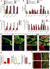
Figure 4. The miR-99/100-dependent heart regeneration pathway is developmentally conserved in mammals but fails to activate upon injury
(A–B) qRT-PCR analysis showing the expression of miR-99/100 and Let-7a/c (A), their protein targets and key cardiac transcription factors (B) during human cardiac differentiation (n=5). Human embryonic stem cell line H1 (H1 ESC); left ventricle (LV and right ventricle (RV). (C–D) qRT-PCR analysis showing the expression of miR-99/100 and Let-7a/c (C), their protein targets and key cardiac transcription factors (D) during mouse development and adult stages (n=5). Mouse embryonic stem cells (mESC). (E–G) FISH/immunofluorescence (E, F) and quantitative analysis (G) demonstrating low levels of miR-99/100 and high levels of FNTβ and SMARCA5 in E11 murine embryonic hearts as opposed to adult hearts, which showed high levels of miR-99/100, and almost undetectable expression of FNTβ and SMARCA5. (H) FISH/immunofluorescence in adult mouse heart before (upper panels) and after myocardial infarction (lower panels) highlighted a failure to downregulate miR-99/100 upon injury in the murine heart. Data are represented as mean ± s.e.m. *p<0.05. (E, F): n = 6 animals; (H): n= 5 animals. In all cases, three different sections per animal were used for quantitative analyses (G). See also Figure S4.
Figure 5
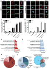
Figure 5. Forced down-regulation of miR-99/100 and Let-7a/c suffices to promote dedifferentiation and proliferation of adult murine cardiomyocytes in culture
(A) Representative pictures showing that pmiRZip control vector-transduced adult murine cardiomyocytes spontaneously disorganized sarcomeric structures in vitro, but did not dedifferentiate to express FNTβ, SMARCA5, GATA4 or the proliferative marker, PCNA. (B) Upon lentiviral transduction with antimiR-99/100, adult cardiomyocytes dedifferentiate and express PCNA, GATA4 and both miR targets. (C, D) Quantitative analysis of FNTβ and SMARCA5 (C), or GATA4 and PCNA (D) positive adult cardiomyocytes after different anti-miR treatments. (E–F) Most relevant upregulated (E) and downregulated (F) functional gene ontology processes observed during in vitro cardiomyocyte dedifferentiation with antimiRs. (G–H) Clusters of genes indicating the most relevant upregulated (G) and downregulated (H) groups involved in cardiomyocyte dedifferentiation. As expected, epigenetic remodeling and developmental processes appeared as highly represented. (I) Comparative proteomic analysis of the heart during regenerative stages. Adult regenerating zebrafish hearts (uninjured, 3 dpa, 7dpa), neonatal regeneration-permissive mouse hearts (P0, P7, adult) and anti-miR treated adult myocardial mouse tissue (control, anti-miR-99/100, anti-Let-7a/c, anti-miR-99/100+anti-Let-7a/c) were collected and processed for semi-quantitative mass spectrometry to determine their translational profiles. The Venn diagram depicts differentially regulated protein candidates for each stage and species after a cross-comparison to find common protein effectors involved in the maintenance or transition to a regeneration-permissive state. Data are represented as mean +/− s.e.m. *p<0.05. n = 3 independent experiments. See also Figure S5 and Tables S2 and S3.
Figure 6
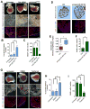
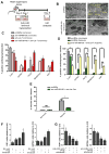
Figure 6. Myocardial tissue can be induced to a partially dedifferentiated proliferative state by anti-miR treatment
(A) Diagram describing organotypic culture experiments: ventricles were obtained from wild-type mice, cut into slices and put in culture conditions. At 24 h, they were transduced with the antimiR constructs and evaluated by immunohistochemistry (IHC) analyses for cardiomyocte proliferation/dedifferentiation, after 7 days. (B) Representative pictures showing that sarcomeric disorganization was readily observed by electron microscopy after induced dedifferentiation in adult cardiac tissue. (C) Quantitative analyses of normoxic organotypic cultures demonstrating significant increases in FNTβ and SMARCA5, enhanced numbers of dedifferentiated cardiomyocytes -determined by Cx43 and GATA4 expression- and significantly increased numbers of proliferating cells, upon anti-miR treatment. (D) Quantitative evaluations of hypoxic organotypic cultures demonstrating cardiomyocyte dedifferentiation upon anti-miR treatment as indicated by GATA4 re-expression, reduced number of Cx43+ cells and increased Histone 3 phosphorylation (H3P). (E) Histomorphometric evaluation of the damaged myocardium in normoxic and hypoxic conditions after Masson’s trichrome staining. (F) Lentiviral overexpression of Fntb, Smarca5 or both is sufficient to impose a dedifferentiated profile in neonatal murine cardiomyocytes. (G) In primary neonatal murine cardiomyocytes, knock-down of miR 99/100 and Let-7a/c is insufficient to promote a dedifferentiated state in the absence of Fntb/Smarca5 (knocked-down with siRNAs), suggesting these two proteins are mostly responsible for the effects of the miRs. In G, dashed lines represent the baseline values in the untreated condition (absolute values being 8% H3P+ and 2.5% GATA4+ cells). Data are represented as mean +/− s.e.m. *p<0.05. n = 4 independent experiments/condition, 3 sections/experiment/condition were used for quantitative analyses. See also Figure S6.
Figure 7
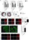
Figure 7. miR-99/100 and Let-7 silencing is sufficient to induce heart regeneration in a murine model of myocardial infarction
(A) AAV2/9 (a serotype showing cardiomyocyte specificity) GFP-mediated anti-miR-99/100 and anti-Let-7 in vivo delivery in a mouse model of myocardial infarction (MI) resulted in the significant improvement of fractional shortening (FS, left panel), ejection fraction (EF, middle panel) and left ventricular anterolateral wall thickness (LVAW, right panel) at 14 and 90 days post-infarction (dpi) as compared to AAV2/9-GFP scrambled injected animals. (B, C) Reduced infarct size in anti-miR treated animals was confirmed by Masson’s trichromic staining at 90 dpi. Representative pictures are depicted in B, and the quantitative analysis is showed in C. (D–F) Immunofluorescent analysis of cardiac tissue at 18 dpi indicating that functional recovery was accompanied by re-expression of FNTβ (D) and SMARCA5 (E), as well as cardiomyocyte dedifferentiation as indicated by GATA4 re-expression (F). (G) Quantitative evaluation of cTnT+ cardiomyocytes (CMs) expressing GATA4 upon anti-miR treatment in vivo. (H–I) Qualitative (H) and quantitative (I) analysis demonstrated a significant increase in the number of BrdU positive cardiomyocytes upon anti-miR delivery. (J) Representative pictures demonstrating cytokinesis in cTnT+ cardiomyocytes from anti-miRs treated animals compared to control animals, as evaluated by anillin and aurora B kinase staining. Data are represented as mean ± s.e.m. *p<0.05. Arrowheads: cells of interest. n = 8 animals/group (14dpi); n = 7 animals/group (90dpi). In all cases, three different sections per animal were utilized for quantitative analyses. See also Figure S7.

Comment in

Similar articles

Cited by

References

    1. Abad M, Mosteiro L, Pantoja C, Cañamero M, Rayon T, Ors I, Graña O, Megías D, Domínguez O, Martínez D, et al. Reprogramming in vivo produces teratomas and iPS cells with totipotency features. Nature. 2013;502:340–345. - PubMed
    1. Addis RC, Epstein Ja. Induced regeneration-the progress and promise of direct reprogramming for heart repair. Nat Med. 2013;19:829–836. - PMC - PubMed
    1. Aguirre A, Sancho-Martinez I, Izpisua Belmonte JC. Reprogramming toward heart regeneration: stem cells and beyond. Cell Stem Cell. 2013;12:275–284. - PubMed
    1. Andersen DC, Ganesalingam S, Jensen CH, Sheikh SP. Do Neonatal Mouse Hearts Regenerate following Heart Apex Resection? Stem Cell Reports. 2014;2:406–413. - PMC - PubMed
    1. Bersell K, Arab S, Haring B, Kühn B. Neuregulin1/ErbB4 signaling induces cardiomyocyte proliferation and repair of heart injury. Cell. 2009;138:257–270. - PubMed

Publication types

Associated data